2. www.AJOG.org General Gynecology Research
imaging examinations, therapeutic pro-
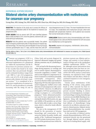
FIGURE 1
cedures, blood loss, and findings at
Transvaginal sonogram of the cesarean scar pregnancy follow-up.
In all patients, the gestational age was
estimated according to the last men-
strual period and ultrasonographic exam-
inations, and serum -human chorionic
gonadotrophin (hCG) concentration was
determined before treatment. The diagno-
ses of CSP were based on symptoms, clin-
ical manifestations, history of prior cesar-
ean section, serum -hCG concentration,
and special presentation on transvaginal
ultrasonography.
The criteria of ultrasound diagnosis
include the following: (1) an empty uter-
ine cavity and cervical canal; (2) a gesta-
tional sac located at the anterior wall of
the isthmic portion, separated from the
endometrial cavity or fallopian tube; (3)
a gestational sac embedded within the
myometrium and the fibrous tissue of
the cesarean section scar at the lower
uterine segment, with an absence of de-
fect in the myometrium between the
bladder and the sac; and (4) and a high-
velocity–low-impedance vascular flow
surrounding the gestation sac.1,2,14 All 46
cases matched these criteria (Figure 1).
The UAE procedure was performed by
Transvaginal sonogram of the cesarean scar pregnancy, showing the empty uterine cavity and experienced radiologists. After local an-
the empty cervical canal and the gestational sac implanted into the previous cesarean section esthesia, catheterization was carried out
scar at the anterior uterine wall and protruding toward the urinary bladder, with rich surrounded via the right femoral artery with a
vascularity. 5F-Yashiro catheter (Terumo, Tokyo, Ja-
Shen. Bilateral uterine artery chemoembolization with methotrexate for cesarean scar pregnancy. Am J Obstet Gynecol 2012.
pan) that was advanced into the uterine
arteries on both sides; digital subtraction
arteriography (AXIOM-Artis-FA; Sie-
subsequent blockage of the feeding vessel M ATERIALS AND M ETHODS mens AG, Munich, Germany) was then
by occlusive agents that are injected The research protocol was approved by performed to confirm that catheters
through the delivery catheter. Because the institutional review board of West were correctly inserted, and 25 mg of
this involves both chemotherapy and tis- China Second University Hospital, Sich- MTX was injected bilaterally; and finally
sue ischemia, it permits a higher concen- uan University. Informed consent was both uterine arteries were embolized
tration of MTX to target the gestational obtained from all patients, and all avail- with gelatin sponge particles (0.5-1.0
foci for a longer period of time and thus able information on the treatments was mm). Subsequently, postembolization
produces more effective embryocide, presented to the patients, including the angiography was performed to validate
with much less systemic toxic effects, risks and benefits of the therapy, poten- that the vascularity of the gestational sac
than embolization alone. To date, only a tial complications, and alternatives. was completely obstructed (Figure 2).
few reports that describe uterine artery Between March 2008 and March 2010, Twenty-four to 72 hours later, the pa-
chemoembolization with MTX for CSP 46 patients were diagnosed with CSP in tients were carefully examined using
treatment11-13 are available. our hospital. We reviewed the clinic re- transvaginal ultrasound, and their serum
We retrospectively reviewed our man- cords of all these patients, including pa- -hCG levels were assessed. In patients
agement with bilateral uterine arterial tient age, gravidity and parity, clinical with persistent vaginal bleeding and/or a
chemoembolization with MTX of 46 presentation, weeks of gestation, the persistent gestational mass larger than 5
cases of CSP over a 2 year period and time interval between the last cesarean cm, suction curettage was performed
analyzed complications and quality of section and cesarean scar pregnancy, under transabdominal ultrasound guid-
life after treatment. clinical findings, results of ultrasound ance after ultrasonic confirmation of the
NOVEMBER 2012 American Journal of Obstetrics & Gynecology 386.e2
3. Research General Gynecology www.AJOG.org
absence of blood flow to the CSP region
FIGURE 2
to remove the retained products of con-
Angiograms of a patient with CSP who received transcatheter UAE
ception and blood clot. If massive hem-
orrhage occurred during investigation or
curettage, an emergency hysterectomy
or local CSP resection was carried out.
Patients were hospitalized during
treatment. Serum -hCG levels, blood
loss, adverse effects (including fever,
nausea and vomiting, abdominal or pel-
vic pain, and abnormal liver or renal
function), and length of hospital stay
were recorded and summarized. Serum
-hCG levels were determined before
the intervention, on day 1 after therapy,
every 3 days until discharged from the
hospital, and then every week until re-
covery to normality. At the same time,
the sizes of the retained gestational prod-
ucts were measured by ultrasound and
clinical status (bleeding pattern and re-
sumption of menses) were assessed.
Follow-up was arranged until the se-
rum -hCG concentration dropped to
normal and pregnancy remnants could
not be detected through ultrasound.
Women who had massive, active vaginal
bleeding and stable serum -hCG con-
centration after UAE were diagnosed as
having their treatment failed and that re-
quired repeat embolization or partial/ Digital subtraction angiograms of a patient with CSP who received transcatheter uterine arterial
subtotal hysterectomy. embolization. A and B, Angiography before embolization. The uterus is enlarged, bilateral uterine
Successful UAE treatment was defined artery is hypertrophied and tortuous, and the gestational sac is surrounded by numerous artery
as a complete recovery without severe branches. C and D, Angiography after embolization. Both uterine arteries are obstructed and the
adverse effects or complications and vascularity of the gestational sac completely disappeared.
CSP, cesarean scar pregnancies; UAE, uterine arteries embolization.
without a need for repeat embolization
Shen. Bilateral uterine artery chemoembolization with methotrexate for cesarean scar pregnancy. Am J Obstet Gynecol 2012.
or hysterectomy.
All data are expressed as mean Ϯ SD.
Statistical analysis was performed using presentation was 55.5 Ϯ 2.4 (37-97) days tion as primary treatment, followed by
the Student t test and a 2 test by the SPSS (Table). suction curettage after 72 hours. The time
19.0 statistical package (SPSS Inc, Chi- Twenty-five women were initially di- of hospital stay was 10.1 Ϯ 1.0 (4-28) days.
cago, IL). agnosed with CSP on admission to our The time for serum -hCG normalization
hospital. The main complaints were ir- was 32.0 Ϯ 5.5 (7-134) days. The time for
regular vaginal bleeding (19 of 25, total lesion disappearance was 32.7 Ϯ 4.0
R ESULTS 76.0%) and mild lower abdominal pain (5-58) days. All these patients experienced
Forty-six cases of CSP were diagnosed (7 of 25, 28.0%). The serum -hCG a rapid, uneventful recovery.
over a 2 year period. The average age of concentration was 28,220.2 Ϯ 7104.4 The remaining 21 patients received
the 46 patients was 32.7 Ϯ 6.0 (21-44) (161.6-181,880) mIU/mL. By ultrosonog- suction curettage at their first visit to
years. The average gravidity was 5.0 Ϯ raphy, the largest diameter of the CSP mass other hospitals because of misdiagnosis
1.6 (2-8) and the average parity was was 1.0-7.6 cm, the embryo within the ges- for inevitable miscarriage or missed
1.09 Ϯ 0.28 (1-3). Four women had un- tational sac could be seen in 18 patients, abortion. They were transferred to our
dergone 2 previous cesarean deliveries. and 8 had fetal cardiac activity; in the re- hospital owing to massive hemorrhage
The average interval from the last cesar- maining 7 patients, only a yolk sac was during the operation. On admission to
ean section was 63.5 Ϯ 8.2 (4-252) identified. All these 25 women underwent our hospital, bilateral uterine artery che-
months. The average gestational age at bilateral uterine artery chemoemboliza- moembolization was performed with
386.e3 American Journal of Obstetrics & Gynecology NOVEMBER 2012