
Recomendados
Más contenido relacionado
La actualidad más candente
La actualidad más candente (20)
Similar a Skull copy
Similar a Skull copy (20)
Más de Ayshah Hashimi
Último
Último (20)
Skull copy
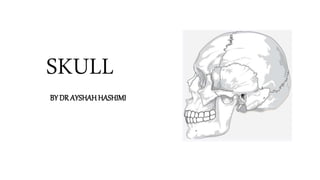
- 1. SKULL BY DR AYSHAH HASHIMI
- 2. • The skull is a bony structure that forms the head. It supports the structures of the face and provides a protective cavity for the brain • The skull is made up of a 22 fused flat bones • Contains many foramina, fossae, processes, and several cavities or sinuses • Bones in skull develops from intramembranous ossification • The joint in the skull are mostly sutures and few are 1° cartilaginous • Only temporomandibular joint is synovial in nature that permits us to speak, eat, drink and laugh • Skull lodges brain, cochlear and vestibular apparatus, retina, olfactory mucosa and taste buds
- 3. Skull is divided into two parts • Cranium/Braincase/Calvaria/skullcap • 8 bones forms the cranium 2 Parietal 2 Temporal Frontal Occipital Sphenoid Ethmoid Bone Frontal Bone Parietal Bone Temporal Bone Sphenoid Occipital Bone Ethmoid
- 4. • Facial skeleton composed of 14 bones 2 Maxilla 2 Zygomatic 2 Nasal 2 Lacrimal 2 Palatine 2 Inferior Nasal Concha Mandible Vomer
- 5. Anatomy of major bones of skull • Frontal bone • Parietal bone • Occipital bone • Temporal bone • Sphenoid bone • Ethmoid bone • Mandible • Maxilla • Zygomatic
- 6. Frontal Bone • Creates the smooth curvature of the forehead • Protects the frontal lobe of the brain • Involved in the three regions of the head, such are the squamous part(forehead), the orbital part and the nasal part Squamous Part forms the major portion of the bone and forms the forehead Orbital part forms the roof of the orbit and ethmoidal sinus Nasal part forms the stem of the nose
- 7. Squamous Part comprises of • Frontal sinuses present superior and medially to orbit • Glabella (elevated surface above the nasal root) • Zygomatic processes of frontal bone • Supraorbital foramen • Supraorbital notch Frontal sinus Glabella Zygomatic processes Supraorbital foramen Supraorbital notch
- 8. Orbital part includes anterior and posterior ethmoidal foramen Nasal part comprises of nasal process that’s ends below in a sharp spine Anterior Ethmoidal Foramen Posterior Ethmoidal Foramen
- 9. Surface Landmarks In Frontal Bone Externally frontal bone includes- Frontal eminence, superciliary arch, glabella, supraorbital margin(frontal notch, supraorbital notch), zygomatic process, temporal line, temporal surface
- 10. Parietal Bone • Located on each side of the skull behind the frontal bone • Irregular quadrilateral in shape- four angles, four margins, and two surfaces • Angles - frontal angle, sphenoidal angle, occipital angle, mastoid angle, • Margins/Border- frontal, occipital, squamous, sagittal • External surface features- parietal tuber, superior temporal line, inferior temporal line, parietal foramen • Internal surface has grooves for sinuses
- 11. Occipital Bone • The occipital bone is an unpaired bone which covers the back of the head • Convex externally and concave internally • Protects cerebellum • It has four parts: the basilar part, two condylar parts and the squamous part • All four are arranged around a large opening, the foramen magnum SQUAMOUS PART CONDYLAR PART BASILAR PART
- 12. THE BASILAR PART • Anterior to the foramen magnum • Anteriorly it fuses with the sphenoid bone to form the clivus • Pharyngeal tubercle found on the inferior surface of the basilar part CLIVUS
- 13. Condylar Parts • Located lateral to the foramen magnum • kidney shaped prominences • Articulate with the first cervical vertebra (atlanto-occipital joint) • Posterior to condyles are the condylar canals where the condylar emissary veins passes Condylar Canals
- 14. Squamous Part • External occipital protuberance in the midline • Highest nuchal line • Superior nuchal line • Inferior nuchal line
- 15. Temporal Bone • Pair of bilateral • Many number of openings and canals • Houses the structures forming the middle and inner ear • It has 4 parts- squamous, petrous , mastoid and tympanic part
- 16. Squamous Part • Antero-superior portion of the temporal bone • Zygomatic process- anterior projection from the squamous part of temporal bone (articulate with the temporal process of the zygomatic bone to form the zygomatic arch) • Below the process is a mandibular fossa that articulate with the mandible to form temporomandibular joint • Articular tubercle is an elevation in front of the mandibular fossa External Acoustic Meatus Zygomatic Arch Zygomatic Process Mandibular Fossa Temporomandibular Joint Articular Tubercle
- 17. Mastoid Part • Most posterior part of the temporal bone • Downward conical projection called the Mastoid Process • Mastoid process consists of mastoid air cells (reservoir of air, equalize pressure within the middle ear) and mastoid antrum • Various muscles are attached to it (SCM etc. ) • Mastoid notch is present on inferior surface of the mastoid process
- 18. Petrous Part • Pyramid shaped mass of bone located between the sphenoid and occipital bones within the cranial cavity • It has a base, an apex and three surfaces (anterior, posterior, inferior), three borders (anterior, superior, posterior) • The acoustic labyrinth is located within the petrous part (houses the inner ear) • Trigeminal impression on anterior margin • Tegmen Tympani forms the roof of the middle ear Petrous Part
- 19. Tympanic Part • Curved plate immediately below the origin of the zygomatic process • Forms the anterior wall, floor and part of the posterior wall of the external acoustic meatus • The styloid process is a narrow, pointed projection that extends downwards and anteriorly from the inferior surface of the temporal bone
- 20. Sphenoid Bone • Butterfly-shaped • It consists of a body, paired greater wings and lesser wings, and two pterygoid processes • Various foramen found- F. Rotundum, F. Ovale, F. Spinosum. Optic Canal and Superior orbital fissure Body Greater Wing Lesser Wing Pterygoid Processes
- 21. Body of Sphenoid Bone • Six surfaces: superior, posterior, anterior, inferior, and two lateral surfaces • Superior surface- comprises of Sella turcica that lodge pituitary gland
- 22. Lesser wings • Forms lateral wall of Optic canal (transmit 2nd optic nerve and ophthalmic artery) • superior orbital fissure-slit-like’ gap between the lesser and greater wings (transmit ophthalmic vein and 5thnerve, 6th abducent nerve, 3rd oculomotor nerve, 4th trochlear nerve) Greater wings • Foramen rotundum, which transmits the 5th maxillary nerve • Foramen ovale, which allows the passage of the 5th mandibular nerve, accessory meningeal artery, lesser petrosal nerve and emissary vein (mnemonic "MALE") • Foramen spinosum
- 23. Pterygoid Processes • Extensions of the basal surface of the sphenoid body • Processes contain two canals known as the pterygoid canal and pharyngeal canal • Pterygoid canal transmit major petrosal nerve, deep petrosal nerve and vessels • Pharyngeal canal transmit pharyngeal nerve Pterygoid Canal
- 24. Ethmoid Bone • Single porous bone • The ethmoid bone has four parts: Cribriform plate Perpendicular plate Ethmoidal labyrinth (2)
- 25. Ethmoidal labyrinths Features consists of - • Ethmoidal Air Cells (Anterior, Middle, Posterior) • Orbital Plate • Superior Nasal Concha • Middle Nasal Concha Perpendicular plate • Forms the nasal septum
- 26. Cribriform Plate of Ethmoid • Forms the roof of the nasal cavity • Has small openings which transmit the fibers of the olfactory nerves (CN I) from the olfactory epithelium (nasal cavity) to the brain (cranial cavity) • A small vertical protrusion on top of the plate is called as Crista galli
- 27. MANDIBLE • Also known as the lower jaw • Located inferiorly in the facial skeleton • Largest and strongest bone of the face • Doesn’t articulate with its adjacent skull bones via sutures • Articulates to the temporal bone, forming the Temporomandibular joint (TMJ) • Movement of the lower jaw opens and closes the mouth and also allows for the chewing of food. • Muscles of mastication attach to it
- 28. Features Consist of • Body • Ramus • Foramina
- 29. Ramus • Extends cranially from the angle of the mandible, away from the body at an angle of 110 degrees • Each ramus contains the following bony landmarks Angle of mandible Coronoid process Mandibular notch Condylar process Head Neck Pterygoid fovea • 2 surfaces External - masseteric tuberosity Internal- mandibular foramen, mandibular canal, mylohyoid groove, pterygoid tuberosity Mandibular Notch
- 31. Body Alveolar process: holds the teeth Base External surface • Mandibular symphysis: small ridge in the midline • Mental protuberance: forms the shape of the chin • Mental tubercle • Mental foramen (located below the second premolar) Internal surface • Mental spine (attachment of genioglossus and geniohyoid muscles.) • Digastric fossa (attachment of digastric muscle) • Mylohyoid line (mylohyoid muscle) • Sublingual fovea (contains sublingual gland) • Submandibular fovea (submandibular gland)
- 32. Maxilla Bone • Upper jaw bone • Paired bone Features • Body • Four processes Frontal process Zygomatic process Palatine process Alveolar process • Involved in the formation of the orbit, nose and palate, holds the upper teeth • Plays an important role for mastication and communication
- 33. Processes • Frontal process • Zygomatic process • Palatine process • Alveolar process Frontal process Zygomatic process Palatine process Alveolar process
- 34. Body • Central portion of the maxilla • Four surfaces Anterior Nasal notch Infra-orbital margin Infra-orbital foramen Canine fossa Orbital (floor of the orbit) Nasal (lateral wall of the nasal cavity) Maxillary sinus Infratemporal Maxillary tuberosity Alveolar foramina Alveolar canals Nasal notch Infra-orbital margin Infra-orbital margin Infra-orbital foramen Infra-orbital foramen Canine fossa Maxillary tuberosity Maxillary tuberosity Alveolar foramina Maxillary sinus
- 35. Zygomatic Bone • Also called as cheek bone, malar bone • Articulates with four bones Maxilla Temporal bone Sphenoid Frontal bone
- 36. Surfaces • Orbital • Temporal • Lateral (zygomaticofacial foramen) Processes • frontal process • temporal process
Notas del editor
- Structures that pass through this foramen magnum include: the brainstem (medulla oblongata), spinal branch of accessory nerve, anterior and posterior spinal arteries, vertebral artery, spinal vein
- Angle of mandible: angle forming between the body and ramus of the mandible Coronoid process: muscular process located anteriorly, temporalis muscle attaches to it Masseteric tuberosity is for masseter muscle attachment Mandibular canal is a bony passage that transmits the inferior alveolar artery and nerve Mylohyoid groove is passage for the mylohyoid nerve and the mylohyoid branch of the inferior alveolar artery Pterygoid tuberosity gives attachment to medial pterygoid muscle