2018 BDSRA Storch CLN7
•
1 recomendación•214 vistas

Using Mouse Models to develop new therapies for CLN7 disease

Recomendados
Más contenido relacionado
Similar a 2018 BDSRA Storch CLN7
Similar a 2018 BDSRA Storch CLN7 (20)
Más de Batten Disease Support and Research Association
Más de Batten Disease Support and Research Association (20)
Último
Último (20)
2018 BDSRA Storch CLN7
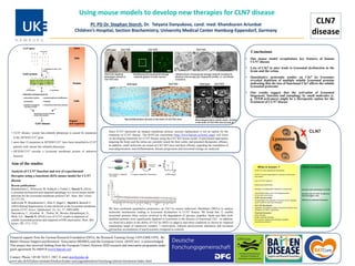
- 1. CLN7 disease Using mouse models to develop new therapies for CLN7 disease PI: PD Dr. Stephan Storch, Dr. Tatyana Danyukova, cand. med. Khandsuren Ariunbat Children’s Hospital, Section Biochemistry, University Medical Center Hamburg-Eppendorf, Germany Financial support from the German Research Foundation (DFG), the Research Training Group 1459 (GRK1459), the Batten Disease Support and Research Association (BDSRA) and the European Union (BATCure) is acknowledged. This project has received funding from the European Union's Horizon 2020 research and innovation programme under grant agreement No 666918 (www.batcure.eu). Contact: Phone +49 40 7410 5 1967, E-mail storch@uke.de www.uke.de/kliniken-institute/kliniken/kinder-und-jugendmedizin/forschung/sektion-biochemie/index.html Conclusions • Our mouse model recapitulates key features of human CLN7 disease • Loss of Cln7 in mice leads to lysosomal dysfunction in the brain and the retina • Quantitative proteomic studies on Cln7 ko lysosomes revealed depletion of multiple soluble lysosomal proteins indicating that the loss of functional Cln7 affects the soluble lysosomal proteome • Our results suggest that the activation of lysosomal biogenesis, function and autophagy by small molecules (e. g. TFEB activators) might be a therapeutic option for the treatment of CLN7 disease • CLN7 disease, variant late-infantile phenotype is caused by mutations in the MFSD8/CLN7 gene • more than 35 mutations in MFSD8/CLN7 have been identified in CLN7 patients with variant late infantile phenotype • MFSD8/CLN7 encodes a lysosomal membrane protein of unknown function Aim of the studies Analysis of CLN7 function and test of experimental therapies using a knockout (KO) mouse model for CLN7 disease Recent publications: Brandenstein L, Schweizer M, Sedlacik J, Fiehler J, Storch S. (2016) Lysosomal dysfunction and impaired autophagy in a novel mouse model deficient for the lysosomal membrane protein Cln7. Hum. Mol. Genet. 25:777-791. Jankowiak W, Brandenstein L, Dulz S, Hagel C, Storch S, Bartsch U. (2016) Retinal degeneration in mice deficient in the lysosomal membrane protein CLN7. Invest. Ophthalmol. Vis. Sci. 57: 4989-4998. Danyukova, T., Ariunbat , K., Thelen, M., Brocke-Ahmadinejad, N., Mole, S.E., Storch, S. (2018) Loss of CLN7 results in depletion of soluble lysosomal proteins and impaired mTOR reactivation. Hum. Mol. Genet., 27: 1711-1722. Since CLN7 represents an integral membrane protein, enzyme replacement is not an option for the treatment of CLN7 disease. The BATCure consortium (http://www.batcure.eu/home_page) will focus on developing treatments for CLN7 disease using the Cln7 KO mouse model. Experimental approaches targeting the brain and the retina are currently tested for their safety and potential therapeutic efficacy. In addition, small molecules are tested on Cln7 KO mice and their efficacy regarding the retardation of neurodegeneration, neuroinflammation, disease progression and lysosomal storage are analyzed. We have perfomed quantitative proteomics on Cln7 ko mouse embryonic fibroblasts (MEFs) to analyse molecular mechanisms leading to lysosomal dysfunction in CLN7 disease. We found that 12 soluble lysosomal proteins (blue circles) involved in the degradation of glycans, peptides, lipids and fatty acid- modified proteins were significantly depleted in lysosomes in the absence of functional Cln7. In addition, we observed a defect in the ability of Cln7 ko MEFs to adapt to starvation conditions as shown by impaired mammalian target of rapamycin complex 1 reactivation, reduced autolysosome tubulation and increased perinuclear accumulation of autolysosomes compared to controls.