Anatomy and physiology of lens
•Download as PPTX, PDF•
4 likes•1,788 views

prepared during lens session

Recommended
More Related Content
What's hot
What's hot (20)
Similar to Anatomy and physiology of lens
Similar to Anatomy and physiology of lens (20)
More from Bipin Bista
More from Bipin Bista (20)
Recently uploaded
Recently uploaded (20)
Anatomy and physiology of lens
- 1. ANATOMY AND PHYSIOLOGY OF LENS Bipin Bista Resident Ophthalmology
- 2. INTRODUCTION Emanation theory of vision Celsus (25BC-AD 50) drew the lens in the center of the eyeball with an empty space called locus vacuus anterior to it True position of the lens was illustrated by the Italian anatomist Fabricus ab Aquapendente in1600 and Swiss physician Felix Plater (1536-1614) postulated to be the part responsible for sight 20 million of people (51%) are suffering from cataract (WHO 2010 A.D) Prevalence of lens disorder and continuing developments in their management make the basic and clinical science about lens an important subject.
- 3. EMBRYOLOGY At 25 days of gestation , 2 lateral evaginations (Optic vesicles) form the forebrain, or diencephalon . As the OV enlarges and extend laterally, they become closely apposed and adherent to surface ectoderm (single layer of cuboidal cell) in 2 patches in either side of the head. Lens Placode : ectodermal cell overlie the optic vesicle become columnar at 27 days and are called LP. For subsequent formation Bone Morphogenetic Protein are required. Lens Pit : appear at 29 days as an indentation (infolding) of the Lens Placode, this pit deepens and invaginates to form the lens vesicle.
- 5. EMBRYOLOGY Lens Vesicle : stalks of cells connecting to the surface ectoderm degenerates by apoptosis separating the lens cells from the surface ectoderm. This resultant sphere, a single layer of cuboidal cells encased in BM (lens capsule) is LV. As the lens vesicle was formed through invagination the apices of the cuboidal cells are oriented toward the lumen of the vesicle , with the base of the cell attached to the capsule around the periphery of the vesicle. At the same time, the optic vesicle is invaginating to form the 2 layered optic cup.
- 6. EMBRYOLOGY Primary lens fibers and the embryonic nucleus : Cells in posterior layer of the lens vesicle stop dividing and begin to elongate they begin to fill the lumen of the lens vesicle, and approximately 40 days of gestation the lumen of the lens vesicle is obliterated .these elongated cells are primary lens fibers which make up the embryonic nucleus which will ultimately the central area. Cells of the anterior lens vesicle remain as a monolayer of cuboidal cells – Lens epithelium responsible for subsequent growth of the lens. Lens capsule develops as a BM elaborated by the lens epithelium anteriorly and by lens
- 7. EMBRYOLOGY Secondary lens fibers : with the proliferation, the epithelial cells near the lens equator elongate and form secondary lens fibers. Layer upon layer the lens fibers are formed anteriorly beneath the epithelium and posteriorly toward the posterior pole. These fibers formed between 2 and 8 months make up fetal nucleus.
- 8. EMBRYOLOGY Lens sutures and fetal nucleus : As the fibers grow anteriorly and posteriorly, and the ends of the fibers meet and interdigitate with the ends of fibers arising of the lens make a pattern – Sutures which are recognisable at about 8 weeks . As the lens continues to grow the pattern of the lens continues to grow the pattern of the lens sutures become complex, resulting in 12 or more sutures.
- 9. TUNICA VASCULOSA LENTIS At about 1 month of gestation , the hyaloid artery enters eye and forms network of capillaries (TVL) on the posterior surface of the lens capsule. These capillaries grow toward the equator of the lens , where they anastomose with a second network of capillaries, Anterior pupillary membrane derived from the ciliary veins and which covers the anterior surface of the lens After complete development at 9 weeks it disappears by an orderly process of Apoptosis.
- 10. ZONULES OF ZINN Secreted by the ciliary epithelium, although insertion into lens capsule is not known. Develops at the end of the third month of gestation. Arrangements : •Main zonular fibers : Pars orbicularis, zonular plexuses, Zonular fork, Zonular limbs •Zonular limbs : Anterior zonular limbs, equatorial limbs, posterior limbs. •Hyaloid zonule •Hyalocapsular zonule •Circumferential zonular girdle : anterior and posterior.
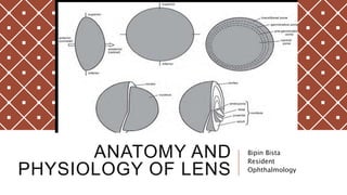
- 11. ANATOMY Transparent, biconvex structure which has no blood supply or innervation after fetal development, depends on aqueous humor for its metabolic wastes. Lens is suspended in position by the zonules of zinn. Lens is composed of the capsule, lens epithelium, cortex, and the nucleus. Anterior and posterior pole are joined by imaginary line called as the optical axis. Line passing from one pole to other are meridians. The greatest circumference comes from the equator of the lens.
- 13. ANATOMY At birth, it measures 6.4 mm equatorially and 3.5 mm anteroposteriorly and weighs 90 mg, adult lens typically measures 9 mm equatorially and 5 mm anteroposteriorly and weighs 255 mg. Capsule : elastic, transparent BM composed of type IV collagen, capable of molding it during accommodative changes. Zonular lamella is the outer layer of capsule and is the point of attachment for zonular fibers. Anterior > posterior Zonular fibers / zonules of fibers : consist of microfibrils composed of elastic tissue, originate from basal laminae of non pigmented epithelium of pars plana and pars plicata. Gets inserted in a continuous fashion, on the capsule in the equatorial region. 1.5 mm onto anterior capsule and 1.25 mm onto posterior. 5-30 µm in diameter shows eosinophilic structures that have positive PAS reaction.
- 14. ANATOMY
- 15. ANATOMY Lens epithelium : single layer , metabolically active and carry biosynthesis of DNA, RNA, protein, and lipid. Also generate ATP to meet energy demands of the lens. These cells are mitotic , premitotic activity occurring the ring around the anterior lens K/A germinative zone. Newly formed cells migrate toward the equator where they differentiate into fibers. With advancement, epithelial cells elongate to form lens epithelial cells, which leads to increase in the mass of cellular proteins. These cells start losing cell organelles which leds the lens to be less absorbant and will have less scattering of light but gets dependent on glycolysis for energy production. Nucleus and Cortex : outermost fibers are recently made and make up cortex, whereas the older fibers get crowded and are usually located in the center. There is no morphological distinctions between the nucleus, epinucleus, and the cortex.
- 16. CONGENITAL ANOMALIES AND ABNORMALITIES Congenital aphakia Lenticonus and lentiglobus Lens coloboma Mittendorf dot Epicapsular star Peters anomaly Microspherophakia Aniridia Cataract
- 17. DEVELOPMENTAL DEFECTS Ectopia lentis Marfan syndrome Homocystinuria Hyperlysinemia Ectopia lentis et pupillae Persistent Fetal Vasculature
- 18. PHYSIOLOGY Continual growth of lens Epithelium and outer cortex : site for highest metabolism Utilises glucose and oxygen for the active transport of electrolytes, carbohydrates and aminoacids into the lens. Communications through gap junction between newer and older cells and also through Major Intrinsic Protein (MIP) Minimise the extracellular space between fiber cells .
- 19. PHYSIOLOGY – MAINTENANCE OF LENS WATER AND CATION BALANCECritical for lens transparency Imbalance in cellular hydration may lead to opacification Normally 66% water and 33% protein Cortex > nucleus (Hydrated) 5% is found between the fibers 20 mM and 120 Mm are the concentrations of sodium and potassium respectively.
- 20. LENS EPITHELIUM : SITE OF ACTIVE TRANSPORT Lens is dehydrated and has higher level of potassium ions and amino acids than surrounding aqueous and vitreous and lower level of sodium and chloride ion Cation balance is result of permeability of lens cell membrane and activity of the sodium pumps that reside within the cell membranes of lens epithelium and each lens fiber Sodium pumps functions by pumping sodium out and potassium in which is dependent on the breakdown of ATP is regulated by the enzyme Na+ ,K+ -ATPase. Inhibition of this enzyme may lead to loss of cation balance and elevated water content in water.
- 21. PUMP LEAK THEORY Referred to combination of active transport and membrane permeability Potassium and various other molecules like AA are actively transported into the lens via the epithelium anteriorly, then diffuse out with concentration gradient through the back of the lens (no active transport mechanism) Sodium flows in through the back of the lens with concentration gradient and then is actively exchanged for potassium by the epithelium. Most of the Na+ , K+ -ATPase are found in the epithelium and the superficial cortical fiber cells
- 22. PUMP LEAK THEORY Electrical potential is generated d/t unequal distribution of electrolytes across the lens cell membranes. Inside of lens is electronegative, approximately -70 mV Potential difference between anterior and posterior surface of lens is - 23mV. Calcium pump plays critical role to lens, lens epithelial cells calcium level is 100 nanomolars maintained by Ca2+ -ATPase Glucose enters through facilated diffusion and waste product leave by simple dissusion.
- 24. ACCOMMODATION Mechanism by which the eye changes focus from distant to near images, is produced by a change in lens shape resulting from the action of the ciliary muscle on the zonular fibers According to the Helmholtz theory of accommodation, most of the accommodative changes in the lens occurs at the central anterior lens surface. There is central anterior bulge with accommodation as the anterior zonular fibers are inserted slightly closer to the visual axis Ciliary muscle is a ring shaped muscle that on contraction has the opposite effect as the muscle contracts the diameter of the muscle ring is reduced , thereby relaxing the tension on the zonular fibers and allowing the lens to become more spherical. When CM relaxes zonular tension increases, zonular tension increases ,lens flattens and diopteric power decreases.
- 25. ACCOMMODATION Stimulated by the known or apparent size and distant of an object or blur, chromatic aberration , or a continual oscillation of ciliary tone. Mediated by parasympathetic fibers of CN III Amplitude of accommodation : amount of change in the eye’s refractive power that is produced by accommodation. Diminishes with age , medications and diseases. Adolescents have accommodative power have 12-16 D of accommodation, whereas adults at age have 4-8 D. After age 50, accommodation decreases to less than 2D.
- 26. PRESBYOPIA Loss of accommodation d/t hardening of the lens . With the age above 40 the rigidity of the lens reduces rigidity, as contraction of ciliary muscle would no longer result in increased convexity and diopteric power of the anterior surface of the lens. - Changes in the elastic property of lens capsule - Sclerosis or hardening of the lens - Weakening of the ciliary
- 27. REFERENCES : - Ophthalmology : Yanoff and Duker : 4th Edition -American academy of ophthalmology : section 11 : 2014-2015
- 28. THANK YOU