
Recommended
Recommended
More Related Content
What's hot
What's hot (20)
Viewers also liked
Viewers also liked (9)
Similar to Gingival recession—can orthodontics be a cure? evidence from a case presentation
Similar to Gingival recession—can orthodontics be a cure? evidence from a case presentation (20)
Recently uploaded
Recently uploaded (20)
Gingival recession—can orthodontics be a cure? evidence from a case presentation
- 1. Case Report Gingival recession—can orthodontics be a cure? Evidence from a case presentation William M. Northwaya ABSTRACT Does orthodontic treatment help or hinder a patient’s periodontal status? What factors affect the periodontium? Can those factors be managed in a way that remedies existing periodontal issues? A 35-year-old woman presented with severe gingival recession and a unilateral Class II malocclusion. The treatment plan was to correct the malocclusion in a way that torques the roots more onto bone and to change her dental hygiene methods. With an extensive review of the literature, this case review attempts to make sense of the enigma of gingival recession and demonstrates an excellent treatment solution to concomitant orthodontic and periodontal problems. (Angle Orthod. 2013;83:1093–1101.) KEY WORDS: Gingival recession; Oscillating toothbrushing; Orthodontic root torque; CBCT; Digital study casts INTRODUCTION Contemporary orthodontics is more than biomechan- ical wizardry; it incorporates a concept of periodontal physiology as well. Perhaps first among the advocates of this breadth of approach was Vanarsdall.1 He taught that normal gingiva has a thick band of keratinized tissue that helps resist attrition and disease. So important is the health of this gingival band that he has advocated that its character should be a diagnostic criterion in determining whether tooth extraction is indicated in an orthodontic patient. A healthy tooth sits on alveolar bone with a healthy gingival apparatus. Dorfman2 wrote that the presence of keratinized tissue might be an indicator that mucogingival prob- lems will be less likely—even in cases where orthodontic proclining of the lower incisors might provide more fullness to the facial profile. Pearson3 tested the impact of tipping lower incisors on recession during orthodontic treatment and found that among his patients there was no correlation between the amount of root apex advancement or retraction and the degree of recession. In 2008, the featured cover article in the Journal of the American Dental Association was an exhaustive review of the literature that found that orthodontics provided little benefit to the periodontium; in fact, the net response was small but detrimental.4 The objective of this article is to discuss the relation- ship between the development of a healthy periodon- tium and the potential impact of orthodontics, to advocate for modifications in orthodontic treatment that might improve the periodontal prognosis, and to present a treatment report that demonstrates concom- itant orthodontic and periodontal benefits. According to Wennstrom et al.5 the gingiva is composed of dense, collagen-rich tissue covered by a keratinized epithelium that extends from the soft tissue margin to the mucogingival line. It comprises the free portion (which corresponds to the probing depth) and the attached portion. Ideally, the tooth will erupt on the crest of the alveolar arch, surrounded by dense, keratinized tissue that will become well established circumferentially. The gingival dimensions will grow as the tooth erupts. Wennstrom references Andlin- Sobocki6 and Bimstein and Eidelman,7 who contended that gingival height will increase with growth. Ainamo and Talari8 stated that ‘‘data on gingival dimensions in adults indicate that there is a tendency of increased apico-coronal width with age.’’ When it comes to response to tooth movement within the arch, Wenn- strom5 stated that ‘‘a more lingual positioning of the tooth results in an increase of the gingival height on the facial aspect with a coronal migration of the soft tissue margin. The opposite will occur when changing to a more facial position in the alveolar process.’’ When the a Private practice, Traverse City, Mich. Corresponding author: Dr William M. Northway, 12776 S West Bay Shore Dr, Traverse City, MI 49684 (e-mail: Northway@umich.edu) Accepted: April 2013. Submitted: January 2013. Published Online: June 7, 2013 G 2013 by The EH Angle Education and Research Foundation, Inc. DOI: 10.2319/012413-76.1 1093 Angle Orthodontist, Vol 83, No 6, 2013
- 2. marginal tissue is apical to the cementoenamel junction (CEJ), it is called gingival recession (GR). Gingival Recession The periodontium is the protecting barrier against disease; once it begins to break down, the tooth becomes susceptible to further destruction and loss. As Daprile et al.9 have shown, in modern society periodontal disease can begin as a result of poor oral hygiene, or it can be the result of exaggerated hygienic measures that abuse the gingiva. It can also result from an acute traumatic episode that does not heal properly. Chronic trauma can cause detachment of the gingival margin in the form of an irregular attachment of a frenum. More recently, there has been discussion of a genetically based susceptibility that is manifest in various types of gingival integrity, known as biotype (thick or thin).10 Although our presumed knowledge of the interde- pendence of orthodontics and GR seems to be ubiquitous, the specifics continue to evolve. Despite many studies on growth and the effects of orthodontics on the dentition, not until relatively recently has reference been made to the periodontium. In 1977, Bernimoulin and Curilovic11 debunked the correlation between GR and tooth mobility and the correlation between tooth mobility and alveolar bone dehiscence. While they found a positive, significant correlation between GR and bone dehiscence, they questioned the impact of trauma from occlusion. More germane to the realm of orthodontics, reces- sion can occur during development in the form of insufficient space for the dentition on an alveolar ridge. As the discrepancy between tooth mass and available space increases, teeth become more crowded and are ultimately pushed off the ridge, compromising the supporting bone. Thus, the likelihood of recession increases. The process begins in the form of dehis- cences or fenestrations. Once these have formed, the absence of alveolar bone makes it more difficult for healthy gingival tissue to persist in the form of a protective barrier. Staufer and Landmeser12 explored the effects of crowding on the dentition and found a degree of correlation among young patients between crowding and ‘‘tooth infractions’’ and ‘‘tooth fractures’’. Among adults crowding correlated with periodontal pockets and GR. They showed that in cases of more than 5 mm of crowding, GR of more than 3.5 mm was 12 times more likely. Richman13 examined 72 teeth from 25 consecutively treated patients with ‘‘facial clinical GR of more than 3 mm.’’ He used cone-beam computed tomography (CBCT) and showed that although all of the teeth were periodontally healthy, they all had significantly prom- inent facial tooth contours and associated alveolar bone dehiscences. He pointed out that conventional orthodontic space analysis ‘‘does not evaluate the buccolingual (sagittal) dimension of the tooth or associated alveolar bone.’’ He developed the radio- graphic supporting bone index (RSBI), which is the sagittal difference between the alveolar bone width, measured 2–3 mm apical to the CEJ, the width of the tooth measured at that level. He divided his sample into three groups, classes A, B, and C, which had varying amounts of bone support and consequential risk. The hypothesis he proposed was that orthodontic movement in the direction of risk would likely exacer- bate the periodontal status and cause GR. In his study, he found that more than 75% of the patients who have received premolar extraction ‘‘demonstrate clinically significant gingival recession, leading him to conclude that these patients were compromised from the outset by reduced RSBI.’’ Managing Recession Trying to become informed on the subject of GR is not unlike joining Alice in her visit to Wonderland. The closer one looks, the less clear the picture becomes. Daprile et al.9 reported on a 5-year study of dental students, wherein the number of subjects with at least one site of recession increased significantly—as did the total number of foci. What’s more, the percentage of affected sites increased with the level of oral hygiene education, and this increase was despite a reduction in harmful dental hygiene habits. Of the 174 fifteen-year-olds in the sample studied by Bjorn et al.,14 an astounding 62% showed some degree of GR on the labial of maxillary teeth. Interestingly, the group with an ‘‘unspecific’’ toothbrushing technique had fewer lesions than the group that used the ‘‘roll or vibratory technique.’’ The authors speculated that the difference might not have been the technique but the fact that the unspecific group was paying less attention to their oral hygiene. Loe et al.15 compared samples from Norway and Sri Lanka and found that more than 60% of Norwegian children had recession by age 20 (compared with 30% in Sri Lanka), and that increased to more than 90% at age 50 (compared with 100% of Sri Lankans by age 40). As the Norwegian recession was largely on the buccal and that of the Sri Lankan cohort was more generally distributed, the researchers conjectured that several factors were involved, that much of the Norwegian recession seemed to be more related to mechanical abrasion associated with excessive hy- giene systems as opposed to recession caused more by periodontal disease in the Sri Lankan sample. Khocht et al.16 also reported an increase in the 1094 NORTHWAY Angle Orthodontist, Vol 83, No 6, 2013
- 3. frequency of recession with age; regression analysis showed that recession increased 3.5% per decade. The researchers attributed the damage to the increased frequency of brushing and especially to hard bristles. Susin et al.17 examined 1460 urban Brazilians ranging in age from 14 to 103 years selected from various geographic areas and stratified by income level. Of these, 83.4% showed recession of 1 mm or more. The prevalence, extent, and severity of reces- sion were each correlated with age. Men older than 30 years showed significantly more recession than females, as did lower socioeconomic groups and those with less regular dental care. Cigarette smoking and the presence of supragingival calculus were statisti- cally significant contributors. The preponderant impact was that of destructive periodontal disease as opposed to mechanical abrasion. Although much has been made during the past few years of periodontal biotype, both Cook et al.18 and Fu et al.19 used CBCT to evaluate the impact and were unable to demonstrate a correlation with GR. Review of More Recent Studies on Toothbrushing Efficacy Sicilia et al.,20 Deery et al.,21 Forrest and Miller,22 and Rajapakse et al.23 have all conducted systematic reviews to examine the effects of various toothbrushes and their efficacy. All reported a reduction of gingival bleeding and inflammation when power toothbrushes were used, but Figure 1. (a–c) Facial images. (d–f) Intraoral images. (g) Lateral cephalogram. (h) Panoramic x-ray. Images taken March 27, 2007. GINGIVAL RECESSION—CAN ORTHODONTICS BE THE CURE? 1095 Angle Orthodontist, Vol 83, No 6, 2013
- 4. they could not state that significant differences were found. Although testing often showed the oscillating- rotating brushes to be more efficient, the myriad of confounding factors (eg, type of toothbrush, bristle stiffness, differences in patient commitment and baseline status, short-term versus long-term study, differences in instruction provided, variability of re- sponse, standardized methods of interpretation of gingival status, simultaneous orthodontic treatment, study design, and statistical management of data) hampered their ability to report statistical significance. Only Forrest and Miller22 and one random clinical trial described by Rajapakse et al.23 showed that rotating-oscillating toothbrushes had a significant benefit over manual toothbrushes. The referenced randomized controlled trial was conducted by the manufacturer.24 The researchers reported that less recession took place when subjects used oscillating- rotating power toothbrushes instead of the American Dental Association ‘‘reference manual toothbrushes.’’ Although recession on buccal surfaces was reduced in both groups, the powered brushes achieved a statis- tically significant greater improvement. CASE REPORT Diagnosis The patient was a 35-year-old woman with an Angle Class II, division 1, subdivision right malocclusion, 70% (6.5 mm) overbite and 6 mm overjet (Figure 1). Her maxillary dental midline was 2 mm to the left of the lower midline and 4.5 mm to the left of her facial midline. The lower right second premolar had been extracted, and the lower right first molar had severe dentinal exposure, possibly requiring endodontic care. Her periodontal status was excellent (no bleeding upon probing and no sulcus depths exceeding 3 mm) except for the presence of severe GR in multiple sites. Her Figure 3. Intraoral images taken 3 weeks after debanding (July 24, 2009). Figure 4. Intraoral images taken after 3 months of settling (October 9, 2009). Figure 2. (a–c) Intraoral images taken on September 28, 2007. 1096 NORTHWAY Angle Orthodontist, Vol 83, No 6, 2013
- 5. toothbrushing style, which tended toward ‘‘compulsive scrubbing’’ with a medium stiff brush, was discussed. She was referred to a periodontist for evaluation and consideration of connective tissue grafting over the sites of recession. The periodontist thought the orthodontics should be completed, and grafting could be used to ‘‘pick up the pieces’’ at the end of treatment. Treatment Plan The treatment plan involved the removal of the upper right first premolar, placement of a treatment restoration and endodontic evaluation of the lower right first molar; and orthodontics to bring the upper anterior dental segment around to the right and to upright the lower right molar. It was planned that the missing lower premolar would be replaced with a bridge at the end of treatment. Importantly, selectively torqued brackets were placed on certain teeth: 22u for the upper left central incisor and 14u on the right lateral. In addition, lower 17u premolar brackets were placed inverted on the upper left canine and first premolar, 0u brackets were placed on the lower left premolar; and a 0u molar band was placed on the lower left first molar (Figure 2). Finally, the patient was given an Oral-B oscillating toothbrush (Oral-B Vitality by Braun Oral-B, Mason, Ohio 45040) with instructions to brush thor- oughly, but gently, no more than two times per day with a nonabrasive gel instead of toothpaste. After 28 months of orthodontic treatment, the patient’s fixed, self-ligating appliances were removed and a tooth positioner was placed for retention (Figure 3). The overbite and overjet had been normal- ized, the dental and facial midlines had been made concentric, and Class I cuspid interdigitation had been established on the right side. At this time, she was referred back to the periodontist who concluded that she did not need grafting. She was also advised to see her family dentist so that a bridge could be placed to fill the lower right second premolar site. Figure 5. (a–c) Facial photos. (d–f) Intraoral images. (g) Panoramic x-ray (September 14, 2012). GINGIVAL RECESSION—CAN ORTHODONTICS BE THE CURE? 1097 Angle Orthodontist, Vol 83, No 6, 2013
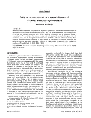
- 6. Three months of wearing the tooth positioner (Figure 4) resulted in settling, especially on the left. She did not have a bridge at that time, and the molars on the right were in an end-on buccal relationship. The gingival heights continued to improve. Three years later (Figure 5), the gingival heights had not changed to a measurable extent, and periodontal probing depths had improved from those recorded 4 months after treatment, returning to pretreatment depths (Figure 6). The strategy used in this treatment was to torque the roots more onto bone. The amount of angular tip was quantified through the use of study casts. Roughly 5– 7u of tip were measured by comparing cusp-tip heights before and after treatment. There was also some expansion, some of which was necessary to conform to arch dimensions during space closure and some for upper and lower arch coordination. To gain insight into the placement of roots on bone (in other words, to determine whether the expansion during treatment offset positive effects of the torque), the study casts Figure 6. Periodontal charting from March 1, 2007, February 10, 2010, and November 12, 2012. Figure 7. Depiction of teeth numbers 44 and 35 as isolated from digital rendering of models at start, final records and 3 years later. 1098 NORTHWAY Angle Orthodontist, Vol 83, No 6, 2013
- 7. were scanned. The before and after widths of the cusp tips, the most buccal contour, and measurements at the CEJ were compared. In this way the ultimate position of the roots could be assessed within the alveolar housing. Figure 7 depicts the widths between the lower right first premolar and the lower left second premolar; the right premolar from each set is lined up on the white line. The images in the top row are from the original set of models. The second row comes from the dental casts taken at final records, and the bottom row is from 3 years later. The white lines detail the original widths at the cusp tips; the second and third rows reflect the proportionate increase in width that took place during treatment. The yellow measurements describe the width between the most buccal point on the teeth, and the pink measurements represent the width between the CEJs. White lines have been drawn through the cusp tips to simulate the amount of torque, and the black lines are an attempted depiction of the long axis of the buccal cusps. Even though the cusp tips were expanded by 3.2 mm during treatment, the most buccal measurements were only widened by 1.7 mm, and the width at the CEJs were constricted by 0.1 mm, resulting in the roots being placed more on bone. The transverse arch widths are presented in Table 1 as measured from the occlusal tip, the most buccal surface and the CEJ. The measurements were taken at the three intervals referenced above; and the differences between intervals are presented. Without three-dimensional radiographic images be- fore treatment, absolute comparisons could not be made to confirm that new bone was grown onto the root where it had not been before. Nevertheless, the Figure 8. Comparisons of three-dimensional radiographic imaging of lower left first and second premolars. The top row shows before and after intraoral photos. Below are paired images of the first premolar (on left); the green lines represent the best effort to depict the distance on a coronal slice between the CEJ and the crest of the buccal alveolar bone. The nine transverse slices are depictions of the presence of alveolar bone at sequential heights along the root with the middle image presenting alveolar bone for the first time. There is a panaramic-type view and the 3-D broad image. The same images are repeated for the second premolar. GINGIVAL RECESSION—CAN ORTHODONTICS BE THE CURE? 1099 Angle Orthodontist, Vol 83, No 6, 2013
- 8. patient underwent CBCT 3 years after appliance removal. These scans were used to evaluate the posttreatment levels of the alveolar crest. Figure 8 compares views of the lower left first and second premolars, the second premolar presenting 3 mm more GR than the first. It is worthy of note that the difference between the two measured distances from the alveolar crest to the CEJ is only 1.3 mm in the CBCT images. The updated images alone cannot confirm the presence of new bone. The extent to which this might represent a false-negative would probably be best determined by a system of higher resolution (with greater radiation exposure) or perhaps direct observation using gingival detachment, neither of which are justified in the opinion of the author for the purpose of answering this question. The patient described in this case presentation has 3 mm more root coverage than she had before orthodontic treatment. The increased root coverage was accomplished by orthodontically moving teeth more onto alveolar bone and by taking more careful oral hygiene measures. In this era of evidence-based approaches to improving the orthodontic specialty, it does not need to be stated that a sample of one is not proof that this will be the result for every patient. On the other hand, orthodontists have an obligation to treat more than just the malocclusion that presents. It is incumbent upon the orthodontic community to evaluate the patient’s entire needs and to try to render as much improvement as possible. If an orthodontic patient presents with GR and orthodontic issues, or, more globally, if a patient presents with an apparent susceptibility toward GR, orthodontists should approach the patient with a more proactive periodontal strategy. ACKNOWLEDGMENTS The author would like to thank Dr Sheldon Peck for invaluable assistance in writing and structuring this article. Also, thanks to Table 1. Measurements from Digital Models Taken at Affected Teeth Original Widths Deband Records Widths Widths 3 Y Later Tip Most Buccal CEJ Tip Most Buccal CEJ Tip Most Buccal CEJ A B C U 3-3 32.49 35.89 35.69 33.12 36.65 35.62 33.20 37.32 36.49 U 5-4 38.75 42.31 42.13 42.21 44.36 44.01 41.71 44.32 43.48 U-6-6 39.14 53.26 52.55 39.88 52.54 52.18 39.60 53.07 52.12 L 3-3 22.76 24.58 28.76 24.40 29.23 28.20 23.35 28.86 28.51 L 4-4 31.71 37.22 36.61 35.05 37.97 37.30 35.26 37.89 37.48 L 4-5 35.29 40.67 40.57 38.49 42.39 40.47 38.55 42.03 40.30 L 6-6 48.53 51.99 51.67 48.01 52.49 51.62 48.64 53.07 51.89 Change from original to deband A to B U 3-3 0.63 0.76 20.07 U 5-4 3.46 2.05 1.88 U-6-6 0.74 20.72 20.37 L 3-3 1.64 4.65 20.56 L 4-4 3.34 0.75 0.69 L 4-5 3.20 1.72 20.10 L 6-6 20.52 0.50 20.05 Change from deband to 3 y later B to C U 3-3 0.08 0.67 0.87 U 5-4 20.50 20.04 20.53 U-6-6 20.28 0.53 20.06 L 3-3 21.05 20.37 0.31 L 4-4 0.21 20.08 0.18 L 4-5 0.06 20.36 20.17 L 6-6 0.63 0.58 0.27 Change from original to 3 y after deband A to C U 3-3 0.71 1.43 0.80 U 5-4 2.96 2.01 1.35 U-6-6 0.46 20.19 20.43 L 3-3 0.59 4.28 20.25 L 4-4 3.55 0.67 0.87 L 4-5 3.26 1.36 20.27 L 6-6 0.11 1.08 0.22 1100 NORTHWAY Angle Orthodontist, Vol 83, No 6, 2013
- 9. Drs. John Hall and Sidney Konigsberg for listening and interacting on the concepts and content; to Drs. Brian Klym and David Gane for their assistance with three-dimensional imaging; and to my wife, Carin, and my incomparable staff for sustaining my love for orthodontics and for helping me to ‘‘get it right.’’ REFERENCES 1. Vanarsdall RL Jr. Periodontal/orthodontic interrelationships. In: Graber TM, Vanarsdall RL Jr, eds. Orthodontics: Current Principles and Techniques. 2nd ed. St Louis, Mo: Mosby; 1995:712–750. 2. Dorfman HS. Mucogingival changes resulting from mandibular incisor tooth movement. Am J Orthod. 1978;74(3):286–297. 3. Pearson LE. Gingival height of lower central incisors orthodontically treated and untreated. Angle Orthod. 1968; 38:337–339. 4. Bollen A-M, Cunha-Cruz J, Bakko DW, Huang GJ, Hujoel PP. The effects of orthodontic therapy on periodontal health: a systematic review of controlled evidence. J Am Dent Assoc. 2008;139:413–422. 5. Wennstrom JL, Lindhe J, Sinclair F, Thilander B. Mucogin- gival therapy. Ann Periodontol. 1996;1:671–701. 6. Andlin-Sobocki A. Changes of facial gingival dimensions in children. A 2-year longitudinal study. J Clin Periodontol. 1993;20:212–218. 7. Bimstein E, Eidelman E. Morphological changes in the attached and keratinized gingiva and gingival sulcus in the mixed dentition period. A 5-year longitudinal study. J Clin Periodontol. 1988;15:175–179. 8. Ainamo J, Talari A. The increase with age of the width of attached gingiva. J Periodont Res. 1976;11:182–188. 9. Daprile G, Gatto MR, Checchi L. The evolution of buccal gingival recessions in a student population: a 5-year follow- up. J Periodontol. 2007;78:611–614. 10. Pontoriero R, Carnevale G. Surgical crown lengthening: a 12-month clinical wound healing study. J Periodontol. 2001; 72:841–848. 11. Bernimoulin J, Curilovic Z. Gingival recession and tooth mobility. J Clin Periodontol. 1977;4:107–114. 12. Staufer K, Landmeser H. Effects of crowding in the lower anterior segment—a risk evaluation depending upon the degree of crowding. J Orofac Orthop. 2004;65:13–25. 13. Richman C. Is gingival recession a consequence of an orthodontic tooth size and/or tooth position discrepancy? Compendium. 2011;32(1):62–69. 14. Bjorn A, Andersson U, Olsson A. Gingival recession in 15- year old pupils. Swed Dent J. 1981;5(4):141–146. 15. Loe H, Anerud A, Boysen H. The natural history of periodontal disease in man: prevalence, severity, and extent of gingival recession. J Periodontol. 1992;63:489– 495. 16. Khocht A, Simon G, Person P, Denepitiya JL. Gingival recession in relation to history of hard toothbrush use. J Periodontol. 1993;64:900–905. 17. Susin C, Haas AN, Oppermann RV, Haugejorden O, Albandar JM. Gingival recession: epidemiology and risk indicators in a representative urban Brazilian population. J Periodontol. 2004;75:1377–1386. 18. Cook DR, Mealey BL, Verrett RG, et al. Relationship between clinical periodontal biotype and labial plate thick- ness: an in vivo study. Int J Periodontics Restorative Dent. 2011;31:345–354. 19. Fu J-H, Yeh C-Y, Chan H-L, Tatarakis N, Leong DJM, Wang H-L. Tissue biotype and its relation to the underlying bone morphology. J Periodontol. 2010;81:569–574. 20. Sicilia A, Arregui I, Gallego M, Cabezas B, Cuesta S. A systematic review of powered vs. manual toothbrushes in periodontal cause-related therapy. J Clin Periodontol. 2002; 39(suppl 3):39–54. 21. Deery C, Heanue M, Deacon S, et al. The effectiveness of manual versus powered toothbrushes for dental health: a systematic review. J Dent. 2004;32(3):197–211. 22. Forrest JL, Miller SA. Part II: manual versus powered toothbrushes: a summary of the Cochrane Oral Health Group’s systematic review. J Dent Hyg. 2004;78:349– 354. 23. Rajapakse PS, McCracken GI, Gwynnett ES, Steen ND, Guentsch A, Heasman PA. Does tooth brushing influence the development and progression of non-inflammatory gingival recession? A systematic review. J Clin Periodontol. 2007;34:1046–1061. 24. Do¨rfer CE, Jorss D, Rau P, Wolff D. The 18-months effect of an oscillating-rotating power toothbrush on recession. J Clin Periodontol. 2006;33(suppl 7):98. GINGIVAL RECESSION—CAN ORTHODONTICS BE THE CURE? 1101 Angle Orthodontist, Vol 83, No 6, 2013