APM Welcome, APM North West Network Conference, Synergies Across Sectors
Biology - Cell Organization & Function
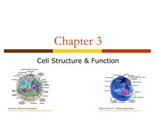
1. Chapter 3
Cell Structure & Function
Picture by Molecular Expressions Picture by Cell II: Cellular Organization
http://micro.magnet.fsu.edu/cells/animalcell.html http://ridge.icu.ac.jp/biobk/BioBookCELL2.html
2. 3.1: Microscopes & Cells
Objectives
1. Define the cell theory
2. Identify the magnification powers of
different types of microscopes
3. Observe how the compound microscope
changes an image
3. Early Microscopes
Anton von Leeuwenhoek
(1600s / Dutch) Picture by Answers.com
http://www.answers.com/topic/anton-van-leeuwenhoek
Used glass lenses to create instrument that
magnified images of very small objects
Light rays bend to make larger image
Earliest light microscope
Picture by Mr. Cantor’s Biology Blog
http://cantorsbiologyblog.blogspot.com/2012/01/second-semester-begins-with-cells.html
4. Discovery of Cells
Leeuwenhoek used microscope to look at
drops of pond water
Filled with tiny living things
Called them “animalcules”
Picture by Lens On Leeuwenhoek
http://lensonleeuwenhoek.net/lenses.htm
5. Robert Hooke
English physicist
Used microscope to observe Picture by History of the Microscope
http://www.history-of-the-microscope.org/robert-hooke-m
Flowers
Insects
Spider webs
Slices of cork Picture by Dare To Unravel
http://daretounravel.blogspot.com/2011/01/mi
Concluded
Wood parts of plants had rectangular chambers
Called them cells
6. Theodor Schwann
1839, German Biologist
Picture by Biographical Outlines
http://www.merke.ch/biografien/biologen_en/schwann.php
Found some animal tissues resemble
cellular tissue of plants
Eventually concluded
Animals are made up of cells
Picture by Biographical Outlines
http://www.merke.ch/biografien/biologen_en/schwann.php
7. The Cell Theory
Robert Brown (Scottish Biologist)
Found object in center of cell (nucleus)
Picture by Wikipedia
http://en.wikipedia.org/wiki/Robert_Brown_(botanist)
Matthias Schleiden (German Biologist)
Suggested nucleus plays role in cell
reproduction
Picture by Biographical Outlines
http://www.merke.ch/biografien/biologen_en/schleiden.php
8. The Cell Theory
Rudolf Virchow
1855 German Physician
Picture by Wikipedia
http://en.wikipedia.org/wiki/Rudolf_Virchow
Proposed that animal and plant cells are
produced only by the division of cells that
already exist
Picture by The Encyclopedia of Science
http://www.daviddarling.info/encyclopedia/P/plant_cell.html
9. The Cell Theory
1. All living things are composed of cells
2. Cells are the smallest working units of
living things
3. All cells come from preexisting cells
Picture by Mrs. Olsen’s 5th Grade Class Page
http://www.ruediger.leon.k12.fl.us/olsonmd/Class%20Pictures/Forms/DispForm.aspx?ID=2&RootFolder=%2Folsonmd%2FClass%20Pictures%2Fclip%20art
10. Modern Microscopes
1) Compound Light Microscope
Contains more than 1 lens
Magnifies up to 1000 times
Parts of Microscope Handout
Picture by Wikipedia
http://wiki.district87.org/index.php/Compound_Light_Microscopes
11. Modern Microscopes
2. Electron Microscope
Uses magnets to focus beam of electrons to
examine sample
Magnifies 1000 times more than light
microscope
Picture by Ego TV
http://egotvonline.com/2012/03/13/25-everyday-objects-under-an-electron-microscope/
Picture by UK Electron Microscope Facility
http://www.engr.uky.edu/~bjhinds/facil/emf
12. Types of Electron Microscopes
Transmission Electron Microscope (TEM)
Sends electrons through sample
Puts image on fluorescent screen
Scanning Electron Microscope (SEM)
Electron scans sample’s surface
Puts image on TV screen
Picture by Electron Microscope
http://egotvonline.com/2012/03/13/25-everyday-objects-under-an-electron-microscope/
13. Electron Microscope Limitations
Sample must be in vacuum
Be nonliving
Samples for TEM need to be thin slices
SEM doesn’t show internal structure
Picture by Electron Microscope
http://egotvonline.com/2012/03/13/25-everyday-objects-under-an-electron-microscope/
14. Scanning Probe Microscope
Trace surface of sample with tiny tip
(probe)
Very tiny objects like atoms and molecules
Picture by Principles of Scanning Probe Microscopy
http://www.physics.leidenuniv.nl/sections/cm/ip/group/Principle_of_SPM.htm
15. 3.2: Cell Boundaries
Objectives
1. Discuss the roles of the cell membrane and
cell wall
2. Describe passive transport and active
transport
16. Cell Membrane
Outer boundary that separates and
protects cell from its surroundings
Must allow certain substances to come in
Must allow waste to exit
Descried as selectively permeable
“The bouncer” or “Main gate”
Picture by The Biology Corner
http://www.biologycorner.com/bio1/cell.html
17. Lipid Bilayer
Cell membrane is made up of lipid molecules
Known as phospholipids
Polar end = head (attract water)
Nonpolar end = tail (repel water)
Picture by TutorVista.com
http://www.tutorvista.com/biology/membrane-lipid-bilayer
18. Lipid Bilayer
Line up in double-layer pattern
Known as lipid bilayer
Lipid Bilayer
Provides cell membrane with tough flexible
barrier
Protects cells
Picture by TutorVista.com
http://www.tutorvista.com/biology/membrane-lipid-bilayer
19. Other Cell Membrane Components
Most cell membranes have
proteins in lipid bilayer
Have carbohydrates
attached
Protein purpose
Move material across cell
membrane
Picture by Cell Membrane Wiki
Protect membrane http://torresbioclan.pbworks.com/w/page/22377072/cell%20m
Carbohydrates act as “ID”
cards
20. Cell Wall
Porous membrane located outside cell membrane
Found in plant cells, algae, and bacteria
NOT found in animal cells
Supports and protects cells
Allow substance to pass in and out
Made of:
Carbohydrates called cellulose
Give plants their strength
Proteins
Picture by Molecular Expressions
http://micro.magnet.fsu.edu/cells/plantcell.html
21. Passive Transport
Process of moving substances in and out
of a cell
Does NOT use energy
Picture by Cell Organization & Functions
http://www.williamsclass.com/SeventhScienceWork/CellsOrganization.htm
22. Diffusion
Process by which substances spread
through a liquid or gas
Move from region of high concentration to area
of low concentration
Example: food coloring in water
In cells…
Liquids and small lipids diffuse directly across
cell membrane
Picture by Biology Corner
http://www.biologycorner.com/bio1/diffusion.html
23. Facilitated Diffusion
Cell membrane also contains protein channels
Allows larger substance to pass through
Facilitated Diffusion
Diffusion that occurs with the aided help of a protein
channel
Each protein channel is specified for a certain substance
Picture by Our Virtual Classroom
http://ccaoscience.wordpress.com/notes/transport-protein/
24. Osmosis
The diffusion of water through a
selectively permeable membrane
Water diffuses from areas of high
concentration to areas of low
concentration
Picture by Free Drinking Water
http://www.freedrinkingwater.com/resource-a-complete-resource-guide-to-osmosis.htm
25. Osmotic Pressure
When water moves by osmosis, it
produces pressure
Enough to destroy cell
Three ways to control:
1) cell wall
2) pump out water
3) bathe cells in blood Picture by Non Ideal Solutions
http://www.chem.ufl.edu/~itl/4411/colligative/lec_i.html
26. Osmotic Pressure
Cell Wall
Strong tough wall prevents
cell from expanding
Counter acts osmotic pressure
Pump Out
Cells use contractile vacuole
to pump out water
Picture by Non Ideal Solutions
http://www.chem.ufl.edu/~itl/4411/colligative/lec_i.html
27. Osmotic Pressure
Bathe Cells in Blood
Mostly in large animals
Blood cells have same concentration of
dissolved substances
Help absorb excess water
Picture by A Sweet Life
http://asweetlife.org/a-sweet-life-staff/articles/interview-dr-zachary-bloomgarden-on-the-hba1c-assay/8020/
28. Active Transport
Movement of a substance against a
concentration difference
Requires A LOT of energy
Transports:
Large molecules
Food
Whole cells
Picture by G11-BioA -2011
http://g11-bioa-2011-12.wikispaces.com/(d)+Active+transport
29. 3.3: Inside The Cell
Objectives
1. Describe the composition and function of
the nucleus
2. List and describe the organelles of the
cytoplasm
3. Interpret the changes observed when a
paramecium takes in food
30. Nucleus
Large, dense structure contained in cells
Known as control center
“Main office of factory”
Picture by Plant Cell
http://library.thinkquest.org/06aug/01942/plcells/nuclues.htm
31. Nucleus
Organisms can be classified into 2 categories
1. Eukaryotes
Organisms cells HAVE nuclei
Include unicellular and multicellular
2. Prokaryotes
Organisms that do NOT contain nuclei
Small single celled organisms
Picture by Bacterial/Prokaryotic Phylogeny
http://www.bacterialphylogeny.info/eukaryotes.html
32. Role of Nucleus
Contains nearly all of cell’s DNA
Has instructions to make proteins / molecules
Form material called chromatin
Spreads through nucleus
When cell divides
Chromatin condense into chromosomes
Acts as “blueprint of factory”
Picture by Animal Port
http://www.animalport.com/animal-cells.html
33. Structures In The Nucleus
Nucleolus
Small dense region inside nucleus
Where ribosomes are made
Picture by Molecular Expressions
http://micro.magnet.fsu.edu/cells/nucleus/nucleolus.html
Nuclear Envelope
Membrane that surrounds the nucleus
Has pores to transport materials in and out
34. Cytoplasm
Fluid outside nucleus held in by cell
membrane
Holds all other cell organelles
These are small structures that perform specialized
functions in the cell
Allows for movement
Known as the “factory floor”
Picture by Daylilies
http://www.daylilies.org/ahs_dictionary/cytoplasm.html
35. Ribosomes
Tiny particles made of RNA and protein
Site for protein synthesis
Gets instructions from nucleus
Known as “the workers”
Picture by Cellupedia
http://library.thinkquest.org/C004535/eukaryotic_cells.html
36. Endoplasmic Reticulum (ER)
Processing and transporting of proteins
and other macromolecules
Network of membranes
2 types
1. Smooth ER… NO ribosomes attached to surface
2. Rough ER… ribosomes attached to surface
Act as the subway
Picture by Molecular Expressions
http://micro.magnet.fsu.edu/cells/endoplasmicreticulum/endoplasmicreticulum.html
37. Golgi Apparatus
Act as packaging center
Modify and add components to proteins
Attach carbohydrates or lipids
Picture by Molecular Expressions
http://micro.magnet.fsu.edu/cells/golgi/golgiapparatus.html
38. Lysosomes
Sac-like membrane that gets rid of waste
Filled with chemicals and enzymes
Can also break down and gets rid of
damaged organelles
Known as the “garbage man”
Picture by Molecular Expressions
http://publications.nigms.nih.gov/insidethecell/images/ch1_lysosome.jpg
39. Cytoskeleton
Act as a supporting framework for cell
Found in eukaryotic cells
Components
Microtubules
Mircofillaments
Picture by Cellupedia
http://library.thinkquest.org/C004535/cytoskeleton.html
These are hollow tubes of protein that provide
framework to support cell
40. Vacuoles
Sac-like structure used for storage
Animals
Store proteins, fats, and carbohydrates
Plants
Store water and dissolved salts
Provide support
Known as “the warehouse”
Picture by Molecular Expressions
http://micro.magnet.fsu.edu/cells/plants/vacuole.html
41. Mitochondria
Produces energy from a chemical fuel
Organic molecules like glucose or other sugars
Found in eukaryotic cells
Plants and animals
Contain own DNA
Picture by Molecular Expressions
http://micro.magnet.fsu.edu/cells/mitochondria/mitochondria.html
Known as “powerhouse of cell”
42. Chloroplasts
Organelle that produces energy from
sunlight
Found in plants ONLY and some algae
Aids in the process of photosynthesis
Green due to chlorophyll pigment
Also contains own DNA
Picture by eTeaching Program
https://www.etap.org/demo/grade7_science/instruction2tutor.html
43. 3.4: The Origin of the Eukaryotic Cell
Objectives
1. Define the endosymbiont hypothesis
44. Eukaryotic vs. Prokaryotic
Eukaryotes Prokaryotes
Nuclues Have NONE of these
Mitochondria
Chloroplasts
Other organelles WHY???
Picture by BioCoach Activity
http://www.phschool.com/science/biology_place/biocoach/cells/common.html
45. The Work of Lynn Margulis
Focused on mitochondria and chloroplasts
Used on DNA to make compounds
Both surrounded by two membranes
Both reproduced separately from rest of cell
Mitochondria from mitochondria
Chloroplasts from chloroplasts
Why did this happen???
Picture by The Alien Next Door Blog
http://sfgirl-thealiennextdoor.blogspot.com/2010/06/celebrating-womanhood-i-am-woman-i-am.html
46. The Endosymbiont Hypothesis
Billions of years
ago…
Eukaryotic cells arose
as a combination of
different prokaryotic
cells
Cells consumed other
cells but still each
survived
Picture by Molecular Expressions
http://www.tokresource.org/tok_classes/biobiobio/biomenu/options_folder/D1_life_origins/index.htm
47. Margulis’s Model
Stated mitochondria and chloroplasts had
ancestors that were free-living organisms
Were consumed by larger cells
Became organelles in larger cells
Picture by The Endosymbiotic Hypothesis
http://endosymbiotichypothesis.wordpress.com/
48. Further Evidence
Chloroplast DNA
Similar to DNA in prokaryotic cells
In Mitochondria & Chloroplasts
Contain own ribosomes to make own proteins
Ribosomes similar to prokaryotic ribosomes
Picture by Molecular Expressions
http://micro.magnet.fsu.edu/cells/mitochondria/mitochondria.html