SlideShare una empresa de Scribd logo
1 de 112
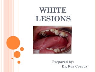
WHITE
LESIONS


   Prepared by:
        Dr. Rea Corpuz
White Lesions

 lesions of the oral mucosa,
  which are white results
  from a

    thickened layer of keratin

    epithelial hyperplasia

    intracellular epithelial
     edema

    reduced vascularity of subjacent connective
White Lesions

 white or yellow lesions
  may also be due to fibrous
  exudate covering an:

    ulcer
    submucosal deposit
    surface debris
    fungal colonies
White Lesions

 (1) Leukoedema

 (2) Leukoplakia

 (3) Lichen Planus

 (4) Candidiasis

 (5) White Sponge Nevus

 (6) Nicotine Stomatitis
White Lesions

 (7) Geographic Tongue

 (8) Hairy Tongue

 (9) Dental Lamina Cyst

 (10) Fordyce’s Disease

 (11) Perleche
(1) Leukoedema

 generalized opacification
  of buccal mucosa that is
  regarded as a variation
  of normal

 can be identified in majority
  of population
(1) Leukoedema

 Etiology & Pathogenesis

    to date, cause has not
     been established

    smoking
    chewing tobacco          none
    alcoholo ingestion        are
    bacterial infection       proven
    salivary condition        cause
    electrochemical interactions
     have been implicated
(1) Leukoedema

 Clinical Features

    usual discovered as
     incidental finding

    asymptomatic

    symmetrically distributed
     in buccal mucosa
(1) Leukoedema

 Clinical Features

    appear as gray-white,
     diffuse, filmy or milky
     surface

    more exaggerated cases,
     whitish cast with surface
     textural changes

      • wrinkling
      • or corrugations
(1) Leukoedema

 Clinical Features

    with stretching of buccal
     mucosa, opaque changes
     dissipate

    more apparent in non-whites,
     especially African-American
(1) Leukoedema

 Treatment

   NO treatment is necessary

   since there is no malignant
    potential

   if there is any doubt about
    diagnosis, a biopsy can
    be performed
(2) Leukoplakia

 also known as Leukokeratosis;
  Erythroplakia

 Leuko= white

 Plakia = patch

 defined by World Health Organization
  (WHO) as a white patch or plaque
  that cannot be characterized
  clinically or pathologically as
  any other disease
(2) Leukoplakia
Leukoplakia

 clinical term indicating a
  white patch or plaque of
  oral mucosa

 cannot be rubbed off

 cannot be characterized clinically
  as any other disease

 biopsy is mandatory to
  establish a definitive diagnosis
(2) Leukoplakia

 Mild or Thin Leukoplakia

 Homogenous or
  Thick Leukoplakia

 Granular or Nodular
  Leukoplakia

 Verrucous or Verruciform
  Leukoplakia
(2) Leukoplakia

 Proliferative Verrucous
  Leukoplakia (PVL)

 Erythroleukoplakia or
  Speckled Leukoplakia
(2) Leukoplakia
(2) Leukoplakia

 Mild or Thin Leukoplakia

    seldom shows dysplasia
     on biopsy

    may disappear or continue
     unchanged
(2) Leukoplakia

 Homogenous or Thick Leukoplakia

   for tobacco smokers who do
     not reduce their habit

   2/3 of such lesions slowly
     extend laterally, become
     thicker + acquire distinctly
     white appearance
(2) Leukoplakia

 Homogenous or Thick Leukoplakia

   affected mucosa may become
     leathery to palpation

   fissures may deepen

   become more numerous

   most thick, smooth lesions
     remain indefinitely at this
     stage
(2) Leukoplakia

 Homogenous or Thick Leukoplakia

   some, perhaps as many
     as 1/3, regress or disappear
(2) Leukoplakia

 Granular or Nodular Leukoplakia

   few become even more
     severe

   develop increased surface
     irregularities
(2) Leukoplakia

 Verrucous or Verruciform
  Leukoplakia

    lesions that demonstrate
     sharp or blunt projections
(2) Leukoplakia

 Proliferative Verrucous
  Leukoplakia (PVL)

    high risk form of
     leukoplakia

    development of multiple
     keratotic plaques

    with roughened
     surface projections
(2) Leukoplakia

 Proliferative Verrucous
  Leukoplakia (PVL)

    tend to slowly spread

    involve additional oral
     mucosal sites

    gingiva is frequently involved

    although other sites may be
     affected as well
(2) Leukoplakia

 Proliferative Verrucous
  Leukoplakia (PVL)

    as lesions progress, there
     may go through a stage
     indistinguishable

    transform into full-fledged
     squamous cell carcinoma
     (usually within 8 years of
      initial PVL diagnosis)
(2) Leukoplakia

 Proliferative Verrucous
  Leukoplakia (PVL)

    lesions rarely regress
     despite therapy

    strong female predilection

    minimal association with
     tobacco use
(2) Leukoplakia

 Erythroplakia

   leukoplakia may become
     dysplastic

   even invasive, with no change
     in its clinical appearance

   however, some lesions eventually
     demonstrate scattered patches
     of redness called erythroplakia
(2) Leukoplakia

 Erythroleukoplakia or
  Speckled Leukoplakia

    such areas usually represent
     sites in which epithelial
     cells are so immature
     or atrophic that they can no
     longer produce keratin
(2) Leukoplakia

 Erythroleukoplakia or
  Speckled Leukoplakia

    intermixed red-and-white
     lesion

    pattern of leukoplakia
     that frequently reveals
     advanced dysplasia on
     biopsy
(2) Leukoplakia

 Etiology & Prognosis

    many cases are etiologically
     related to use of tobacco
     in smoked or smokeless
     forms and may regress
     after discontinuation
     of tobacco use
(2) Leukoplakia

 Etiology & Prognosis

    other factors, such as

      • alcohol abuse         may have
      • trauma                 a role in
      • C. albicans infection etiology
(2) Leukoplakia

 Etiology & Prognosis

    nutritional factors have
     been cited as important,
     especially iron deficiency
     anemia
(2) Leukoplakia

 Clinical Features

    associated with middle-aged
     + older population

    vast majority of cases occur
     after age of 40 years
(2) Leukoplakia

 Site of Occurence

    Vestibule
    Buccal
    Palate
    Alveolar Ridge
    Lip
    Tongue
    Floor
(2) Leukoplakia

 leukoplakia of lips + tongue
  also exhibits relative high
  percentage of dysplastic or
  neoplastic change
(2) Leukoplakia

 Treatment & Prognosis

   absence of dysplastic or
    atypical epithelial changes

     • periodic examinations +
      rebiopsy of new suspicious
      areas are recommended
(2) Leukoplakia

 Treatment & Prognosis

   if diagnosis as moderate to
     severe dysplasia

     • excision is obligatory

   for large lesions, grafting
     procedures may be necessary after
     surgery

   may recur after complete removal
(3) Lichen Planus

 chronic mucocutaneous
  disease of unknown cause

 relatively common

 typically presents as bilateral
  white lesions

 occasionally with associated
  ulcers
(3) Lichen Planus

 Pathogenesis

   although cause is unknown

   generally considered to be
     a immunologically mediated
     process

   resembles hypersensitivity
     reaction
(3) Lichen Planus

 Clinical Features

    disease of middle age

    affects men + women in
     nearly equal numbers

    children rarely affected
(3) Lichen Planus

 Clinical Features

    Types:

      • Reticular
      • Erosive (ulcerative)
      • Plaque
      • Papular
      • Erythematous (atrophic)
(3) Lichen Planus

 Clinical Features

    Reticular Form

      • most common type

      • numerous interlacing white
       keratotic lines or striae
       (Wickham’s striae)

          produces anular or lacy
           pattern
(3) Lichen Planus

 Clinical Features

    Reticular Form

      • buccal mucosa is the site
       most commonly involved

      • may also be noted on:

          tongue
          gingiva – less common
          lips
(3) Lichen Planus

 Clinical Features

    Plaque Form

      • resembles leukoplakia

      • but has multifocal distribution

      • range from slightly elevated
       to smooth and flat
(3) Lichen Planus

 Clinical Features

    Plaque Form

      • primary sites are

          dorsum of tongue
          buccal mucosa
(3) Lichen Planus

 Clinical Features

    Erythematous Form

      • red patches

      • with very fine white
       striae

      • attached gingiva commonly
       involved
(3) Lichen Planus

 Clinical Features

    Erythematous Form

      • patchy distribution often
       in four quadrants

      • patient may complain of

          burning
          sensitivity
          generalized discomfort
(3) Lichen Planus

 Clinical Features

    Erosive Form

      • central area of lesion is
       ulcerated

      • fibrinous plaque or
       pseudomembrane covers
       ulcer

      • changing patterns of involvement
        from week to week
(3) Lichen Planus

 Treatment

   although it cannot be
    generally cured

   some drugs can provide
    satisfactory control

   corticosteroids are the single
    most useful group of drugs
    in the management of lichen
    planus
(3) Lichen Planus

 Treatment

   corticosteroid

     • ability to modulate
      inflammation + immune
      response
(3) Lichen Planus

 Treatment

   topical application +
    local injection of steroids
    have been used successfully
    in controlling but not
    curing this disease
(4) Candidiasis
 common oppurtunistic
  oral mycotic infection

 develops in the presence of
  one of several predisposing
  factors

   • immunodeficiency
   • endocrine disturbances
   • hypoparathyroidism
   • diabetes mellitus
   • poor oral hygiene
   • xerostomia
(4) Candidiasis

 caused by Candida albicans

 infection with this organism
  is usually superficial, affecting
  the outer aspects of involved
  oral mucosa or skin
(4) Candidiasis

 in severely debilitated +
  immunocompromised patients
  such as patients with AIDS

    infection may extend into
     alimentary tract (candidal
     esophagitis

    bronchopulmonary tract

    and other organ system
(4) Candidiasis

 Clinical Features

    most common form is
     acute pseudomembranous
     also known, as thrush

      • young infants + elderly
       are commonly affected
(4) Candidiasis

 Clinical Features

    oral lesion of acute
     candidiasis (thrush)

      • white
      • soft plaques that sometime
        grow centrifugally + merge
      • wiping plaques with gauze
         sponge leaves a painful,
         eroded, eryhtematous or
         ulcerated surface
(4) Candidiasis

 Clinical Features

    Chronic Erythematous
     Candidiasis

      • commonly seen on
       geriatric individuals

      • who wear complete
        maxillary denture
(4) Candidiasis

 Clinical Features

    Chronic Erythematous
     Candidiasis

      • distinct predilection for
       palatal mucosa as
       compared with mandibular
       alveolar arch
(4) Candidiasis

 Clinical Features

    Chronic Erythematous
     Candidiasis

      • chronic low-grade
       resulting from poor
       prosthesis fit

      • failure to remove
       appliance at night
(4) Candidiasis

 Clinical Features

    Chronic Erythematous
     Candidiasis

      • bright red

      • relative little
        keratinization
(4) Candidiasis

 Clinical Features

    Hyperplastic Candidiasis

      • may involve dorsum of
        tongue

      • pattern referred to as
       median rhomboid
       glossitis
(4) Candidiasis

 Clinical Features

    Hyperplastic Candidiasis

      • usually asymptomatic

      • usually discovered on
       routine oral
       examination
(4) Candidiasis

 Clinical Features

    Hyperplastic Candidiasis

      • found anterior to
       circumvallate
       papillae

      • oval or rhomboid
        outline
(4) Candidiasis

 Clinical Features

    Hyperplastic Candidiasis

      • may have smooth,
        nodular or fissured
        surface

      • range in color from
        white to more red
(4) Candidiasis

 Clinical Features

    Mucocutaneous Candidiasis

      • long standing

      • persistent candidiasis of

          oral mucosa
          skin
          vaginal mucosa
(4) Candidiasis

 Clinical Features

    Mucocutaneous Candidiasis

      • often resistant to treatment

      • begins as a pseudomembranous
        type of candidiasis

      • soon followed by nail +
       cutaneous involvement
(4) Candidiasis

 Treatment

   majority of infections may
    be simply treated with
    topical applications of
    nystatin suspension

     • nystatin cream or
      ointment often effective
      when applied directly to
      denture-bearing surface itself
(4) Candidiasis

 Treatment

   topical applications of either
    nystatin or clotrimazole
    should be continued for at
    least 1 week beyond
    disappearance of clinical
    manifestations of disease
(4) Candidiasis

 Treatment

   Hyperplastic Candidiasis

     • topical + systemic antifungal
      agents may not be effective
      at completely removing
      lesions

         surgical management
         may be necessary
(4) Candidiasis

 Treatment

   Chronic Mucocutaneous
    Candidiasis associated
    with immunosuppression

     • topical agents may not
       be effective
(4) Candidiasis

 Treatment

   Chronic Mucocutaneous
    Candidiasis associated
    with immunosuppression

     • systemic administration
      of medications:

         Ketoconazole
         Fluconazole
         Itraconazole
(5) White Sponge Nevus

 autosomal-dominant condition

 due to point mutations for
  genes coding for keratin 4
  and/or 13.

 affects oral mucosa bilaterally

 NO treatment is required
(5) White Sponge Nevus

 Clinical Features

   • asymptomatic

   • folded white lesions

   • may affect several mucosal
    sites

   • lesions tend to be thickened
    + spongy consitency
(5) White Sponge Nevus

 Clinical Features

   • presentation intraorally
    is almost always bilateral
    + symmetric

   • usually appears early in
    life, typically before
    puberty
(5) White Sponge Nevus

 Clinical Features

   • usually observed in buccal
    mucosa

   • tongue + vestibular mucosa
    may be involved
(5) White Sponge Nevus

 Treatment

  • NO treatment necessary
   since it is asymptomatic
   + benign
(6) Nicotine Stomatitis

 common tobacco-related
  form of keratosis

 typically associated with pipe
  + cigar smoking

 with positive correlation
   between intensity of smoking
   + severity of condition
(6) Nicotine Stomatitis

 combination of tobacco
  carcinogens + heat is markedly
  intensified in reverse smoking
  (lit end positioned inside the
   mouth)

 adding a significant risk for
  malignant conversion
(6) Nicotine Stomatitis

 Clinical Features

    palatal mucosa initially
     responds with an erythematous
     change follwed by
     keratinization
(6) Nicotine Stomatitis

 Clinical Features

    subsequent to opacification
     or keratinization of palate

      • red dots surrounded by
        white keratotic rings
        appear

          dot represent inflammation
          of salivary gland excretory
          duct
(6) Nicotine Stomatitis

 Treatment

   condition rarely evolves into
    malignancy

   except in individuals who
    reverse smoke

   discontinuation of tobacco
    habit
(7) Geographic Tongue

 also known as erythema migrans,
  benign migratory glossitis

 prevalent among whites +
  blacks

 strongly associated with fissure
  tongue

 inversely associated with cigarette
  smoking
(7) Geographic Tongue

 emotional stress may enhance
  the process
(7) Geographic Tongue

 Clinical Features

    affects women slightly more
     than men

    children occasionally may
     be affected

    characterized initially by
     presence of atrophic patches
     surrounded by elevated
     keratotic margins
(7) Geographic Tongue

 Clinical Features

    desquamated areas appear
     red + may be slightly
     tender

    followed over a period of
     days or weeks, pattern
     changes

    appearing to move across
     dorsum of tongue
(7) Geographic Tongue

 Clinical Features

    most patients are asymptomatic

    occasionally patients complain
     of irritation or tenderness

    especially in relation to
     consumption of spicy foods
     + alcoholic beverages
(7) Geographic Tongue

 Clinical Features

    lesions periodically disappear

    recur for no apparent reason
(7) Geographic Tongue

 Treatment

   NO treatment is required
    because of self-limiting +
    usually asymptomatic nature
    of this condition
(7) Geographic Tongue

 Treatment

   when symptoms occur,

     • topical steroids especially
       ones containing antifungal
       agent

         helpful in reducing symptoms
(7) Geographic Tongue

 Treatment

   mouth clean using mouthrinse
    composed of sodium
    bicarbonate in water

   reassure patients that condition
    is totally benign
(8) Hairy Tongue

 clinical term referring to a
  condition of filiform papillae
  overgrowth on dorsal surface
  of tongue

 there are numerous initiating
  or predisposing factors for
  hairy tongue
(8) Hairy Tongue

 broad spectrum antibiotics
  such as penicillin + systemic
  cortiocosteroids are often
  identified in clinical
  history of patients with
  this condition
(8) Hairy Tongue

 oxygenating mouthrinses
  containing:

   hydrogen peroxide
   sodium perborate
   carbamide peroxide

       have been cited as
       possible etiologic agents
(8) Hairy Tongue

 Clinical Features

    clinical alteration translates
     to hyperplasia of filiform
     papillae; result is

      • thick           serves to trap
      • matted surface bacteria, fungi,
                        foreign materials
(8) Hairy Tongue

 Clinical Features

    extensive elongation of
     papillae occurs,

      • gagging             may be
      • tickiling sensation felt
(8) Hairy Tongue

 Clinical Features

    color may range from white
     to tan to deep brown
     depending on:

      • diet
      • oral hygiene
      • composition of bacteria
       inhabiting papillary surface
(8) Hairy Tongue

 Treatment

   brush/scrape tongue with
    baking soda

   maintain good oral hygiene

   emphasize to patients that
    this process is entirely benign
(8) Hairy Tongue

 Treatment

   self-limiting

   tongue should return to
    normal after institution
    of physical debridement
    + proper oral hygiene
(9) Dental Lamina Cyst

 also known as Gingival Cyst of
  New Born or Bohn’s nodules

 appear as multiple nodules
  along alveolar ridge in neonates
(9) Dental Lamina Cyst

 similar epithelial inclusion
  cysts may occur along midline
  of palate (palatine cyst of
  new born or Epstein’s pearls)

    developmental origin
    derived from epithelium
    included in fusion line
     between palatal shelves +
     nasal processes
    no treatment; resolve spontaneously
(9) Dental Lamina Cyst

 Treatment

   not necessary because nearly
    all spontaneously rupture
    before patient is 3 months
    of age
(10) Fordyce’s Granules

 represents ectopic sebaceous
  glands or sebaceous
  choristomas

    normal tissue in abnormal
     location

 regarded as developmental

 considered a variation of normal
(10) Fordyce’s Granules

 multiple

 often seen in aggregates

 sites of predilection include

   • buccal mucosa
   • vermillion of upper lip
(10) Fordyce’s Granules

 lesions generally are symmetrical
  distributed

 tend to become obvious after
  puberty

 maximal expression occurring
  between 20-30 years of age
(10) Fordyce’s Granules

 lesions are asymptomatic

 discovered during routine
  oral examination
(10) Fordyce’s Granules

 Treatment

   No treatment is indicated

     • glands are normal in
       character

     • do not cause any untoward
       effects
(11) Perleche

 also known as Angular
  Cheilitis

 inflammation + atrophy
  of skin of folds at angles
  of mouth
(11) Perleche

 may be due to:

    excessive lip licking

    thumb sucking

    sagging of facial skin
    in edentulous or elderly
    persons
(11) Perleche

 may be due to:

    prolonged contact with
     saliva results in
     maceration

    with possible secondary
     infection by Candida
     or staphylococci
(11) Perleche

 Clinical Features

    skin at angles of mouth
     has erythematous fissures

    often with exudate + crust

    further licking to moisten
     inflamed area exacerbates
     the problem
(11) Perleche

 Treatment

   applying antimicrobial
    creams

   followed by low-potency
    steroid creams until
    symptoms resolve

   protective lip balm may help
    prevent recurrence
References:
 Books
   Cawson, R.A: Cawson’s Essentials of Oral
       Oral Pathology and Oral Medicine,
       8th Edition
        • (page 165-167 )
   Neville, et. al: Oral and Maxillofacial Pathology
        3rd Edition
        • (pages 388- 397; 590-592; 819-820)
  Regezi, et. al: Oral Pathology: Clinical Pathologic
       Correlations, 5th Edition
        • (pages 73-105; 241-242; 296-299; 394)

Más contenido relacionado

La actualidad más candente

Peripheral and central giant cell granuloma
Peripheral and central giant cell granulomaPeripheral and central giant cell granuloma
Peripheral and central giant cell granulomaRijuwana77
 
ORAL VESICULOBULLOUS LESION
ORAL VESICULOBULLOUS LESIONORAL VESICULOBULLOUS LESION
ORAL VESICULOBULLOUS LESIONSHREYADAS49
 
Oral White lesions
Oral White lesionsOral White lesions
Oral White lesionsIAU Dent
 
Adenomatoid odontogenic tumour and others
Adenomatoid odontogenic tumour and othersAdenomatoid odontogenic tumour and others
Adenomatoid odontogenic tumour and othersKhin Soe
 
Priya seminar on ulcerative,vesicular and bullous lesions
Priya seminar on ulcerative,vesicular and bullous lesionsPriya seminar on ulcerative,vesicular and bullous lesions
Priya seminar on ulcerative,vesicular and bullous lesionspriyadershini rangari
 
Pulp polyp and gingival polyp
Pulp polyp and gingival polypPulp polyp and gingival polyp
Pulp polyp and gingival polypAkankshasingh546
 
Pigmented lesions of oral cavity
Pigmented lesions of oral cavityPigmented lesions of oral cavity
Pigmented lesions of oral cavityPraveena Veena
 
Oral lichen planus seminar
Oral lichen planus seminarOral lichen planus seminar
Oral lichen planus seminarPRIYA GIMEKAR
 
Acute Necrotising Ulcerative Gingivitis
Acute Necrotising Ulcerative GingivitisAcute Necrotising Ulcerative Gingivitis
Acute Necrotising Ulcerative Gingivitisshabeel pn
 
28.regional odontodysplasia
28.regional odontodysplasia28.regional odontodysplasia
28.regional odontodysplasiaNehal Vithlani
 
Amelogenesis Imperfecta
Amelogenesis ImperfectaAmelogenesis Imperfecta
Amelogenesis Imperfectashabeel pn
 
Non keratotic white lesions of the oral cavity
Non keratotic white lesions of the oral cavityNon keratotic white lesions of the oral cavity
Non keratotic white lesions of the oral cavityMammootty Ik
 

La actualidad más candente (20)

Peripheral and central giant cell granuloma
Peripheral and central giant cell granulomaPeripheral and central giant cell granuloma
Peripheral and central giant cell granuloma
 
ORAL VESICULOBULLOUS LESION
ORAL VESICULOBULLOUS LESIONORAL VESICULOBULLOUS LESION
ORAL VESICULOBULLOUS LESION
 
Oral White lesions
Oral White lesionsOral White lesions
Oral White lesions
 
Oral pyogenic granuloma
Oral pyogenic granulomaOral pyogenic granuloma
Oral pyogenic granuloma
 
Adenomatoid odontogenic tumour and others
Adenomatoid odontogenic tumour and othersAdenomatoid odontogenic tumour and others
Adenomatoid odontogenic tumour and others
 
Oral Lichen Planus (OLP)
Oral Lichen Planus (OLP)Oral Lichen Planus (OLP)
Oral Lichen Planus (OLP)
 
red and white lesions of oral cavity
red and white lesions of oral cavityred and white lesions of oral cavity
red and white lesions of oral cavity
 
Priya seminar on ulcerative,vesicular and bullous lesions
Priya seminar on ulcerative,vesicular and bullous lesionsPriya seminar on ulcerative,vesicular and bullous lesions
Priya seminar on ulcerative,vesicular and bullous lesions
 
Pulp polyp and gingival polyp
Pulp polyp and gingival polypPulp polyp and gingival polyp
Pulp polyp and gingival polyp
 
Oral Lichen Planus
Oral Lichen PlanusOral Lichen Planus
Oral Lichen Planus
 
Pigmented lesions of oral cavity
Pigmented lesions of oral cavityPigmented lesions of oral cavity
Pigmented lesions of oral cavity
 
Pre cancerous lesion and condition
Pre cancerous lesion and conditionPre cancerous lesion and condition
Pre cancerous lesion and condition
 
Oral lichen planus seminar
Oral lichen planus seminarOral lichen planus seminar
Oral lichen planus seminar
 
Acute Necrotising Ulcerative Gingivitis
Acute Necrotising Ulcerative GingivitisAcute Necrotising Ulcerative Gingivitis
Acute Necrotising Ulcerative Gingivitis
 
White sponge nevus
White sponge nevusWhite sponge nevus
White sponge nevus
 
ANUG
ANUGANUG
ANUG
 
28.regional odontodysplasia
28.regional odontodysplasia28.regional odontodysplasia
28.regional odontodysplasia
 
Amelogenesis Imperfecta
Amelogenesis ImperfectaAmelogenesis Imperfecta
Amelogenesis Imperfecta
 
Red and White Lesions
Red and White LesionsRed and White Lesions
Red and White Lesions
 
Non keratotic white lesions of the oral cavity
Non keratotic white lesions of the oral cavityNon keratotic white lesions of the oral cavity
Non keratotic white lesions of the oral cavity
 

Destacado (19)

White Lesions
White LesionsWhite Lesions
White Lesions
 
Diseases of oral cavity
Diseases of oral cavityDiseases of oral cavity
Diseases of oral cavity
 
Tuberculosis and dentistry
Tuberculosis and dentistryTuberculosis and dentistry
Tuberculosis and dentistry
 
Bacterial infections (4)
Bacterial infections (4)Bacterial infections (4)
Bacterial infections (4)
 
Acute systemic lupus erythematosus
Acute systemic lupus erythematosusAcute systemic lupus erythematosus
Acute systemic lupus erythematosus
 
Clinical features of systemic lupus erythematosus
Clinical features of systemic lupus erythematosusClinical features of systemic lupus erythematosus
Clinical features of systemic lupus erythematosus
 
Lupus
Lupus Lupus
Lupus
 
Common dermatologic disorders systemic lupus erythematosus
Common dermatologic disorders systemic lupus erythematosusCommon dermatologic disorders systemic lupus erythematosus
Common dermatologic disorders systemic lupus erythematosus
 
Systemic lupus erythematosus
Systemic lupus erythematosusSystemic lupus erythematosus
Systemic lupus erythematosus
 
Autoimmune Diseases.
Autoimmune Diseases.Autoimmune Diseases.
Autoimmune Diseases.
 
Systemic Lupus Erythematosus
Systemic Lupus ErythematosusSystemic Lupus Erythematosus
Systemic Lupus Erythematosus
 
What is lupus
What is lupusWhat is lupus
What is lupus
 
Oral manifestations of systemic diseases
Oral manifestations of systemic diseasesOral manifestations of systemic diseases
Oral manifestations of systemic diseases
 
Oral candidiasis
Oral candidiasisOral candidiasis
Oral candidiasis
 
Oral candidiasis
Oral candidiasis Oral candidiasis
Oral candidiasis
 
Hematologic disorders
Hematologic disordersHematologic disorders
Hematologic disorders
 
Oral ulcersppt
Oral ulcerspptOral ulcersppt
Oral ulcersppt
 
Dental pain
Dental painDental pain
Dental pain
 
Candidiasis
CandidiasisCandidiasis
Candidiasis
 

Similar a White lesions (2)

Leukoplakia
LeukoplakiaLeukoplakia
LeukoplakiaAnnu Mj
 
Oral Potentially Malignant Disorders
Oral Potentially Malignant DisordersOral Potentially Malignant Disorders
Oral Potentially Malignant DisordersShreyaSinha676143
 
4 keratoses and related disorders of oral mucosa ii
4  keratoses and related disorders of oral mucosa ii4  keratoses and related disorders of oral mucosa ii
4 keratoses and related disorders of oral mucosa iiRmz Study
 
5 dermatological causes of white patches
5  dermatological causes of white patches5  dermatological causes of white patches
5 dermatological causes of white patchesRmz Rabadi
 
precancerous lesions in oral cavity
precancerous lesions in oral cavityprecancerous lesions in oral cavity
precancerous lesions in oral cavitysuleman khan
 
Premalignant lesion
Premalignant lesionPremalignant lesion
Premalignant lesiondhuha khalid
 
6th lecture lichen-planus
6th lecture lichen-planus6th lecture lichen-planus
6th lecture lichen-planusLama K Banna
 
Red and White Lesions of the.pptx
Red and White Lesions of the.pptxRed and White Lesions of the.pptx
Red and White Lesions of the.pptx52581
 
Oral lichen planus
Oral lichen planusOral lichen planus
Oral lichen planusIman Zubair
 
Lichenoid dermatoses.potx
Lichenoid dermatoses.potxLichenoid dermatoses.potx
Lichenoid dermatoses.potxIbrahim Farag
 
Precancerous lesions of oral cavity
Precancerous lesions of oral cavityPrecancerous lesions of oral cavity
Precancerous lesions of oral cavityDr. Bibina George
 
lichen planus and lichenoid reaction 4 .ppt
lichen planus and lichenoid reaction 4 .pptlichen planus and lichenoid reaction 4 .ppt
lichen planus and lichenoid reaction 4 .pptmadhusudhan reddy
 

Similar a White lesions (2) (20)

Lichen planus
Lichen planusLichen planus
Lichen planus
 
Leukoplakia
LeukoplakiaLeukoplakia
Leukoplakia
 
Leukoplakia
LeukoplakiaLeukoplakia
Leukoplakia
 
Oral Potentially Malignant Disorders
Oral Potentially Malignant DisordersOral Potentially Malignant Disorders
Oral Potentially Malignant Disorders
 
Leukoplakia
LeukoplakiaLeukoplakia
Leukoplakia
 
Lichen planus
Lichen planus Lichen planus
Lichen planus
 
Oral mucosal lesions
Oral mucosal lesionsOral mucosal lesions
Oral mucosal lesions
 
4 keratoses and related disorders of oral mucosa ii
4  keratoses and related disorders of oral mucosa ii4  keratoses and related disorders of oral mucosa ii
4 keratoses and related disorders of oral mucosa ii
 
5 dermatological causes of white patches
5  dermatological causes of white patches5  dermatological causes of white patches
5 dermatological causes of white patches
 
precancerous lesions in oral cavity
precancerous lesions in oral cavityprecancerous lesions in oral cavity
precancerous lesions in oral cavity
 
Premalignant lesion
Premalignant lesionPremalignant lesion
Premalignant lesion
 
6th lecture lichen-planus
6th lecture lichen-planus6th lecture lichen-planus
6th lecture lichen-planus
 
Red and White Lesions of the.pptx
Red and White Lesions of the.pptxRed and White Lesions of the.pptx
Red and White Lesions of the.pptx
 
Oral lichen planus
Oral lichen planusOral lichen planus
Oral lichen planus
 
Lichenoid dermatoses.potx
Lichenoid dermatoses.potxLichenoid dermatoses.potx
Lichenoid dermatoses.potx
 
Oral Pathology
Oral PathologyOral Pathology
Oral Pathology
 
Salivary Gland.pptx
Salivary Gland.pptxSalivary Gland.pptx
Salivary Gland.pptx
 
Precancerous lesions of oral cavity
Precancerous lesions of oral cavityPrecancerous lesions of oral cavity
Precancerous lesions of oral cavity
 
Pre cancerous lesions & conditions
Pre cancerous lesions & conditionsPre cancerous lesions & conditions
Pre cancerous lesions & conditions
 
lichen planus and lichenoid reaction 4 .ppt
lichen planus and lichenoid reaction 4 .pptlichen planus and lichenoid reaction 4 .ppt
lichen planus and lichenoid reaction 4 .ppt
 

White lesions (2)

  • 1. WHITE LESIONS Prepared by: Dr. Rea Corpuz
  • 2. White Lesions  lesions of the oral mucosa, which are white results from a  thickened layer of keratin  epithelial hyperplasia  intracellular epithelial edema  reduced vascularity of subjacent connective
  • 3. White Lesions  white or yellow lesions may also be due to fibrous exudate covering an:  ulcer  submucosal deposit  surface debris  fungal colonies
  • 4. White Lesions  (1) Leukoedema  (2) Leukoplakia  (3) Lichen Planus  (4) Candidiasis  (5) White Sponge Nevus  (6) Nicotine Stomatitis
  • 5. White Lesions  (7) Geographic Tongue  (8) Hairy Tongue  (9) Dental Lamina Cyst  (10) Fordyce’s Disease  (11) Perleche
  • 6. (1) Leukoedema  generalized opacification of buccal mucosa that is regarded as a variation of normal  can be identified in majority of population
  • 7. (1) Leukoedema  Etiology & Pathogenesis  to date, cause has not been established  smoking  chewing tobacco none  alcoholo ingestion are  bacterial infection proven  salivary condition cause  electrochemical interactions have been implicated
  • 8. (1) Leukoedema  Clinical Features  usual discovered as incidental finding  asymptomatic  symmetrically distributed in buccal mucosa
  • 9. (1) Leukoedema  Clinical Features  appear as gray-white, diffuse, filmy or milky surface  more exaggerated cases, whitish cast with surface textural changes • wrinkling • or corrugations
  • 10. (1) Leukoedema  Clinical Features  with stretching of buccal mucosa, opaque changes dissipate  more apparent in non-whites, especially African-American
  • 11. (1) Leukoedema  Treatment  NO treatment is necessary  since there is no malignant potential  if there is any doubt about diagnosis, a biopsy can be performed
  • 12. (2) Leukoplakia  also known as Leukokeratosis; Erythroplakia  Leuko= white  Plakia = patch  defined by World Health Organization (WHO) as a white patch or plaque that cannot be characterized clinically or pathologically as any other disease
  • 13. (2) Leukoplakia Leukoplakia  clinical term indicating a white patch or plaque of oral mucosa  cannot be rubbed off  cannot be characterized clinically as any other disease  biopsy is mandatory to establish a definitive diagnosis
  • 14. (2) Leukoplakia  Mild or Thin Leukoplakia  Homogenous or Thick Leukoplakia  Granular or Nodular Leukoplakia  Verrucous or Verruciform Leukoplakia
  • 15. (2) Leukoplakia  Proliferative Verrucous Leukoplakia (PVL)  Erythroleukoplakia or Speckled Leukoplakia
  • 17. (2) Leukoplakia  Mild or Thin Leukoplakia  seldom shows dysplasia on biopsy  may disappear or continue unchanged
  • 18. (2) Leukoplakia  Homogenous or Thick Leukoplakia  for tobacco smokers who do not reduce their habit  2/3 of such lesions slowly extend laterally, become thicker + acquire distinctly white appearance
  • 19. (2) Leukoplakia  Homogenous or Thick Leukoplakia  affected mucosa may become leathery to palpation  fissures may deepen  become more numerous  most thick, smooth lesions remain indefinitely at this stage
  • 20. (2) Leukoplakia  Homogenous or Thick Leukoplakia  some, perhaps as many as 1/3, regress or disappear
  • 21. (2) Leukoplakia  Granular or Nodular Leukoplakia  few become even more severe  develop increased surface irregularities
  • 22. (2) Leukoplakia  Verrucous or Verruciform Leukoplakia  lesions that demonstrate sharp or blunt projections
  • 23. (2) Leukoplakia  Proliferative Verrucous Leukoplakia (PVL)  high risk form of leukoplakia  development of multiple keratotic plaques  with roughened surface projections
  • 24. (2) Leukoplakia  Proliferative Verrucous Leukoplakia (PVL)  tend to slowly spread  involve additional oral mucosal sites  gingiva is frequently involved  although other sites may be affected as well
  • 25. (2) Leukoplakia  Proliferative Verrucous Leukoplakia (PVL)  as lesions progress, there may go through a stage indistinguishable  transform into full-fledged squamous cell carcinoma (usually within 8 years of initial PVL diagnosis)
  • 26. (2) Leukoplakia  Proliferative Verrucous Leukoplakia (PVL)  lesions rarely regress despite therapy  strong female predilection  minimal association with tobacco use
  • 27. (2) Leukoplakia  Erythroplakia  leukoplakia may become dysplastic  even invasive, with no change in its clinical appearance  however, some lesions eventually demonstrate scattered patches of redness called erythroplakia
  • 28. (2) Leukoplakia  Erythroleukoplakia or Speckled Leukoplakia  such areas usually represent sites in which epithelial cells are so immature or atrophic that they can no longer produce keratin
  • 29. (2) Leukoplakia  Erythroleukoplakia or Speckled Leukoplakia  intermixed red-and-white lesion  pattern of leukoplakia that frequently reveals advanced dysplasia on biopsy
  • 30. (2) Leukoplakia  Etiology & Prognosis  many cases are etiologically related to use of tobacco in smoked or smokeless forms and may regress after discontinuation of tobacco use
  • 31. (2) Leukoplakia  Etiology & Prognosis  other factors, such as • alcohol abuse may have • trauma a role in • C. albicans infection etiology
  • 32. (2) Leukoplakia  Etiology & Prognosis  nutritional factors have been cited as important, especially iron deficiency anemia
  • 33. (2) Leukoplakia  Clinical Features  associated with middle-aged + older population  vast majority of cases occur after age of 40 years
  • 34. (2) Leukoplakia  Site of Occurence  Vestibule  Buccal  Palate  Alveolar Ridge  Lip  Tongue  Floor
  • 35. (2) Leukoplakia  leukoplakia of lips + tongue also exhibits relative high percentage of dysplastic or neoplastic change
  • 36. (2) Leukoplakia  Treatment & Prognosis  absence of dysplastic or atypical epithelial changes • periodic examinations + rebiopsy of new suspicious areas are recommended
  • 37. (2) Leukoplakia  Treatment & Prognosis  if diagnosis as moderate to severe dysplasia • excision is obligatory  for large lesions, grafting procedures may be necessary after surgery  may recur after complete removal
  • 38. (3) Lichen Planus  chronic mucocutaneous disease of unknown cause  relatively common  typically presents as bilateral white lesions  occasionally with associated ulcers
  • 39. (3) Lichen Planus  Pathogenesis  although cause is unknown  generally considered to be a immunologically mediated process  resembles hypersensitivity reaction
  • 40. (3) Lichen Planus  Clinical Features  disease of middle age  affects men + women in nearly equal numbers  children rarely affected
  • 41. (3) Lichen Planus  Clinical Features  Types: • Reticular • Erosive (ulcerative) • Plaque • Papular • Erythematous (atrophic)
  • 42. (3) Lichen Planus  Clinical Features  Reticular Form • most common type • numerous interlacing white keratotic lines or striae (Wickham’s striae)  produces anular or lacy pattern
  • 43. (3) Lichen Planus  Clinical Features  Reticular Form • buccal mucosa is the site most commonly involved • may also be noted on:  tongue  gingiva – less common  lips
  • 44. (3) Lichen Planus  Clinical Features  Plaque Form • resembles leukoplakia • but has multifocal distribution • range from slightly elevated to smooth and flat
  • 45. (3) Lichen Planus  Clinical Features  Plaque Form • primary sites are  dorsum of tongue  buccal mucosa
  • 46. (3) Lichen Planus  Clinical Features  Erythematous Form • red patches • with very fine white striae • attached gingiva commonly involved
  • 47. (3) Lichen Planus  Clinical Features  Erythematous Form • patchy distribution often in four quadrants • patient may complain of  burning  sensitivity  generalized discomfort
  • 48. (3) Lichen Planus  Clinical Features  Erosive Form • central area of lesion is ulcerated • fibrinous plaque or pseudomembrane covers ulcer • changing patterns of involvement from week to week
  • 49. (3) Lichen Planus  Treatment  although it cannot be generally cured  some drugs can provide satisfactory control  corticosteroids are the single most useful group of drugs in the management of lichen planus
  • 50. (3) Lichen Planus  Treatment  corticosteroid • ability to modulate inflammation + immune response
  • 51. (3) Lichen Planus  Treatment  topical application + local injection of steroids have been used successfully in controlling but not curing this disease
  • 52. (4) Candidiasis  common oppurtunistic oral mycotic infection  develops in the presence of one of several predisposing factors • immunodeficiency • endocrine disturbances • hypoparathyroidism • diabetes mellitus • poor oral hygiene • xerostomia
  • 53. (4) Candidiasis  caused by Candida albicans  infection with this organism is usually superficial, affecting the outer aspects of involved oral mucosa or skin
  • 54. (4) Candidiasis  in severely debilitated + immunocompromised patients such as patients with AIDS  infection may extend into alimentary tract (candidal esophagitis  bronchopulmonary tract  and other organ system
  • 55. (4) Candidiasis  Clinical Features  most common form is acute pseudomembranous also known, as thrush • young infants + elderly are commonly affected
  • 56. (4) Candidiasis  Clinical Features  oral lesion of acute candidiasis (thrush) • white • soft plaques that sometime grow centrifugally + merge • wiping plaques with gauze sponge leaves a painful, eroded, eryhtematous or ulcerated surface
  • 57. (4) Candidiasis  Clinical Features  Chronic Erythematous Candidiasis • commonly seen on geriatric individuals • who wear complete maxillary denture
  • 58. (4) Candidiasis  Clinical Features  Chronic Erythematous Candidiasis • distinct predilection for palatal mucosa as compared with mandibular alveolar arch
  • 59. (4) Candidiasis  Clinical Features  Chronic Erythematous Candidiasis • chronic low-grade resulting from poor prosthesis fit • failure to remove appliance at night
  • 60. (4) Candidiasis  Clinical Features  Chronic Erythematous Candidiasis • bright red • relative little keratinization
  • 61. (4) Candidiasis  Clinical Features  Hyperplastic Candidiasis • may involve dorsum of tongue • pattern referred to as median rhomboid glossitis
  • 62. (4) Candidiasis  Clinical Features  Hyperplastic Candidiasis • usually asymptomatic • usually discovered on routine oral examination
  • 63. (4) Candidiasis  Clinical Features  Hyperplastic Candidiasis • found anterior to circumvallate papillae • oval or rhomboid outline
  • 64. (4) Candidiasis  Clinical Features  Hyperplastic Candidiasis • may have smooth, nodular or fissured surface • range in color from white to more red
  • 65. (4) Candidiasis  Clinical Features  Mucocutaneous Candidiasis • long standing • persistent candidiasis of  oral mucosa  skin  vaginal mucosa
  • 66. (4) Candidiasis  Clinical Features  Mucocutaneous Candidiasis • often resistant to treatment • begins as a pseudomembranous type of candidiasis • soon followed by nail + cutaneous involvement
  • 67. (4) Candidiasis  Treatment  majority of infections may be simply treated with topical applications of nystatin suspension • nystatin cream or ointment often effective when applied directly to denture-bearing surface itself
  • 68. (4) Candidiasis  Treatment  topical applications of either nystatin or clotrimazole should be continued for at least 1 week beyond disappearance of clinical manifestations of disease
  • 69. (4) Candidiasis  Treatment  Hyperplastic Candidiasis • topical + systemic antifungal agents may not be effective at completely removing lesions  surgical management may be necessary
  • 70. (4) Candidiasis  Treatment  Chronic Mucocutaneous Candidiasis associated with immunosuppression • topical agents may not be effective
  • 71. (4) Candidiasis  Treatment  Chronic Mucocutaneous Candidiasis associated with immunosuppression • systemic administration of medications:  Ketoconazole  Fluconazole  Itraconazole
  • 72. (5) White Sponge Nevus  autosomal-dominant condition  due to point mutations for genes coding for keratin 4 and/or 13.  affects oral mucosa bilaterally  NO treatment is required
  • 73. (5) White Sponge Nevus  Clinical Features • asymptomatic • folded white lesions • may affect several mucosal sites • lesions tend to be thickened + spongy consitency
  • 74. (5) White Sponge Nevus  Clinical Features • presentation intraorally is almost always bilateral + symmetric • usually appears early in life, typically before puberty
  • 75. (5) White Sponge Nevus  Clinical Features • usually observed in buccal mucosa • tongue + vestibular mucosa may be involved
  • 76. (5) White Sponge Nevus  Treatment • NO treatment necessary since it is asymptomatic + benign
  • 77. (6) Nicotine Stomatitis  common tobacco-related form of keratosis  typically associated with pipe + cigar smoking  with positive correlation between intensity of smoking + severity of condition
  • 78. (6) Nicotine Stomatitis  combination of tobacco carcinogens + heat is markedly intensified in reverse smoking (lit end positioned inside the mouth)  adding a significant risk for malignant conversion
  • 79. (6) Nicotine Stomatitis  Clinical Features  palatal mucosa initially responds with an erythematous change follwed by keratinization
  • 80. (6) Nicotine Stomatitis  Clinical Features  subsequent to opacification or keratinization of palate • red dots surrounded by white keratotic rings appear  dot represent inflammation of salivary gland excretory duct
  • 81. (6) Nicotine Stomatitis  Treatment  condition rarely evolves into malignancy  except in individuals who reverse smoke  discontinuation of tobacco habit
  • 82. (7) Geographic Tongue  also known as erythema migrans, benign migratory glossitis  prevalent among whites + blacks  strongly associated with fissure tongue  inversely associated with cigarette smoking
  • 83. (7) Geographic Tongue  emotional stress may enhance the process
  • 84. (7) Geographic Tongue  Clinical Features  affects women slightly more than men  children occasionally may be affected  characterized initially by presence of atrophic patches surrounded by elevated keratotic margins
  • 85. (7) Geographic Tongue  Clinical Features  desquamated areas appear red + may be slightly tender  followed over a period of days or weeks, pattern changes  appearing to move across dorsum of tongue
  • 86. (7) Geographic Tongue  Clinical Features  most patients are asymptomatic  occasionally patients complain of irritation or tenderness  especially in relation to consumption of spicy foods + alcoholic beverages
  • 87. (7) Geographic Tongue  Clinical Features  lesions periodically disappear  recur for no apparent reason
  • 88. (7) Geographic Tongue  Treatment  NO treatment is required because of self-limiting + usually asymptomatic nature of this condition
  • 89. (7) Geographic Tongue  Treatment  when symptoms occur, • topical steroids especially ones containing antifungal agent  helpful in reducing symptoms
  • 90. (7) Geographic Tongue  Treatment  mouth clean using mouthrinse composed of sodium bicarbonate in water  reassure patients that condition is totally benign
  • 91. (8) Hairy Tongue  clinical term referring to a condition of filiform papillae overgrowth on dorsal surface of tongue  there are numerous initiating or predisposing factors for hairy tongue
  • 92. (8) Hairy Tongue  broad spectrum antibiotics such as penicillin + systemic cortiocosteroids are often identified in clinical history of patients with this condition
  • 93. (8) Hairy Tongue  oxygenating mouthrinses containing:  hydrogen peroxide  sodium perborate  carbamide peroxide  have been cited as possible etiologic agents
  • 94. (8) Hairy Tongue  Clinical Features  clinical alteration translates to hyperplasia of filiform papillae; result is • thick serves to trap • matted surface bacteria, fungi, foreign materials
  • 95. (8) Hairy Tongue  Clinical Features  extensive elongation of papillae occurs, • gagging may be • tickiling sensation felt
  • 96. (8) Hairy Tongue  Clinical Features  color may range from white to tan to deep brown depending on: • diet • oral hygiene • composition of bacteria inhabiting papillary surface
  • 97. (8) Hairy Tongue  Treatment  brush/scrape tongue with baking soda  maintain good oral hygiene  emphasize to patients that this process is entirely benign
  • 98. (8) Hairy Tongue  Treatment  self-limiting  tongue should return to normal after institution of physical debridement + proper oral hygiene
  • 99. (9) Dental Lamina Cyst  also known as Gingival Cyst of New Born or Bohn’s nodules  appear as multiple nodules along alveolar ridge in neonates
  • 100. (9) Dental Lamina Cyst  similar epithelial inclusion cysts may occur along midline of palate (palatine cyst of new born or Epstein’s pearls)  developmental origin  derived from epithelium  included in fusion line between palatal shelves + nasal processes  no treatment; resolve spontaneously
  • 101. (9) Dental Lamina Cyst  Treatment  not necessary because nearly all spontaneously rupture before patient is 3 months of age
  • 102. (10) Fordyce’s Granules  represents ectopic sebaceous glands or sebaceous choristomas  normal tissue in abnormal location  regarded as developmental  considered a variation of normal
  • 103. (10) Fordyce’s Granules  multiple  often seen in aggregates  sites of predilection include • buccal mucosa • vermillion of upper lip
  • 104. (10) Fordyce’s Granules  lesions generally are symmetrical distributed  tend to become obvious after puberty  maximal expression occurring between 20-30 years of age
  • 105. (10) Fordyce’s Granules  lesions are asymptomatic  discovered during routine oral examination
  • 106. (10) Fordyce’s Granules  Treatment  No treatment is indicated • glands are normal in character • do not cause any untoward effects
  • 107. (11) Perleche  also known as Angular Cheilitis  inflammation + atrophy of skin of folds at angles of mouth
  • 108. (11) Perleche  may be due to:  excessive lip licking  thumb sucking  sagging of facial skin in edentulous or elderly persons
  • 109. (11) Perleche  may be due to:  prolonged contact with saliva results in maceration  with possible secondary infection by Candida or staphylococci
  • 110. (11) Perleche  Clinical Features  skin at angles of mouth has erythematous fissures  often with exudate + crust  further licking to moisten inflamed area exacerbates the problem
  • 111. (11) Perleche  Treatment  applying antimicrobial creams  followed by low-potency steroid creams until symptoms resolve  protective lip balm may help prevent recurrence
  • 112. References:  Books  Cawson, R.A: Cawson’s Essentials of Oral Oral Pathology and Oral Medicine, 8th Edition • (page 165-167 )  Neville, et. al: Oral and Maxillofacial Pathology 3rd Edition • (pages 388- 397; 590-592; 819-820) Regezi, et. al: Oral Pathology: Clinical Pathologic Correlations, 5th Edition • (pages 73-105; 241-242; 296-299; 394)