SlideShare a Scribd company logo
1 of 78
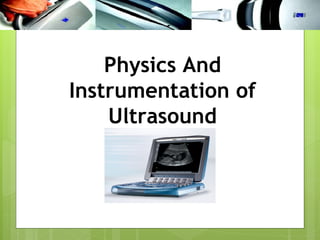
Physics And
Instrumentation of
    Ultrasound
WHAT DO YOU
UNDERSTAND ABOUT
ULTRASOUND ?
Bats navigate using ultrasound
Bats make high-pitched chirps which are too high for
humans to hear. This is called ultrasound
Like normal sound, ultrasound echoes off objects
The bat hears the echoes and works out what caused
them
•Dolphins also navigate with ultrasound
•Submarines use a similar method called sonar
•We can also use ultrasound to look inside the body…
•   Ultrasound

    –   Cyclic sound pressure with a
        frequency greater than the upper
        limit of human hearing.


           • Human Ear  Audible
             Range Frequency?
The human ear can only
The human ear can
only respond to the
audible frequency range
~ 20Hz - 20kHz to the
audible frequency range ~
20Hz - 20kHz
Medical sonography
(ultrasonography)
 Ultrasound-based     diagnostic imaging
    technique used to visualize muscles and
    internal organs, their size, structures and
    possible pathologies or lesions.

   APPLICATIONS?
   ADVANTAGES & DISADVANTAGES?
Diagnostic applications
•   Cardiology
•   Gynaecology & Obstetrics
•   Ophthalmology
•   Abdomen
•   Urology- to determine, for example, the amount of fluid
    retained in a patient's bladder.
•   Musculoskeletal - tendons, muscles, and nerves
•   Vascular - arteries and veins
•   Interventional biopsy - emptying fluids, intrauterine
    transfusion
Therapeutic applications
•   Therapeutic applications use
    ultrasound to bring heat or
    agitation into the body.

•   Therefore much higher energies
    are used than in diagnostic
    ultrasound.
ULTRASOUND PHYSICS
Format
 What is sound/ultrasound?
 How is ultrasound produced
 Transducers - properties
 Effect of Frequency
 Image Formation
 Interaction of ultrasound with   tissue
 Acoustic impedance
 Image appearance
Sound?
 Sound  is a mechanical, longitudinal wave that travels
 in a straight line

 Sound   requires a medium through which to travel
CATEGORIES OF SOUND
 Infrasound (subsonic) below 20Hz
 Audible sound 20-20,000Hz
 Ultrasound above 20,000Hz
 Nondiagnostic medical applications
  <1MHz
 Medical diagnostic ultrasound >1MHz
In 1826 Daniel
Colladon, a Swiss
physicist, and Charles
Sturm, a French
mathematician,
accurately measured its
speed in water. Using a
long tube to listen
underwater (as Leonardo
da Vinci suggested in
1490), they recorded how
fast the sound of a
submerged bell traveled
across Lake Geneva.
Their result--1,435
meters per
second in water of 1.8
degrees Celsius (35
degrees Fahrenheit)--was
only 3 meters per second
off from the speed
accepted today.
Compression wave
Acoustic Variables
•   Period
•   Wavelength
•   Amplitude
•   Frequency
•   Velocity
Acoustic Variables
Acoustic Variables
Amplitude, A (m)
 The maximum displacement that occurs in an
 acoustic variable.
Why we use different frequency?
Basic Ultrasound Physics
                         Amplitude


oscillations/sec = frequency - expressed in
Hertz (Hz)
What is Ultrasound?
 Ultrasound    is a mechanical, longitudinal wave with a
    frequency exceeding the upper limit of human hearing,
    which is 20,000 Hz or 20 kHz.

   Medical Ultrasound 2MHz to 16MHz
ULTRASOUND – How is it produced?
Produced by passing an electrical current through a
  piezoelectrical (material that expands and contracts
  with current) crystal
Human Hair

                        Single
                        Crystal


Microscopic view of scanhead
In ultrasound, the following events
     happen:
1. The ultrasound machine transmits high-
   frequency (1 to 12 megahertz) sound pulses into
   the body using a probe.
2. The sound waves travel into the body and hit a
   boundary between tissues (e.g. between fluid
   and soft tissue, soft tissue and bone).
3. Some of the sound waves reflect back to the
   probe, while some travel on further until they
   reach another boundary and then reflect back
   to the probe .
4. The reflected waves are detected by the probe
   and relayed to the machine.
1.   The machine calculates the distance from
     the probe to the tissue or organ
     (boundaries) using the speed of sound in
     tissue (1540 m/s) and the time of the each
     echo's return (usually on the order of
     millionths of a second).

6. The machine displays the distances and
   intensities of the echoes on the screen,
   forming a two dimensional image.
Piezoelectric material
 ACapplied to a piezoelectric crystal
 causes it to expand and contract –
 generating ultrasound, and vice versa

 Naturally   occurring - quartz

 Synthetic   - Lead zirconate titanate
 (PZT)
Ultrasound Production
 Transducer  produces ultrasound pulses (transmit 1%
  of the time)
 These elements convert electrical energy into a
  mechanical ultrasound wave


 Reflectedechoes return to the scanhead which
 converts the ultrasound wave into an electrical
 signal
Piezoelectric Crystals
  Thethickness of the crystal determines the
  frequency of the scanhead


         Low Frequency   High Frequency
             3 MHz           10 MHz
Frequency also affects the QUALITY of the
 The frequency
                vs. Resolution
  ultrasound image
   The HIGHER the frequency, the BETTER the
    resolution
 The LOWER the frequency, the LESS the resolution
 A 12 MHz transducer has very good resolution, but
  cannot penetrate very deep into the body
 A 3 MHz transducer can penetrate deep into the
  body, but the resolution is not as good as the 12
  MHz


         Low Frequency     High Frequency
             3 MHz             12 MHz
Broadband vs. Narrowband
 Amplitude


             Frequency
Broadband vs. Narrowband
Nerve Visualisation:
 5-10 MHz
 6-13 MHz
 By altering the transmit frequencies one transducer
  replaces several transducers
 View a range of superficial to deep structures without
  changing transducers
Transducer Design


Size, design and
frequency
depend upon the
examination
Image Formation
Electrical signal produces ‘dots’ on the screen

 Brightness of the dots is proportional to the
  strength of the returning echoes
 Location of the dots is determined by travel
  time. The velocity in tissue is assumed constant
  at 1540m/sec
           Distance = Velocity
                           Time
Image Formation


‘B’ mode
Interactions of Ultrasound with Tissue

      Reflection
      Refraction
      Transmission
      Attenuation
Interactions of Ultrasound with Tissue
     Reflection
       The ultrasound reflects off tissue and returns to
        the transducer, the amount of reflection depends on
        differences in acoustic impedance
       The ultrasound image is formed from reflected echoes


  transducer
Refraction
     reflective
                              refraction
Scattered
echoes


     Incident

     Angle of incidence = angle of reflection
Interactions of Ultrasound with Tissue
  Transmission
     Some of the ultrasound waves continue deeper into
      the body
     These waves will reflect from deeper tissue
      structures


  transducer
Interactions of Ultrasound with Tissue
  Attenuation
     Defined - the deeper the wave travels in the
      body, the weaker it becomes -3 processes:
      reflection, absorption, refraction
     Air (lung)> bone > muscle > soft tissue >blood >
      water
Interactions of Ultrasound with
              Tissue
• Acoustic impedance (AI) is dependent on the density of
  the material in which sound is propagated
     - the greater the impedance the denser the material.
• Reflections comes from the interface of different AI’s
    • greater ∆ of the AI = more signal reflected
    • works both ways (send and receive directions)
         Transducer


                      Medium 1   Medium 2   Medium 3
Interaction of Ultrasound
           with Tissue

• Greater the AI, greater the returned signal
    • largest difference is solid-gas interface
    • we don’t like gas or air
    • we don’t like bone for the same reason
                    GEL!!
• Sound is attenuated as it goes deeper into the body
• Z (Rayls) = Density (kg/m³) x Speed (m/s)
• Incident beam has
  normal incidence 90
  degree
  (perpendicular
  incidence) on the
  tissue interface, the
  magnitude of
  reflection can be
  calculated (IRC)

• α  Z values
Attenuation & Gain
 Sound is attenuated by tissue
 More tissue to penetrate = more
  attenuation of signal
 Compensate by adjusting gain based on
  depth
    near field / far field
    AKA: TGC
Ultrasound Gain

 Gain   controls
     receiver gain only
     does NOT change power output
     think: stereo volume
 Increasegain = brighter
 Decrease gain = darker
Balanced Gain
 Gain
     settings are important to obtaining adequate
 images.
                                        bad far field
bad near field


                      balanced
Reflected Echo’s
 Strong   Reflections = White dots
  Diaphragm, tendons, bone
               ‘Hyperechoic’
Reflected Echo’s


Weaker Reflections =
Grey dots


    Most solid organs,
    thick fluid – ‘isoechoic’
Reflected Echo’s
 No   Reflections = Black dots
  Fluid within a cyst, urine, blood
 ‘Hypoechoic’ or echofree
What determines how far ultrasound
 waves can travel?

 The   FREQUENCY of the transducer
    The HIGHER the frequency, the LESS it can penetrate
    The LOWER the frequency, the DEEPER it can penetrate
    Attenuation is directly related to frequency
Ultrasound Beam Depth
• Need to image at proper depth
• Can’t control depth of beam
   • keeps going until attenuated
• You can control the depth of displayed data
Ultrasound Beam Profile
 Beam comes out as a slice
 Beam Profile
    Approx. 1 mm thick
    Depth displayed – user controlled
 Image    produced is “2D”
    tomographic slice
    assumes no thickness
 You   control the aim


                                         1mm
Goal of an Ultrasound System
  The ultimate goal of any ultrasound
  system is to make like tissues look the
  same and unlike tissues look different
Accomplishing this goal depends
upon...

  Resolving   capability of the system
   axial/lateral resolution
   spatial resolution
   contrast resolution
   temporal resolution

  Processing   Power
   ability
          to capture, preserve and display the
    information
Types of Resolution
  Axial   Resolution
      specifies how close together two objects can be along
       the axis of the beam, yet still be detected as two
       separate objects
      frequency (wavelength) affects axial resolution –
       frequency resolution
Types of Resolution
 Lateral   Resolution
     the ability to resolve two adjacent objects that are
      perpendicular to the beam axis as separate objects
     beamwidth affects lateral resolution
Types of Resolution
 Spatial   Resolution
     also called Detail Resolution

     the combination of AXIAL and LATERAL resolution -
      how closely two reflectors can be to one another
      while they can be identified as different reflectors
Types of Resolution
 Temporal    Resolution
     the ability to accurately locate the position of moving
      structures at particular instants in time
     also known as frame rate
Types of Resolution
 Contrast   Resolution
     the ability to resolve two adjacent objects of similar
      intensity/reflective properties as separate objects -
      dependant on the dynamic range
Liver metastases
Ultrasound Applications
Visualisation Tool:
Nerves, soft tissue masses
Vessels - assessment of position, size,
patency
Ultrasound Guided Procedures in real
time – dynamic imaging; central venous
access, nerve blocks
Imaging

Know your anatomy – Skin, muscle,
tendons, nerves and vessels
Recognise normal appearances –
compare sides!
Skin, subcutaneous tissue
           Epidermis


           Loose connective tissue and subcutaneous fat
           is hypoechoic


           Muscle interface


           Muscle fibres interface


           Bone
Transverse scan – Internal Jugular Vein and
         Common Carotid Artery
Summary

•Imaging tool – Must have the knowledge to
understand how the image is formed


•Dynamic technique


•Acquisition and interpretation dependant
upon the skills of the operator.
Ultrasound Physics and Applications

More Related Content

What's hot

Doppler Ultrasonography And Advancements in USG
Doppler Ultrasonography And Advancements in USGDoppler Ultrasonography And Advancements in USG
Doppler Ultrasonography And Advancements in USGSudil Paudyal
 
Introduction to ultarsound machine and physics
Introduction to ultarsound machine and physicsIntroduction to ultarsound machine and physics
Introduction to ultarsound machine and physicsmanishyadav513
 
Bioeffect Of Ultra Sound
Bioeffect Of  Ultra SoundBioeffect Of  Ultra Sound
Bioeffect Of Ultra SoundShatha M
 
Principle of usg imaging, construction of transducers
Principle of usg imaging, construction of transducersPrinciple of usg imaging, construction of transducers
Principle of usg imaging, construction of transducersDev Lakhera
 
Tissue harmonic imaging
Tissue harmonic imaging Tissue harmonic imaging
Tissue harmonic imaging MAMTA PANDA
 
Ultrasound transducer doppler ppt pdf pk
Ultrasound transducer doppler ppt pdf pkUltrasound transducer doppler ppt pdf pk
Ultrasound transducer doppler ppt pdf pkDr pradeep Kumar
 
Ultrasound imaging
Ultrasound imagingUltrasound imaging
Ultrasound imagingAnjan Dangal
 
Physics of Ultrasound Imaging
Physics of Ultrasound ImagingPhysics of Ultrasound Imaging
Physics of Ultrasound Imagingu.surgery
 
Ultrasound imaging
Ultrasound imagingUltrasound imaging
Ultrasound imagingAbid Hussain
 
Ultrasound artifacts
Ultrasound artifactsUltrasound artifacts
Ultrasound artifactsansaripv
 
Ultrasound &amp; properties
Ultrasound &amp;  propertiesUltrasound &amp;  properties
Ultrasound &amp; propertiesMahesh Kumar
 
ULTRASOUND IMAGING PRINCIPLES
ULTRASOUND IMAGING PRINCIPLESULTRASOUND IMAGING PRINCIPLES
ULTRASOUND IMAGING PRINCIPLESINDIA ULTRASOUND
 
Interactions of ultrasound with matter
Interactions of ultrasound with matterInteractions of ultrasound with matter
Interactions of ultrasound with matterSwapnil Shetty
 
Types of Ultrasound Probes
Types of Ultrasound ProbesTypes of Ultrasound Probes
Types of Ultrasound Probeshafsatalat1
 
Physical Principles Of Ultrasound
Physical Principles Of UltrasoundPhysical Principles Of Ultrasound
Physical Principles Of Ultrasoundu.surgery
 
Ultrasound physics and image optimization1 (1)
Ultrasound physics and image optimization1 (1)Ultrasound physics and image optimization1 (1)
Ultrasound physics and image optimization1 (1)Prajwith Rai
 

What's hot (20)

Doppler Ultrasonography And Advancements in USG
Doppler Ultrasonography And Advancements in USGDoppler Ultrasonography And Advancements in USG
Doppler Ultrasonography And Advancements in USG
 
Introduction to ultarsound machine and physics
Introduction to ultarsound machine and physicsIntroduction to ultarsound machine and physics
Introduction to ultarsound machine and physics
 
Bioeffect Of Ultra Sound
Bioeffect Of  Ultra SoundBioeffect Of  Ultra Sound
Bioeffect Of Ultra Sound
 
Principle of usg imaging, construction of transducers
Principle of usg imaging, construction of transducersPrinciple of usg imaging, construction of transducers
Principle of usg imaging, construction of transducers
 
Tissue harmonic imaging
Tissue harmonic imaging Tissue harmonic imaging
Tissue harmonic imaging
 
Ultrasound physics
Ultrasound physicsUltrasound physics
Ultrasound physics
 
Ultrasound transducer doppler ppt pdf pk
Ultrasound transducer doppler ppt pdf pkUltrasound transducer doppler ppt pdf pk
Ultrasound transducer doppler ppt pdf pk
 
Ultrasound imaging
Ultrasound imagingUltrasound imaging
Ultrasound imaging
 
Physics of Ultrasound Imaging
Physics of Ultrasound ImagingPhysics of Ultrasound Imaging
Physics of Ultrasound Imaging
 
Dual Energy CT
Dual Energy CTDual Energy CT
Dual Energy CT
 
Ultrasound imaging
Ultrasound imagingUltrasound imaging
Ultrasound imaging
 
Basics Physics of ultrasound
Basics Physics of ultrasoundBasics Physics of ultrasound
Basics Physics of ultrasound
 
Ultrasound artifacts
Ultrasound artifactsUltrasound artifacts
Ultrasound artifacts
 
Ultrasound &amp; properties
Ultrasound &amp;  propertiesUltrasound &amp;  properties
Ultrasound &amp; properties
 
ULTRASOUND IMAGING PRINCIPLES
ULTRASOUND IMAGING PRINCIPLESULTRASOUND IMAGING PRINCIPLES
ULTRASOUND IMAGING PRINCIPLES
 
Ct instrument
Ct instrumentCt instrument
Ct instrument
 
Interactions of ultrasound with matter
Interactions of ultrasound with matterInteractions of ultrasound with matter
Interactions of ultrasound with matter
 
Types of Ultrasound Probes
Types of Ultrasound ProbesTypes of Ultrasound Probes
Types of Ultrasound Probes
 
Physical Principles Of Ultrasound
Physical Principles Of UltrasoundPhysical Principles Of Ultrasound
Physical Principles Of Ultrasound
 
Ultrasound physics and image optimization1 (1)
Ultrasound physics and image optimization1 (1)Ultrasound physics and image optimization1 (1)
Ultrasound physics and image optimization1 (1)
 

Viewers also liked

ultrasound transducers and resolution
ultrasound transducers and resolutionultrasound transducers and resolution
ultrasound transducers and resolutionVallabhaneni Bhupal
 
Understanding ultrasound
Understanding ultrasoundUnderstanding ultrasound
Understanding ultrasoundMohammad Amir
 
Ultrasound Physics
Ultrasound PhysicsUltrasound Physics
Ultrasound Physicsu.surgery
 
01 basic principles of ultrasound & basic term
01 basic principles of ultrasound & basic term01 basic principles of ultrasound & basic term
01 basic principles of ultrasound & basic termJuan Alcatruz
 
Basic gynae ultrasound
Basic gynae ultrasoundBasic gynae ultrasound
Basic gynae ultrasoundobsgynhsnz
 
Basic Obstetric Ultrasound
Basic Obstetric UltrasoundBasic Obstetric Ultrasound
Basic Obstetric UltrasoundDoctorsask
 
Vibration advance longi and trans equation.
Vibration advance   longi and trans equation.Vibration advance   longi and trans equation.
Vibration advance longi and trans equation.Khalis Karim
 
The Cranial nerves
The Cranial nerves The Cranial nerves
The Cranial nerves mac os
 
Physics of ultrasound
Physics of ultrasoundPhysics of ultrasound
Physics of ultrasoundanmyamas
 
Approach to gynaecology patient
Approach to gynaecology patientApproach to gynaecology patient
Approach to gynaecology patientobsgynhsnz
 
The Role of Ultrasound in Obstetric and Gynaecology
The Role of Ultrasound in Obstetric and GynaecologyThe Role of Ultrasound in Obstetric and Gynaecology
The Role of Ultrasound in Obstetric and GynaecologyMohammad Amir
 
Point of Care Ultrasound - Hyperechoic Future in Medical School?
Point of Care Ultrasound - Hyperechoic Future in Medical School?Point of Care Ultrasound - Hyperechoic Future in Medical School?
Point of Care Ultrasound - Hyperechoic Future in Medical School?cbyrne2014
 
Ankle Mobility Presentation
Ankle Mobility PresentationAnkle Mobility Presentation
Ankle Mobility PresentationBrent Rasmussen
 
Point of Care Ultrasound - Hyperechoic Future in Family Practice?
Point of Care Ultrasound - Hyperechoic Future in Family Practice?Point of Care Ultrasound - Hyperechoic Future in Family Practice?
Point of Care Ultrasound - Hyperechoic Future in Family Practice?cbyrne2014
 
Presentation1.pptx, ultrasound study of the spleen and pancreas.
Presentation1.pptx, ultrasound study of the spleen and pancreas.Presentation1.pptx, ultrasound study of the spleen and pancreas.
Presentation1.pptx, ultrasound study of the spleen and pancreas.Abdellah Nazeer
 

Viewers also liked (20)

Basics of Ultrasound
Basics of UltrasoundBasics of Ultrasound
Basics of Ultrasound
 
ultrasound transducers and resolution
ultrasound transducers and resolutionultrasound transducers and resolution
ultrasound transducers and resolution
 
Understanding ultrasound
Understanding ultrasoundUnderstanding ultrasound
Understanding ultrasound
 
Ultrasound physics
Ultrasound physicsUltrasound physics
Ultrasound physics
 
Ultrasound Physics
Ultrasound PhysicsUltrasound Physics
Ultrasound Physics
 
01 basic principles of ultrasound & basic term
01 basic principles of ultrasound & basic term01 basic principles of ultrasound & basic term
01 basic principles of ultrasound & basic term
 
Basic gynae ultrasound
Basic gynae ultrasoundBasic gynae ultrasound
Basic gynae ultrasound
 
Basic Obstetric Ultrasound
Basic Obstetric UltrasoundBasic Obstetric Ultrasound
Basic Obstetric Ultrasound
 
Vibration advance longi and trans equation.
Vibration advance   longi and trans equation.Vibration advance   longi and trans equation.
Vibration advance longi and trans equation.
 
Doppler fisica
Doppler fisicaDoppler fisica
Doppler fisica
 
The Cranial nerves
The Cranial nerves The Cranial nerves
The Cranial nerves
 
Physics of ultrasound
Physics of ultrasoundPhysics of ultrasound
Physics of ultrasound
 
Approach to gynaecology patient
Approach to gynaecology patientApproach to gynaecology patient
Approach to gynaecology patient
 
The Role of Ultrasound in Obstetric and Gynaecology
The Role of Ultrasound in Obstetric and GynaecologyThe Role of Ultrasound in Obstetric and Gynaecology
The Role of Ultrasound in Obstetric and Gynaecology
 
Physics of echo i.tammi raju
Physics of echo i.tammi rajuPhysics of echo i.tammi raju
Physics of echo i.tammi raju
 
Point of Care Ultrasound - Hyperechoic Future in Medical School?
Point of Care Ultrasound - Hyperechoic Future in Medical School?Point of Care Ultrasound - Hyperechoic Future in Medical School?
Point of Care Ultrasound - Hyperechoic Future in Medical School?
 
Ankle Mobility Presentation
Ankle Mobility PresentationAnkle Mobility Presentation
Ankle Mobility Presentation
 
Point of Care Ultrasound - Hyperechoic Future in Family Practice?
Point of Care Ultrasound - Hyperechoic Future in Family Practice?Point of Care Ultrasound - Hyperechoic Future in Family Practice?
Point of Care Ultrasound - Hyperechoic Future in Family Practice?
 
U ltrasound physiscs
U ltrasound physiscsU ltrasound physiscs
U ltrasound physiscs
 
Presentation1.pptx, ultrasound study of the spleen and pancreas.
Presentation1.pptx, ultrasound study of the spleen and pancreas.Presentation1.pptx, ultrasound study of the spleen and pancreas.
Presentation1.pptx, ultrasound study of the spleen and pancreas.
 

Similar to Ultrasound Physics and Applications

Applications of Ultrasound in Medicine
Applications of Ultrasound in MedicineApplications of Ultrasound in Medicine
Applications of Ultrasound in MedicinePranay Dutta
 
Presentation1, basic principle of ultrasound.
Presentation1, basic principle of ultrasound.Presentation1, basic principle of ultrasound.
Presentation1, basic principle of ultrasound.Abdellah Nazeer
 
BIS CHAPTER ONE.pptx
BIS CHAPTER ONE.pptxBIS CHAPTER ONE.pptx
BIS CHAPTER ONE.pptxTirusew1
 
Basics of ultrasound machine
Basics of ultrasound machineBasics of ultrasound machine
Basics of ultrasound machineLokender Yadav
 
BASICS OF US & DUPLEX (NEW).ppt
BASICS OF US & DUPLEX (NEW).pptBASICS OF US & DUPLEX (NEW).ppt
BASICS OF US & DUPLEX (NEW).pptmohamedabushanab1
 
Principles of Ultrasound Physics.pptx
Principles of Ultrasound Physics.pptxPrinciples of Ultrasound Physics.pptx
Principles of Ultrasound Physics.pptxGemechisAsefa1
 
Physics of ultrasound and echocardiography
Physics of ultrasound and echocardiographyPhysics of ultrasound and echocardiography
Physics of ultrasound and echocardiographyjeetshitole
 
Basic of ultrasound
Basic of ultrasoundBasic of ultrasound
Basic of ultrasoundAsad Moosa
 
Emergency us physics and instrumentation
Emergency us physics  and instrumentationEmergency us physics  and instrumentation
Emergency us physics and instrumentationdrnikahmad
 
Ultrasound Imaging_2023.pdf
Ultrasound Imaging_2023.pdfUltrasound Imaging_2023.pdf
Ultrasound Imaging_2023.pdfghadaElbanby1
 
Ultrasonography final
Ultrasonography finalUltrasonography final
Ultrasonography finalravikumarbme
 
basicsofultrasound-170807183128 (1).pdf
basicsofultrasound-170807183128 (1).pdfbasicsofultrasound-170807183128 (1).pdf
basicsofultrasound-170807183128 (1).pdflittlealphonsa
 
basicsofultrasound-170807183128 (1).pdf
basicsofultrasound-170807183128 (1).pdfbasicsofultrasound-170807183128 (1).pdf
basicsofultrasound-170807183128 (1).pdfMohamedAli690824
 
8159968.ppt
8159968.ppt8159968.ppt
8159968.pptYtchechy
 
ultra sound.pptx
ultra sound.pptxultra sound.pptx
ultra sound.pptxAliMRiyath
 
Usg applications in anaesthesia dr gs
Usg applications in anaesthesia dr gsUsg applications in anaesthesia dr gs
Usg applications in anaesthesia dr gsGowri Shankar
 

Similar to Ultrasound Physics and Applications (20)

Applications of Ultrasound in Medicine
Applications of Ultrasound in MedicineApplications of Ultrasound in Medicine
Applications of Ultrasound in Medicine
 
Presentation1, basic principle of ultrasound.
Presentation1, basic principle of ultrasound.Presentation1, basic principle of ultrasound.
Presentation1, basic principle of ultrasound.
 
BIS CHAPTER ONE.pptx
BIS CHAPTER ONE.pptxBIS CHAPTER ONE.pptx
BIS CHAPTER ONE.pptx
 
Basics of ultrasound machine
Basics of ultrasound machineBasics of ultrasound machine
Basics of ultrasound machine
 
BASICS OF US & DUPLEX (NEW).ppt
BASICS OF US & DUPLEX (NEW).pptBASICS OF US & DUPLEX (NEW).ppt
BASICS OF US & DUPLEX (NEW).ppt
 
Principles of Ultrasound Physics.pptx
Principles of Ultrasound Physics.pptxPrinciples of Ultrasound Physics.pptx
Principles of Ultrasound Physics.pptx
 
Physics of ultrasound and echocardiography
Physics of ultrasound and echocardiographyPhysics of ultrasound and echocardiography
Physics of ultrasound and echocardiography
 
Basic of ultrasound
Basic of ultrasoundBasic of ultrasound
Basic of ultrasound
 
Emergency us physics and instrumentation
Emergency us physics  and instrumentationEmergency us physics  and instrumentation
Emergency us physics and instrumentation
 
PRINCIPLES OF ULTRASONOGRAPHY
PRINCIPLES OF ULTRASONOGRAPHYPRINCIPLES OF ULTRASONOGRAPHY
PRINCIPLES OF ULTRASONOGRAPHY
 
Ultrasound Imaging_2023.pdf
Ultrasound Imaging_2023.pdfUltrasound Imaging_2023.pdf
Ultrasound Imaging_2023.pdf
 
Ultrasonography final
Ultrasonography finalUltrasonography final
Ultrasonography final
 
Paper
PaperPaper
Paper
 
basicsofultrasound-170807183128 (1).pdf
basicsofultrasound-170807183128 (1).pdfbasicsofultrasound-170807183128 (1).pdf
basicsofultrasound-170807183128 (1).pdf
 
basicsofultrasound-170807183128 (1).pdf
basicsofultrasound-170807183128 (1).pdfbasicsofultrasound-170807183128 (1).pdf
basicsofultrasound-170807183128 (1).pdf
 
PHYSICS REPORT.pptx
PHYSICS REPORT.pptxPHYSICS REPORT.pptx
PHYSICS REPORT.pptx
 
Us physics (4)
Us physics (4)Us physics (4)
Us physics (4)
 
8159968.ppt
8159968.ppt8159968.ppt
8159968.ppt
 
ultra sound.pptx
ultra sound.pptxultra sound.pptx
ultra sound.pptx
 
Usg applications in anaesthesia dr gs
Usg applications in anaesthesia dr gsUsg applications in anaesthesia dr gs
Usg applications in anaesthesia dr gs
 

Recently uploaded

Accessible design: Minimum effort, maximum impact
Accessible design: Minimum effort, maximum impactAccessible design: Minimum effort, maximum impact
Accessible design: Minimum effort, maximum impactdawncurless
 
APM Welcome, APM North West Network Conference, Synergies Across Sectors
APM Welcome, APM North West Network Conference, Synergies Across SectorsAPM Welcome, APM North West Network Conference, Synergies Across Sectors
APM Welcome, APM North West Network Conference, Synergies Across SectorsAssociation for Project Management
 
Software Engineering Methodologies (overview)
Software Engineering Methodologies (overview)Software Engineering Methodologies (overview)
Software Engineering Methodologies (overview)eniolaolutunde
 
Organic Name Reactions for the students and aspirants of Chemistry12th.pptx
Organic Name Reactions  for the students and aspirants of Chemistry12th.pptxOrganic Name Reactions  for the students and aspirants of Chemistry12th.pptx
Organic Name Reactions for the students and aspirants of Chemistry12th.pptxVS Mahajan Coaching Centre
 
Web & Social Media Analytics Previous Year Question Paper.pdf
Web & Social Media Analytics Previous Year Question Paper.pdfWeb & Social Media Analytics Previous Year Question Paper.pdf
Web & Social Media Analytics Previous Year Question Paper.pdfJayanti Pande
 
Separation of Lanthanides/ Lanthanides and Actinides
Separation of Lanthanides/ Lanthanides and ActinidesSeparation of Lanthanides/ Lanthanides and Actinides
Separation of Lanthanides/ Lanthanides and ActinidesFatimaKhan178732
 
Measures of Central Tendency: Mean, Median and Mode
Measures of Central Tendency: Mean, Median and ModeMeasures of Central Tendency: Mean, Median and Mode
Measures of Central Tendency: Mean, Median and ModeThiyagu K
 
Russian Call Girls in Andheri Airport Mumbai WhatsApp 9167673311 💞 Full Nigh...
Russian Call Girls in Andheri Airport Mumbai WhatsApp  9167673311 💞 Full Nigh...Russian Call Girls in Andheri Airport Mumbai WhatsApp  9167673311 💞 Full Nigh...
Russian Call Girls in Andheri Airport Mumbai WhatsApp 9167673311 💞 Full Nigh...Pooja Nehwal
 
A Critique of the Proposed National Education Policy Reform
A Critique of the Proposed National Education Policy ReformA Critique of the Proposed National Education Policy Reform
A Critique of the Proposed National Education Policy ReformChameera Dedduwage
 
Kisan Call Centre - To harness potential of ICT in Agriculture by answer farm...
Kisan Call Centre - To harness potential of ICT in Agriculture by answer farm...Kisan Call Centre - To harness potential of ICT in Agriculture by answer farm...
Kisan Call Centre - To harness potential of ICT in Agriculture by answer farm...Krashi Coaching
 
CARE OF CHILD IN INCUBATOR..........pptx
CARE OF CHILD IN INCUBATOR..........pptxCARE OF CHILD IN INCUBATOR..........pptx
CARE OF CHILD IN INCUBATOR..........pptxGaneshChakor2
 
1029 - Danh muc Sach Giao Khoa 10 . pdf
1029 -  Danh muc Sach Giao Khoa 10 . pdf1029 -  Danh muc Sach Giao Khoa 10 . pdf
1029 - Danh muc Sach Giao Khoa 10 . pdfQucHHunhnh
 
Advanced Views - Calendar View in Odoo 17
Advanced Views - Calendar View in Odoo 17Advanced Views - Calendar View in Odoo 17
Advanced Views - Calendar View in Odoo 17Celine George
 
JAPAN: ORGANISATION OF PMDA, PHARMACEUTICAL LAWS & REGULATIONS, TYPES OF REGI...
JAPAN: ORGANISATION OF PMDA, PHARMACEUTICAL LAWS & REGULATIONS, TYPES OF REGI...JAPAN: ORGANISATION OF PMDA, PHARMACEUTICAL LAWS & REGULATIONS, TYPES OF REGI...
JAPAN: ORGANISATION OF PMDA, PHARMACEUTICAL LAWS & REGULATIONS, TYPES OF REGI...anjaliyadav012327
 
POINT- BIOCHEMISTRY SEM 2 ENZYMES UNIT 5.pptx
POINT- BIOCHEMISTRY SEM 2 ENZYMES UNIT 5.pptxPOINT- BIOCHEMISTRY SEM 2 ENZYMES UNIT 5.pptx
POINT- BIOCHEMISTRY SEM 2 ENZYMES UNIT 5.pptxSayali Powar
 
The Most Excellent Way | 1 Corinthians 13
The Most Excellent Way | 1 Corinthians 13The Most Excellent Way | 1 Corinthians 13
The Most Excellent Way | 1 Corinthians 13Steve Thomason
 

Recently uploaded (20)

Mattingly "AI & Prompt Design: The Basics of Prompt Design"
Mattingly "AI & Prompt Design: The Basics of Prompt Design"Mattingly "AI & Prompt Design: The Basics of Prompt Design"
Mattingly "AI & Prompt Design: The Basics of Prompt Design"
 
Accessible design: Minimum effort, maximum impact
Accessible design: Minimum effort, maximum impactAccessible design: Minimum effort, maximum impact
Accessible design: Minimum effort, maximum impact
 
APM Welcome, APM North West Network Conference, Synergies Across Sectors
APM Welcome, APM North West Network Conference, Synergies Across SectorsAPM Welcome, APM North West Network Conference, Synergies Across Sectors
APM Welcome, APM North West Network Conference, Synergies Across Sectors
 
Software Engineering Methodologies (overview)
Software Engineering Methodologies (overview)Software Engineering Methodologies (overview)
Software Engineering Methodologies (overview)
 
Organic Name Reactions for the students and aspirants of Chemistry12th.pptx
Organic Name Reactions  for the students and aspirants of Chemistry12th.pptxOrganic Name Reactions  for the students and aspirants of Chemistry12th.pptx
Organic Name Reactions for the students and aspirants of Chemistry12th.pptx
 
Web & Social Media Analytics Previous Year Question Paper.pdf
Web & Social Media Analytics Previous Year Question Paper.pdfWeb & Social Media Analytics Previous Year Question Paper.pdf
Web & Social Media Analytics Previous Year Question Paper.pdf
 
Código Creativo y Arte de Software | Unidad 1
Código Creativo y Arte de Software | Unidad 1Código Creativo y Arte de Software | Unidad 1
Código Creativo y Arte de Software | Unidad 1
 
Separation of Lanthanides/ Lanthanides and Actinides
Separation of Lanthanides/ Lanthanides and ActinidesSeparation of Lanthanides/ Lanthanides and Actinides
Separation of Lanthanides/ Lanthanides and Actinides
 
Measures of Central Tendency: Mean, Median and Mode
Measures of Central Tendency: Mean, Median and ModeMeasures of Central Tendency: Mean, Median and Mode
Measures of Central Tendency: Mean, Median and Mode
 
Russian Call Girls in Andheri Airport Mumbai WhatsApp 9167673311 💞 Full Nigh...
Russian Call Girls in Andheri Airport Mumbai WhatsApp  9167673311 💞 Full Nigh...Russian Call Girls in Andheri Airport Mumbai WhatsApp  9167673311 💞 Full Nigh...
Russian Call Girls in Andheri Airport Mumbai WhatsApp 9167673311 💞 Full Nigh...
 
A Critique of the Proposed National Education Policy Reform
A Critique of the Proposed National Education Policy ReformA Critique of the Proposed National Education Policy Reform
A Critique of the Proposed National Education Policy Reform
 
Kisan Call Centre - To harness potential of ICT in Agriculture by answer farm...
Kisan Call Centre - To harness potential of ICT in Agriculture by answer farm...Kisan Call Centre - To harness potential of ICT in Agriculture by answer farm...
Kisan Call Centre - To harness potential of ICT in Agriculture by answer farm...
 
INDIA QUIZ 2024 RLAC DELHI UNIVERSITY.pptx
INDIA QUIZ 2024 RLAC DELHI UNIVERSITY.pptxINDIA QUIZ 2024 RLAC DELHI UNIVERSITY.pptx
INDIA QUIZ 2024 RLAC DELHI UNIVERSITY.pptx
 
CARE OF CHILD IN INCUBATOR..........pptx
CARE OF CHILD IN INCUBATOR..........pptxCARE OF CHILD IN INCUBATOR..........pptx
CARE OF CHILD IN INCUBATOR..........pptx
 
1029 - Danh muc Sach Giao Khoa 10 . pdf
1029 -  Danh muc Sach Giao Khoa 10 . pdf1029 -  Danh muc Sach Giao Khoa 10 . pdf
1029 - Danh muc Sach Giao Khoa 10 . pdf
 
Advance Mobile Application Development class 07
Advance Mobile Application Development class 07Advance Mobile Application Development class 07
Advance Mobile Application Development class 07
 
Advanced Views - Calendar View in Odoo 17
Advanced Views - Calendar View in Odoo 17Advanced Views - Calendar View in Odoo 17
Advanced Views - Calendar View in Odoo 17
 
JAPAN: ORGANISATION OF PMDA, PHARMACEUTICAL LAWS & REGULATIONS, TYPES OF REGI...
JAPAN: ORGANISATION OF PMDA, PHARMACEUTICAL LAWS & REGULATIONS, TYPES OF REGI...JAPAN: ORGANISATION OF PMDA, PHARMACEUTICAL LAWS & REGULATIONS, TYPES OF REGI...
JAPAN: ORGANISATION OF PMDA, PHARMACEUTICAL LAWS & REGULATIONS, TYPES OF REGI...
 
POINT- BIOCHEMISTRY SEM 2 ENZYMES UNIT 5.pptx
POINT- BIOCHEMISTRY SEM 2 ENZYMES UNIT 5.pptxPOINT- BIOCHEMISTRY SEM 2 ENZYMES UNIT 5.pptx
POINT- BIOCHEMISTRY SEM 2 ENZYMES UNIT 5.pptx
 
The Most Excellent Way | 1 Corinthians 13
The Most Excellent Way | 1 Corinthians 13The Most Excellent Way | 1 Corinthians 13
The Most Excellent Way | 1 Corinthians 13
 

Ultrasound Physics and Applications

  • 2. WHAT DO YOU UNDERSTAND ABOUT ULTRASOUND ?
  • 3. Bats navigate using ultrasound
  • 4. Bats make high-pitched chirps which are too high for humans to hear. This is called ultrasound Like normal sound, ultrasound echoes off objects The bat hears the echoes and works out what caused them •Dolphins also navigate with ultrasound •Submarines use a similar method called sonar •We can also use ultrasound to look inside the body…
  • 5. Ultrasound – Cyclic sound pressure with a frequency greater than the upper limit of human hearing. • Human Ear  Audible Range Frequency?
  • 6. The human ear can only The human ear can only respond to the audible frequency range ~ 20Hz - 20kHz to the audible frequency range ~ 20Hz - 20kHz
  • 7. Medical sonography (ultrasonography)  Ultrasound-based diagnostic imaging technique used to visualize muscles and internal organs, their size, structures and possible pathologies or lesions.  APPLICATIONS?  ADVANTAGES & DISADVANTAGES?
  • 8. Diagnostic applications • Cardiology • Gynaecology & Obstetrics • Ophthalmology • Abdomen • Urology- to determine, for example, the amount of fluid retained in a patient's bladder. • Musculoskeletal - tendons, muscles, and nerves • Vascular - arteries and veins • Interventional biopsy - emptying fluids, intrauterine transfusion
  • 9. Therapeutic applications • Therapeutic applications use ultrasound to bring heat or agitation into the body. • Therefore much higher energies are used than in diagnostic ultrasound.
  • 10. ULTRASOUND PHYSICS Format  What is sound/ultrasound?  How is ultrasound produced  Transducers - properties  Effect of Frequency  Image Formation  Interaction of ultrasound with tissue  Acoustic impedance  Image appearance
  • 11. Sound?  Sound is a mechanical, longitudinal wave that travels in a straight line  Sound requires a medium through which to travel
  • 12. CATEGORIES OF SOUND  Infrasound (subsonic) below 20Hz  Audible sound 20-20,000Hz  Ultrasound above 20,000Hz  Nondiagnostic medical applications <1MHz  Medical diagnostic ultrasound >1MHz
  • 13.
  • 14.
  • 15. In 1826 Daniel Colladon, a Swiss physicist, and Charles Sturm, a French mathematician, accurately measured its speed in water. Using a long tube to listen underwater (as Leonardo da Vinci suggested in 1490), they recorded how fast the sound of a submerged bell traveled across Lake Geneva. Their result--1,435 meters per second in water of 1.8 degrees Celsius (35 degrees Fahrenheit)--was only 3 meters per second off from the speed accepted today.
  • 17. Acoustic Variables • Period • Wavelength • Amplitude • Frequency • Velocity
  • 20.
  • 21. Amplitude, A (m) The maximum displacement that occurs in an acoustic variable.
  • 22.
  • 23.
  • 24.
  • 25.
  • 26. Why we use different frequency?
  • 27.
  • 28. Basic Ultrasound Physics Amplitude oscillations/sec = frequency - expressed in Hertz (Hz)
  • 29. What is Ultrasound?  Ultrasound is a mechanical, longitudinal wave with a frequency exceeding the upper limit of human hearing, which is 20,000 Hz or 20 kHz.  Medical Ultrasound 2MHz to 16MHz
  • 30. ULTRASOUND – How is it produced? Produced by passing an electrical current through a piezoelectrical (material that expands and contracts with current) crystal
  • 31. Human Hair Single Crystal Microscopic view of scanhead
  • 32. In ultrasound, the following events happen: 1. The ultrasound machine transmits high- frequency (1 to 12 megahertz) sound pulses into the body using a probe. 2. The sound waves travel into the body and hit a boundary between tissues (e.g. between fluid and soft tissue, soft tissue and bone). 3. Some of the sound waves reflect back to the probe, while some travel on further until they reach another boundary and then reflect back to the probe . 4. The reflected waves are detected by the probe and relayed to the machine.
  • 33. 1. The machine calculates the distance from the probe to the tissue or organ (boundaries) using the speed of sound in tissue (1540 m/s) and the time of the each echo's return (usually on the order of millionths of a second). 6. The machine displays the distances and intensities of the echoes on the screen, forming a two dimensional image.
  • 34. Piezoelectric material  ACapplied to a piezoelectric crystal causes it to expand and contract – generating ultrasound, and vice versa  Naturally occurring - quartz  Synthetic - Lead zirconate titanate (PZT)
  • 35. Ultrasound Production  Transducer produces ultrasound pulses (transmit 1% of the time)  These elements convert electrical energy into a mechanical ultrasound wave  Reflectedechoes return to the scanhead which converts the ultrasound wave into an electrical signal
  • 36. Piezoelectric Crystals  Thethickness of the crystal determines the frequency of the scanhead Low Frequency High Frequency 3 MHz 10 MHz
  • 37. Frequency also affects the QUALITY of the  The frequency vs. Resolution ultrasound image  The HIGHER the frequency, the BETTER the resolution  The LOWER the frequency, the LESS the resolution  A 12 MHz transducer has very good resolution, but cannot penetrate very deep into the body  A 3 MHz transducer can penetrate deep into the body, but the resolution is not as good as the 12 MHz Low Frequency High Frequency 3 MHz 12 MHz
  • 38. Broadband vs. Narrowband Amplitude Frequency
  • 39. Broadband vs. Narrowband Nerve Visualisation:  5-10 MHz  6-13 MHz  By altering the transmit frequencies one transducer replaces several transducers  View a range of superficial to deep structures without changing transducers
  • 40. Transducer Design Size, design and frequency depend upon the examination
  • 41. Image Formation Electrical signal produces ‘dots’ on the screen  Brightness of the dots is proportional to the strength of the returning echoes  Location of the dots is determined by travel time. The velocity in tissue is assumed constant at 1540m/sec Distance = Velocity Time
  • 43. Interactions of Ultrasound with Tissue  Reflection  Refraction  Transmission  Attenuation
  • 44. Interactions of Ultrasound with Tissue  Reflection  The ultrasound reflects off tissue and returns to the transducer, the amount of reflection depends on differences in acoustic impedance  The ultrasound image is formed from reflected echoes transducer
  • 45. Refraction reflective refraction Scattered echoes Incident Angle of incidence = angle of reflection
  • 46. Interactions of Ultrasound with Tissue Transmission  Some of the ultrasound waves continue deeper into the body  These waves will reflect from deeper tissue structures transducer
  • 47. Interactions of Ultrasound with Tissue Attenuation  Defined - the deeper the wave travels in the body, the weaker it becomes -3 processes: reflection, absorption, refraction  Air (lung)> bone > muscle > soft tissue >blood > water
  • 48. Interactions of Ultrasound with Tissue • Acoustic impedance (AI) is dependent on the density of the material in which sound is propagated - the greater the impedance the denser the material. • Reflections comes from the interface of different AI’s • greater ∆ of the AI = more signal reflected • works both ways (send and receive directions) Transducer Medium 1 Medium 2 Medium 3
  • 49. Interaction of Ultrasound with Tissue • Greater the AI, greater the returned signal • largest difference is solid-gas interface • we don’t like gas or air • we don’t like bone for the same reason GEL!! • Sound is attenuated as it goes deeper into the body
  • 50. • Z (Rayls) = Density (kg/m³) x Speed (m/s)
  • 51. • Incident beam has normal incidence 90 degree (perpendicular incidence) on the tissue interface, the magnitude of reflection can be calculated (IRC) • α  Z values
  • 52.
  • 53.
  • 54.
  • 55. Attenuation & Gain  Sound is attenuated by tissue  More tissue to penetrate = more attenuation of signal  Compensate by adjusting gain based on depth  near field / far field  AKA: TGC
  • 56. Ultrasound Gain  Gain controls  receiver gain only  does NOT change power output  think: stereo volume  Increasegain = brighter  Decrease gain = darker
  • 57. Balanced Gain  Gain settings are important to obtaining adequate images. bad far field bad near field balanced
  • 58. Reflected Echo’s  Strong Reflections = White dots Diaphragm, tendons, bone ‘Hyperechoic’
  • 59. Reflected Echo’s Weaker Reflections = Grey dots  Most solid organs,  thick fluid – ‘isoechoic’
  • 60. Reflected Echo’s  No Reflections = Black dots  Fluid within a cyst, urine, blood ‘Hypoechoic’ or echofree
  • 61. What determines how far ultrasound waves can travel?  The FREQUENCY of the transducer  The HIGHER the frequency, the LESS it can penetrate  The LOWER the frequency, the DEEPER it can penetrate  Attenuation is directly related to frequency
  • 62. Ultrasound Beam Depth • Need to image at proper depth • Can’t control depth of beam • keeps going until attenuated • You can control the depth of displayed data
  • 63. Ultrasound Beam Profile  Beam comes out as a slice  Beam Profile  Approx. 1 mm thick  Depth displayed – user controlled  Image produced is “2D”  tomographic slice  assumes no thickness  You control the aim 1mm
  • 64. Goal of an Ultrasound System  The ultimate goal of any ultrasound system is to make like tissues look the same and unlike tissues look different
  • 65. Accomplishing this goal depends upon...  Resolving capability of the system  axial/lateral resolution  spatial resolution  contrast resolution  temporal resolution  Processing Power  ability to capture, preserve and display the information
  • 66. Types of Resolution  Axial Resolution  specifies how close together two objects can be along the axis of the beam, yet still be detected as two separate objects  frequency (wavelength) affects axial resolution – frequency resolution
  • 67. Types of Resolution  Lateral Resolution  the ability to resolve two adjacent objects that are perpendicular to the beam axis as separate objects  beamwidth affects lateral resolution
  • 68. Types of Resolution  Spatial Resolution  also called Detail Resolution  the combination of AXIAL and LATERAL resolution - how closely two reflectors can be to one another while they can be identified as different reflectors
  • 69. Types of Resolution  Temporal Resolution  the ability to accurately locate the position of moving structures at particular instants in time  also known as frame rate
  • 70. Types of Resolution  Contrast Resolution  the ability to resolve two adjacent objects of similar intensity/reflective properties as separate objects - dependant on the dynamic range
  • 72. Ultrasound Applications Visualisation Tool: Nerves, soft tissue masses Vessels - assessment of position, size, patency Ultrasound Guided Procedures in real time – dynamic imaging; central venous access, nerve blocks
  • 73. Imaging Know your anatomy – Skin, muscle, tendons, nerves and vessels Recognise normal appearances – compare sides!
  • 74. Skin, subcutaneous tissue Epidermis Loose connective tissue and subcutaneous fat is hypoechoic Muscle interface Muscle fibres interface Bone
  • 75. Transverse scan – Internal Jugular Vein and Common Carotid Artery
  • 76.
  • 77. Summary •Imaging tool – Must have the knowledge to understand how the image is formed •Dynamic technique •Acquisition and interpretation dependant upon the skills of the operator.