I bought this file from (FB name: Dee Dee). The files are extremely helpful, visit his Facebook account or Facebook page.
https://web.facebook.com/groups/670462807397676/
The next social challenge to public health: the information environment.pptx
Fluids and Electrolytes Compilation.pdf
1. Fluids & electrolytes
KEY ELEMENTS OF F&E
1. Cell Membrane
2. Body Fluid Composition
a. Water
b. Electrolyte
b.1 Anions (-)
b.2 Cations (+)
CELL MEMBRANE
Characteristic:
• Semipermeable membrane
Composition:
• Double phospholipid layer
• Proteins – second major component of the cell
membrane
• Cell coat – long chains of complex carbohydrates
(glycoproteins, glycolipids, and lectins); fxn: in cell-
to-cell recognition and adhesion
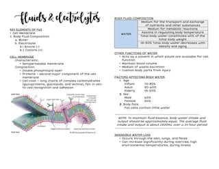
BODY FLUID COMPOSITION
WATER
Medium for the transport and exchange
of nutrients and other substances
Medium for metabolic reactions
Assists in regulating body temperature
Total body water constitutes 60% of the
total body weight
45-50% Total body water decreases with
obesity and aging
OTHER FUNCTIONS OF WATER
• Acts as a solvent in which solute are available for cell
function
• Maintain blood volume
• Medium of waste excretion
• Cushion body parts from injury
FACTORS AFFECTING BODY WATER
1. Age
Infant 70-80%
Adult 50-60%
Elderly 45-50%
2. Sex
Male 60%
Female 50%
3. Body fats
Fat cells contain little water
----------------------------------------------------------
NOTE: To maintain fluid balance, body water intake and
output should be approximately equal. The average fluid
intake and output is about 2500mL over a 24 hour period
----------------------------------------------------------
INSENSIBLE WATER LOSS
• Occurs through the skin, lungs, and feces
• Can increase significantly during: exercise; high
environmental temperatures, during illness
2. ELECTROLYTES
- These are minerals in your body that have an
electric charge. They are in your blood, urine, tissues, and
other body fluids. Electrolytes help balance the amount of
water in the body.
4 MAJOR FUNCTIONS
✔ Assist with regulation of water balance
✔ Regulating and maintaining acid-base balance
✔ Contributing to enzyme reactions
✔ Essential for neuromuscular activity
ELECTROLYTE DISTRIBUTION
Cations Plasma Interstitial ICF
Sodium (Na+) 142 146 15
Potassium (k+) 5 5 150
Calcium (Ca++) 5 3 2
Magnesium (Mg++) 2 1 27
Anions Plasma Interstitial ICF
Chloride (Cl) 102 14 1
Bicarb (HCO3) 27 30 10
Phosphate (HPO4-2) 2 2 100
Sulfate (SO4-2) 1 1 20
Organic Acid 5 8 0
Proteinate (Prot-) 16 1 73
ELECTRO-
LYTE ION
DISTRIBUTION
IN BODY FLUID
BASIC FUNCTIONS
DIETARY
SOURCES
ECF
(mEq/L)
ICF
(mEq/L)
Sodium
(Na+)
135-
154
15-20
• regulates fluid
volume within
ECF
compartment
• regulates
vascular
osmotic
pressure
• controls water
distribution
between ECF
and ICF
compartments
• participates in
conduction of
nerve impulses
• maintains
neuromuscular
excitability
• table salt
• cheese,
milk,
processed
meat,
poultry,
shellfish,
fish, eggs
and foods
preserved
with salt
(i.e. ham
and bacon)
Potassium 3.5-5
150 -
155
• regulates
osmolality of
ICF
• participates in
transmission
of nerve
impulses
• promotes
contraction of
skeletal and
smooth
muscles
• regulates acid-
base balance
by cellular
exchange of
hydrogen ions
• Fruits,
especially
bananas,
oranges,
and dried
fruits
• vegetables
• meats
• nuts
3. ELECTRO-
LYTE ION
DISTRIBUTION
IN BODY FLUID
BASIC FUNCTIONS
DIETARY
SOURCES
Calcium 4.5-5.5 1-2
• provides
strength and
durability to
bones and
teeth
• establishes
thickness and
strength of cell
membranes
• promotes
transmission
of nerve
impulses
• maintains
neuromuscular
excitability
• essential for
blood
coagulation
• activates
enzyme
reactions and
hormone
secretions
• dairy
products
(milk,
cheese, and
yogurt
• sardines,
whole
grains and
green leafy
vegetables
Magnesium 4.5-5.5 27-29
• activates
enzyme
systems,
mainly those
associated
with vit. B
metabolism
and the use of
K, Ca, and CHON
• promotes
regulation of
serum Ca, P, Ca
levels
• promotes
neuromuscular
activity
• green leafy
vegetables,
whole
grains, fish
and nuts
SERUM COMPONENT
VALUES
Conventional
SI
millimoles
Sodium 135-145 mEq/L 135-145 mmol/L
Chloride 98-106 mEq/L 98-106 mmol/L
Bicarbonate 22-26 mEq/L 22-26 mmol/L
Calcium 8.5- 10.0 mEq/L 2.1-2.6 mmol/L
Potassium 3.5-5.0 mEq/L 3.5-5.0 mmol/L
Phosphate/inorganic
phosphorus
1.7-2.6 mEq/L
(2.5-4.5 mg/dl)
0.8-1.5 mmol/L
Magnesium
1.6-2.6 mg/dl
(1.3-2.1 mEq/L)
0.8-1.3 mmol/L
Serum osmolality
275-295
mOsm/kg
(milliosmole)
275-295 mmol/kg
BODY FLUID COMPARTMENT
1. Intracellular
2. Extracellular
a. Intravascular
b. Interstitial
c. Transcellular
BODY FLUID COMPARTMENT
• Body fluid is classified by its location inside or outside
the cells.
• Capillary and cell membranes separate total body
fluids into two main compartments: the intracellular
and extracellular
Interstitial fluid
4. MECHANISM OF FLUID TRANSPORT
1. Active Transport
a. Sodium Potassium Pump
b. Endocytosis
b.1 Phagocytosis
b.2 Pinocytosis
b.3 Receptor-Mediated endocytosis (RME)
c. Exocytosis
2. Passive Transport
a. Osmosis
b. Diffusion
b.1 Facilitated
b.2 Simple
c. Filtration
d. Hydrostatic Pressure
ACTIVE TRANSPORT
Transport substances that are unable to pass by
diffusion:
- They may be too large
- They may not be able to dissolve in the fat core of
the membrane
- They may have to move against a concentration
gradient
Two common forms of active transport:
- Solute pumping
- Bulk transport
SOLUTE PUMPING
Amino acids, some sugars and ions are transported by
solute pumps
ATP energizes protein carriers, and in most cases,
moves substances against concentration gradients
BULK TRANSPORT
Endocytosis
- Extracellular substances are engulfed by being
enclosed in a membranous vesicle
- Types of endocytosis
• Phagocytosis – cell eating; dissolved materials
enter the cell plasma membrane engulfs the
solid material phagocytic vesicle
• Pinocytosis – cell drinking; plasma membrane
folds inward to form a channel allowing
dissolved substances to enter the cell
pinocytic vesicle
• RME – a process by which cells absorb
metabolites, hormones, proteins – and in some
cases viruses – by the inward budding of the
plasma membrane
Exocytosis
- Moves materials out of the cell
- Material is carried in a membranous vesicle
- Vesicle migrates to plasma membrane
- Vesicle combines with plasma membrane
- Material is emptied to the outside
5. PASSIVE TRANSPORT
A. Diffusion
Simple diffusion
- Unassisted process
- Solutes are lipid-soluble materials or small
enough to pass through membrane pores
Facilitated diffusion
- Substances require a protein carrier for passive
transport
B. Filtration
- process by which water and dissolved substances
(solutes) move from an area of high hydrostatic
pressure to an area of low hydrostatic pressure
- usually occurs across capillary membranes
C. Hydrostatic pressure
- created by the pumping action of the heart and
gravity against the capillary wall
- filtration occurs in the glomerulus of the kidneys,
as well as at the arterial end of capillaries
6. D. Osmosis
- process by which water moves across a selectively
permeable membrane from an area of lower solute
concentration to an area of higher solute
concentration
- Osmolality - number of solutes per Liter of fluid
- Osmotic Pressure - power of a solution to draw
water across a membrane
E. Oncotic Pressure
- also called colloid osmotic pressure (COP)
- osmotic pressure is exerted by plasma proteins in
the vessels (e.g., albumin)
- proteins in the bloodstream exert oncotic pressure
to pull fluid out of the interstitial space into the
intravascular space to maintain fluid balance and
osmolality
- average COP is 28mmHg, a pressure that remains
constant across the capillary
- Tonicity: refers to the effect a solution’s osmotic
pressure has on water movement across the cell
membrane of cells within that solution
TONICITY
7. ELECTROLYTE IMBALANCE
ELECTROLYTE
- substances that dissociate in solution to form
charged particles called ions
- cations are positively charged electrolytes
- anions are negatively charged electrolytes
- Function:
• assisting with the regulation of water balance
• regulating and maintaining acid–base balance
• contributing to enzyme reactions
• essential for neuromuscular activity
- Major roles:
• Maintain body fluid osmolality - regulate water
distribution
• Nervous System - Propagation of Action Potential
• Cardiovascular System - Cardiac conduction &
contraction
SODIUM
• one of the most important elements in the body
• accounts for 90% of extracellular fluid cations
(positively charged ions) and is the most abundant
solute in extracellular fluid
• 135 to 145 mEq/L
• maintain proper extracellular fluid osmolality
(concentration)
• imbalances affect the osmolality of ECF and water
distribution between the fluid compartments
• Hyponatremia water is drawn into the cells of the
body, causing them to swell
• Hypernatremia draw water out of body cells,
causing them to shrink
• kidney is the primary regulator of sodium balance in
the body
• excretes or conserves sodium changes in vascular
volume
HYPONATREMIA
• common electrolyte imbalance
• usually results from a loss of sodium from the body
• may also be caused by water gains that dilute
extracellular fluid (ECF)
CLASSIFICATIONS OF HYPONATREMIA
• Hypovolemic - both sodium and water levels decrease
in the extracellular area, but sodium loss is greater
than water loss
Causes: vomiting, diarrhea, fistulas, gastric
suctioning, excessive sweating, cystic fibrosis,
burns, wound drainage, osmotic diuresis, adrenal
insufficiency, diuretic use
• Hypervolemic - both water and sodium levels increase
in the extracellular area, but the water gain is more
impressive
Causes: heart failure, liver failure, nephrotic
syndrome, excessive administration of hypotonic
IV fluids, hyperaldosteronism
• Isovolemic - sodium levels may appear low because
too much fluid is in the body; Patients may not exhibit
signs of fluid volume excess, and total body sodium
remains stable
glucocorticoid deficiency, hypothyroidism, renal
failure
PATHOPHYSIOLOGY (ADH)
8. MANIFESTATIONS (HYPONATREMIA)
• depend on the rapidity of onset, the severity, and the
cause of the imbalance
• If the condition develops slowly, manifestations are
usually not experienced until the serum sodium levels
reach 125 mEq/L
• Poor skin turgor
• Dry mucosa
• Decrease saliva production
• Orthostatic hypotension
• Nausea, abdominal cramping
• Neurologic changes such as: lethargy, confusion, signs
of increase ICP, muscle twitch, seizure
DIAGNOSTIC (HYPONATREMIA)
• Serum osmolality less than 280 mOsm/kg (dilute blood)
• Serum sodium level less than 135 mEq/L (low sodium
level in blood)
• Urine specific gravity less than 1.010
• Elevated hematocrit and plasma protein levels
MEDICAL MANAGEMENT (HYPONATREMIA)
Hypervolemia/Isovolemia
- Fluid restrictions
- Oral sodium supplements
Hyponatremia
- Give isotonic IV fluids (e.g. normal saline)
- Offer High sodium foods
In SEVERE cases:
- Infusion of hypertonic saline solution (3% or 5%
saline)
NURSING MANAGEMENT (HYPONATREMIA)
• Monitor and record vital signs, especially blood
pressure and pulse, and watch for orthostatic
hypotension and tachycardia
• Monitor neurologic status frequently Report any
deterioration in level of consciousness.
• Accurately measure and record intake and output.
• Assess skin turgor at least every 8 hours for signs of
dehydration.
• Restrict fluid intake as ordered (fluid restriction is
the primary treatment for dilutional hyponatremia).
• Administer oral sodium supplements as ordered.
• Provide a safe environment for patients with altered
thought processes
• Seizure precautions
HYPERNATREMIA
• Hypernatremia, a less common problem than
hyponatremia, refers to excess of sodium relative to
the amount of water in the body
PATHOPHYSIOLOGY (HYPERNATREMIA)
• The cells play a role in maintaining sodium balance.
When serum osmolality increases because of
hypernatremia, fluid moves by osmosis from inside the
cell to outside the cell. As fluid leaves the cells, they
become dehydrated and shrink.
MANIFESTATIONS (HYPERNATREMIA)
• Thirst is the first manifestation
• If thirst is not relieved, the primary manifestations
relate to altered neurologic function (lethargy,
weakness, and irritability can progress to seizures,
coma, and death in severe hypernatremia)
• Low grade fever
• Flushed skin
• Dry mucous membranes
• Oliguria
• Orthostatic hypotension
DIAGNOSTIC (HYPERNATREMIA)
Serum sodium level: >145 mEq/L
Serum osmolality: >295 mOsm/kg
MEDICAL MANAGEMENT (HYPERNATREMIA)
• Treatment for hypernatremia varies with the cause.
The underlying disorder must be corrected, and serum
sodium levels and related diagnostic tests must be
monitored.
• Note that the fluids should be given gradually over 48
hours to avoid shifting water into brain cells.
9. NURSING MANAGEMENT (HYPERNATREMIA)
• Intravenous fluid replacement (dextrose 5% in water)
to return serum sodium levels to normal
• Sodium intake restrictions
• Administer diuretics to increase sodium loss
• Monitor and record vital signs, especially blood
pressure and pulse
• Check neurologic status frequently.
• Report any deterioration in the level of consciousness
• Carefully measure and record intake and output.
• Assess skin and mucous membranes for signs of
breakdown and infection
• Monitor serum sodium level and report any increase.
• Assist with oral hygiene. Lubricate the patient’s lips
frequently.
• Provide a safe environment for confused or agitated
patients
POTASSIUM
• major cation (ion with a positive charge) in
intracellular fluid
• affects nerve impulse transmission
• 3.5 to 5.0 mEq/L
• Aldosterone helps regulate potassium elimination by
the kidneys
• Function: (Potassium directly affects how well the
body cells, nerves, and muscles function by:)
- Maintaining cells’ electrical neutrality and
osmolality
- Aiding neuromuscular transmission of nerve
impulses
- Assisting skeletal and cardiac muscle contraction
and electrical conductivity
- Affecting acid-base balance in relationship to the
hydrogen ion.
HYPOKALEMIA
• an abnormally low serum potassium level
• usually results from excess potassium loss
• hospitalized patients may be at risk for hypokalemia
because of inadequate potassium intake
PATHOPHYSIOLOGY (HYPOKALEMIA)
• Inadequate intake and excessive output of potassium
can cause a moderate drop in its level
• Causes:
- Inadequate potassium intake
- Severe GI losses, e.g. suction, lavage, prolonged
vomiting can deplete the body’s potassium supply
as a result potassium levels drop
- Drug associated, e.g. diuretics,(esp. thiazides and
furosemides), corticosteroids, insulin
- Decrease bowel motility
MANIFESTATIONS (HYPOKALEMIA)
• Skeletal muscle weakness, especially in the legs a
sign of a moderate loss of potassium. This also
includes paresthesia and leg cramps
• Decreased bowel sounds, constipation, paralytic ileus
• Weak and irregular pulse
• Orthostatic hypotension and palpitations
• ECG changes, flattened or inverted T wave, prominent
U wave
10. LABORATORY FINDINGS (HYPOKALEMIA)
• The following test results may develop to confirm the
diagnosis of hypokalemia:
- Serum potassium level <3.5 mEQ/L
- Increased 24-hour urine level
- Characteristic ECG changes
MEDICAL MANAGEMENT (HYPOKALEMIA)
• Identify and treat the cause treatment for
hypokalemia focuses on restoring a normal potassium
balance, preventing serious complications, and
removing or treating the underlying causes
• Oral and IV replacements (40-80 meq/day – Kalium
durule or K IV), it can be safely given at the same time
NURSING MANAGEMENT (HYPOKALEMIA)
• Monitor vital signs, especially blood pressure
hypokalemia is commonly associated with
hypovolemia, which can cause orthostatic hypotension
• Check heart rate and rhythm and ECG tracings in
patients with serum potassium level less than 3
meQ/L hypovolemia causes tachyarrthymias
• Assess respiratory rate, depth and pattern
hypokalemia may weaken or paralyze respiratory
muscles. Notify the doctor immediately if respirations
become shallow and rapid
• Monitor serum potassium levels changes in serum
potassium level can lead to serious cardiac
complications
• Administer potassium infusions cautiously
HYPERKALEMIA
• Hyperkalemia can result from inadequate excretion of
potassium, excessively high intake of potassium, or a
shift of potassium from the ICF to the ECF.
Hyperkalemia affects neuromuscular and cardiac
function
CAUSES OF HYPERKALEMIA
• Increased dietary intake
• Blood transfusion (stored blood) serum potassium
level increases longer the blood is stored.
• Rapid IV potassium administration
• Renal insufficiency
• Metabolic acidosis, potassium shift to ECF in exchange
of H ions
PATHOPHYSIOLOGY (HYPERKALEMIA)
• Potassium move from the extracellular to the
intracellular compartment and increases cell
excitability, so that cells respond to stimuli of less
intensity and may actually discharge independently
without a stimulus
MANIFESTATIONS (HYPERKALEMIA)
• Signs and symptoms of hyperkalemia reflect its
effects on neuromuscular and cardiac functioning in
the body
- Nausea, abdominal cramping and diarrhea the
early signs of hyperkalemia due to smooth muscle
hyperactivity
- Muscle weakness that in turn lead to flaccid
paralysis
- Decreased heart rate, irregular pulse, decreased
cardiac output, hypotension
- ECG changes, elevated T wave (Tall or peaked T
wave)
11. LABORATORY FINDINGS (HYPERKALEMIA)
• Serum potassium level >5 mEq/L
• Decreased arterial pH, indicating acidosis
• ECG abnormalities
MEDICAL MANAGEMENT (HYPERKALEMIA)
• Treatment for hyperkalemia is aimed at lowering the
potassium level, treating its cause, stabilizing the
myocardium, and promoting renal and
gastrointestinal excretion of potassium. The severity
of hyperkalemia dictates how it should be treated.
- Diet restrictions
- May administer loop diuretics for mild
hyperkalemia to increase potassium loss from
the body or to resolve any acidosis present
- Sodium polystyrene sulfonate (Kayexalate) a
cation-exchange resin (common treatment for
hyperkalemia)
- Hemodialysis, if patient has renal failure
NURSING INTERVENTIONS (HYPERKALEMIA)
• Assess vital signs
• Monitor the patient’s intake and output Report an
output of less than 30 mL/hour.
• Prepare to administer a slow calcium chloride or
gluconate IV infusion to counteract the myocardial
depressant effects of hyperkalemia
• Keep in mind when giving Kayexalate that serum
sodium levels may rise. Watch for signs of heart
failure.
• Monitor ECG changes
CALCIUM
• 4.5-5.5 mEq/L
• Calcium is a positively charged ion or cation
• found in both extracellular and intracellular fluid
• About 99% of the body’s calcium is found in the bones
and teeth
• Only 1% is found in serum and in soft tissue
• Function:
- Skeletal and heart muscle relaxation, activation,
excitation and contraction
- Maintains cellular permeability
- Promotes blood coagulation
- Nerve impulse transmission
• three forms of calcium in the body:
- 45% is bound to protein, mostly albumin
- 40% is ionized calcium, it is the calcium that is
physiologically active and clinically important for
neuromuscular transmission
- 15% is bound to other substances such as
phosphate, citrate, or carbonate
• system interactions:
- Parathyroid Hormone (PTH) raises the plasma
calcium
- Calcium is dependent upon calcitriol, the most
active from of vitamin D
- Calcitonin, a calcium-lowering hormone produced by
the thyroid gland
HYPOCALCEMIA
• Hypocalcemia can result from decreased total body
calcium stores or low levels of extracellular calcium
with normal amounts of calcium stored in bone. The
systemic effects of hypocalcemia are caused by
decreased levels of ionized calcium in extracellular
fluid.
CAUSES (HYPOCALCEMIA)
• Inadequate intake
• Hypoparathyroidism, resulting from surgery
(parathyroidectomy, thyroidectomy, radical neck
dissection)
• Electrolyte imbalances Low serum albumin (most
common cause)
12. • Malabsorption can result from increased intestinal
motility
• Vitamin D deficiency due to lack sun exposure or
malabsorption
• Massive blood transfusion, liver unable to metabolize
citrate (added to prevent clotting)
• Chronic diarrhea
• Diuretic phase of ARF
• Severe burns
PATHOPHYSIOLOGY (HYPOCALCEMIA)
• Extracellular calcium acts to stabilize neuromuscular
cell membranes. This effect is reduced in
hypocalcemia, increasing neuromuscular irritability.
Thus, electrical activity occurs spontaneously and
continuously.
MANIFESTATIONS (HYPOCALCEMIA)
• Signs and symptoms of hypocalcemia reflect calcium’s
effects on nerve transmission and muscle and heart
functions; therefore, neuromuscular and
cardiovascular findings are most evident
- Anxiety, confusion, and irritability that can
progress to seizures
- Paresthesia, muscle twitching, cramps or tremors
- Diarrhea
- Hyperactive tendon reflexes
- Fractures may occur
- Brittle nails, dry skin and hair
- Decreased cardiac output and subsequent
arrhythmias
- Prolonged QT segment on electrocardiogram (ECG)
----------------------------------------------------------------
TROUSSEAU’S SIGN
- carpal spasm induced by inflating a blood pressure
cuff on the upper arm to above systolic blood
pressure for 2 to 5 minutes due to increase nerve
excitability
CHVOSTEK’S SIGN
- contraction of facial muscles produced by tapping the
facial nerve in front of the ear
----------------------------------------------------------------
LABORATORY FINDINGS (HYPOCALCEMIA)
• Elevated total serum calcium level
• Low Ionized calcium level (ionized calcium
measurement is the definitive method to diagnose
hypocalcemia)
• Low albumin level
• Characteristic ECG changes, prolonged ST segment
MEDICAL MANAGEMENT
• Treatment for hypocalcemia focuses on correcting the
imbalance as quickly and safely as possible. The
underlying cause should be addressed to prevent
recurrence.
- Dietary supplement, e.g. milk, cheese, cereal
- Oral/IV calcium, e.g. Calcium carbonate/gluconate
- Vitamin D supplements, facilitates GI absorption of
calcium
NURSING MANAGEMENT (HYPOCALCEMIA)
• Monitor vital signs
• Place patient on a cardiac monitor, and evaluate for
changes in heart rate and rhythm
• Check for Chvostek’s and Trousseau’s sign
• Serum levels monitoring
13. HYPERCALCEMIA
• Hypercalcemia is a common metabolic emergency that
occurs when serum calcium level rises, or the rate of
calcium entry into extracellular fluid exceeds the rate
of calcium excretion by the kidneys
CAUSES OF HYPERCALCEMIA
• Hyperparathyroidism (most common cause) the body
excretes more PTH than normal, which greatly
strengthens the effects of the hormone
• Metastatic malignancy causes bone destruction as
malignant cells invade the bones and cause the
release of a hormone similar to PTH
• Thiazide diuretics potentiates PTH and decrease
excretion in kidneys
• Increase Vitamin D (can prompt an increase in serum
calcium levels) & prolong use of alkaline antacid
(calcium carbonate)
• Prolong immobility
MANIFESTATIONS (HYPERCALCEMIA)
• Muscle weakness
• Bradycardia
• Shortened QT interval
• Decreased gastrointestinal motility (anorexia, n/v)
• Stone formation
LABORATORY FINDINGS (HYPERCALCEMIA)
• Elevated serum calcium level
• Elevated Ionized calcium level
• Digoxin toxicity if patient is taking digoxin
• Characteristic ECG changes (Shortened QT interval)
MEDICAL MANAGEMENT (HYPERCALCEMIA)
• ECG monitoring
• Treat with IV saline the sodium in the solution is
typically used for hydration in these cases
• Loop diuretics (Lasix) promotes calcium excretion
• Administer Calcitonin (IM/SC)
NURSING MANAGEMENT (HYPERCALCEMIA)
• Be sure to monitor the calcium levels of patients who
are at risk for hypercalcemia, such as those who have
cancer or parathyroid disorders, are immobile, or are
receiving a calcium supplement. For a patient who
develops hypercalcemia, you can take the following
actions:
- Monitor vital signs
- Watch the patient for arrhythmias
- ECG monitoring
- Calcium levels monitoring
- Encourage Mobilization
- Dietary limits of vitamin D/Calcium
MAGNESIUM
• 1.5-2.5 mEq/L
• Promotes enzyme reactions within the cell during
carbohydrate metabolism
• Influences vasodilation and irritability and
contractility of the cardiac muscles, thereby helping
the cardiovascular system function normally
• Aids in neurotransmission and hormone-receptor
binding
• Makes production of parathyroid hormone possible
HYPOMAGNESEMIA
• Hypomagnesemia is a common problem in critically ill
patients. It is may be caused by deficient magnesium
intake, excessive losses, or a shift between the
intracellular and extracellular compartment
CAUSES OF HYPOMAGNESEMIA
• Poor dietary intake of magnesium
• Poor magnesium absorption by the GI tract
• Excessive magnesium loss from the GI tract
• Excessive magnesium loss from the urinary tract
14. PATHOPHYSIOLOGY (HYPOMAGNESEMIA)
• Hypomagnesemia causes increased neuromuscular
excitability which commonly occurs with hypokalemia
and hypocalcemia
MANIFESTATIONS (HYPOMAGNESEMIA)
• Signs and symptoms of hypomagnesemia can range
from mild to life-threatening. Generally speaking, your
patient’s signs and symptoms may resemble those you
would see with a potassium or calcium imbalance.
However, you can’t always count on detecting
hypomagnesemia from clinical findings alone
- Altered level of consciousness, tetany, seizures
- Emotional lability
- Vomiting
- Tremors, twitching, tetany, hyperactive deep
tendon reflexes
- Rapid heart rate
- Chvostek’s and Trousseau’s sign
LABORATORY FINDINGS (HYPOMAGNESEMIA)
• Serum magnesium level below 1.5 mEq/L (possibly with
a below normal serum albumin level)
• Low potassium and calcium level
• Characteristic ECG changes
• Elevated serum levels of digoxin in a patient receiving
the drug
MEDICAL MANAGEMENT (HYPOMAGNESEMIA)
• Treatment for hypomagnesemia depends on the
underlying cause of the condition and the patient’s
clinical findings:
- Dietary management, (green leaf vegetables, nuts,
legumes, seafood, wholegrain, chocolates)
- Intravenous infusion/ intramuscular injection,
before magnesium administration, renal function
should be assessed
NURSING MANAGEMENT (HYPOMAGNESEMIA)
• Assess the patient’s mental status and report
changes
• Evaluate the patient’s neuromuscular status regularly
by checking for hyperactive Deep Tendon Reflexes,
tremors, and tetany.
• Check for Chvostek’s and Trousseau’s sign
• Monitor patients who have lost an excessive amount
of fluid, they are at risk for magnesium deficiency
• Urine output should be monitored at least every 4
hours. Magnesium generally isn’t administered if urine
output is less than 100 mL in 4 hours.
• If patient id receiving digoxin, monitor him closely for
signs and symptoms of digoxin toxicity (such as
nausea, vomiting and bradycardia
• During magnesium replacement, check the cardiac
monitor frequently and assess the patient closely for
signs of magnesium excess, such as hypotension and
respiratory distress. Keep calcium gluconate at the
bedside in case signs occur
HYPERMAGNESEMIA
• Having too much magnesium in the serum can be just
as bad as having too little. Hypermagnesemia occurs
when the body’s serum magnesium level rises above
the normal range. However, hypermagnesemia is
uncommon; typically, the kidneys can rapidly reduce
the amount of excess magnesium in the body.
CAUSES OF HYPERMAGNESEMIA
• Impaired magnesium excretion, e.g. renal dysfunction,
most common cause of hypermagnesemia
• Excessive magnesium intake
PATHOPHYSIOLOGY (HYPERMAGNESEMIA)
• Elevated serum magnesium levels suppress cellular
excitability, resulting to muscular flaccidity and
suppression of electrical impulses
LABORATORY FINDINGS (HYPERMAGNESEMIA)
• Serum magnesium level above 2.5 mEq/L
• ECG changes (prolonged PR interval, widened QRS
complex, tall T wave.
15. MEDICAL MANAGEMENT (HYPERMAGNESEMIA)
• Correct underlying cause
• Intravenous infusion of calcium gluconate, to
antagonize magnesium effect
NURSING MANAGEMENT (HYPERMAGNESEMIA)
• Monitor vital signs
• Checked for flushed skin and diaphoresis
• Check for deep tendon reflexes
• Evaluate for changes in mental status
PHOSPHOROUS
• The primary anion, or negatively charged ion, found in
the intracellular fluid. It’s contained in the body as
phosphate.
• 1.2 to 3.0 mEq/L
• Integral part of acid base buffer system
• Regulate ATP use for muscle contraction, nerve
transmission, electrolyte transport
• Regulates 2,3 DPG (diphosphoglycerate) a compound in
red blood cells that facilitates oxygen delivery from
the red blood cells to the tissues.
• Regulates 2,3 DPG (diphosphoglycerate) a substance in
RBC affecting oxygen affinity
HYPOPHOSPHATEMIA
• Hypophosphatemia occurs when the serum
phosphorous level falls below 1.2 mEq/L. although this
condition generally indicates a deficiency of
phosphorous, it can occur under various
circumstances when total body phosphorous stores
are normal
CAUSES OF HYPOPHOSPHATEMIA
• Respiratory alkalosis one of the most common
cause of hypophosphatemia, can stem from a number
of conditions that produce hyperventilation
• Malabsorption syndromes
• Diuretic use, (Thiazides, Loop diuretics, Acetazolamide)
PATHOPHYSIOLOGY (HYPOPHOSPHATEMIA)
• Most effects of hypophosphatemia result from
depletion of ATP and impaired oxygen delivery to the
cells due to a deficiency of the red blood cell enzyme
2,3-DPG. Severe hypophosphatemia affects virtually
every major organ system
MANIFESTATIONS (HYPOPHOSPHATEMIA)
• Muscle weakness, most common symptom
• malaise, weakened hand grasp, myalgia (pain in the
muscles)
• Rhabdomyolysis (skeletal muscle destruction), can
occur with altered muscle cell activity
• Osteomalacia/ Fracture, due to loss of bone density
• Bruising and bleeding
LABORATORY FINDINGS (HYPOPHOSPHATEMIA)
• serum phosphorous level less than 1.2 mEq/L
• elevated Creatinine Kinase level if Rhabdomyolysis is
present
• X-ray studies that reveal skeletal changes typical of
osteomalacia or bone fractures
• Abnormal electrolytes (decreased magnesium levels
and increased calcium levels)
MEDICAL MANAGEMENT (HYPOPHOSPHATEMIA)
• Treat the underlying cause
• Oral and intravenous supplements
NURSING INTERVENTIONS (HYPOPHOSPHATEMIA)
• Monitor vital signs.
• Assess the patient frequently for evidence of
decreasing muscle strength, such as weak hand
grasps or slurred speech, and document findings
regularly.
• Assist in ambulation and activities of daily living, if
needed, and keep essential objects near the patient to
prevent accidents.
16. HYPERPHOSPHATEMIA
• Hyperphosphatemia is a serum phosphate level
greater than 4.5mg/dL. As with other electrolyte
imbalances, it may be the result of impaired
phosphate excretion, excess intake, or a shift of
phosphate from the intracellular space into
extracellular fluids.
CAUSES OF HYPERPHOSPHATEMIA
• Renal Failure
• Hypoparathyroidism impairs less synthesis of
parathyroid hormone(PTH), when less PTH is
synthesized, less phosphorous is excreted from the
kidneys.
• Respiratory acidosis
• Increase tissue breakdown
PATHOPHYSIOLOGY (HYPERPHOSPHATEMIA)
• Effects of high phosphorous level are actually due to
hypocalcemia; calcium suppresses cellular excitability
MANIFESTATIONS (HYPERPHOSPHATEMIA)
• Hyperphosphatemia causes few symptoms (tetany,
tissue calcification)
• Symptoms occurring result from decrease calcium
secondary to reciprocity
LABORATORY FINDINGS (HYPERPHOSPHATEMIA)
• Serum phosphorous above normal
• Low serum calcium level
MEDICAL MANAGEMENT (HYPERPHOSPHATEMIA)
• Treat underlying cause
• Restrict dietary intake
• Administer phosphate binding antacid, this may
decrease absorption of phosphorous in the
gastrointestinal system
NURSING MANAGEMENT (HYPERPHOSPHATEMIA)
• Monitor vital signs
• Monitor intake and output.( if urine output falls below
30 mL/hour, immediately notify the doctor)
Decreased urine output can seriously affect renal
clearance of excess serum phosphorous
• Monitor serum phosphorous and calcium levels
• Monitor signs of tetany, such as positive Trousseau’s
and Chvostek’s sign
FLUID SHIFTS
FLUID SHIFTS
• Plasma to Interstitial
o Edema – palpable swelling produced by expansion
of the interstitial fluid volume
May be localized or generalized; pitting or
nonpitting, depending on the cause
Can cause fluid shifts in other VULNERABLE
areas of the body termed as third space
shifts
Where can your fluid shift?
PLEURAL SPACE (PERITONEAL SPACE)
• Interstitial to Plasma
o Movement back of edema to circulatory volume
o e.g. excessive administration of hypertonic solution
MAJOR CAUSES OF EDEMATOUS STATE
• Decreased colloid osmotic pressure in the capillary
o e.g. burns, liver failure
• Increased capillary hydrostatic pressure
o e.g. CHF
• Increased capillary permeability
o e.g. burns, allergic reaction
• Lymphatic obstruction or increased interstitial colloid
osmotic pressure
o e.g. surgical removal of lymph structures
MANAGEMENT OF EDEMA
• Diuretic therapy
• Elevating the affected extremity
• Elastic support stockings in the morning
• Albumin IV
17. FLUID IMPAIRMENT
• Two types of fluid imbalance
o Fluid volume deficit
o Fluid volume excess
• Both types can be life threatening
• Often seen in acute care settings
FLUID VOLUME DEFICIT (FVD)
FLUID VOLUME DEFICIT (FVD)
• is a decrease in intravascular, interstitial, and/or
intracellular fluid in the body
• CAUSE: excessive fluid losses, insufficient fluid intake,
or failure of regulatory mechanisms and fluid shifts
within the body
• a relatively common problem that may exist alone or
in combination with other electrolyte or acid–base
imbalances
ISOOSMOLAR FLUID VOLUME DEFICIT
- Occurs when sodium and water are lost in equal
amounts
HYPEROSMOLAR FLUID VOLUME DEFICIT
- Occurs when more fluid is lost than sodium, resulting
in higher serum osmolality than normal
HYPOOSMOLAR FLUID VOLUME DEFICIT
- Occurs when electrolyte loss is greater than fluid
(rare)
ETIOLOGY OF FVD
• Common cause of fluid volume deficit is excessive loss
of GI fluids
o Vomiting
o Diarrhea
o GI suctioning
o Intestinal fistulas
o Intestinal drainage
• Other causes
o Diuretics
o renal disorders
o endocrine disorders
o excessive exercise
o hot environment
o Hemorrhage
o chronic abuse of laxatives and/or enemas
• Other factors
o inadequate fluid intake
o inability to access fluids
o inability to request or to swallow fluids
o oral trauma
o altered thirst mechanisms
PATHOPHYSIOLOGY
Loss of extracellular fluid volume
l
Hypovolemia
l
decreased circulating blood volume
(Electrolytes often are lost along with fluid – ISOOSMOLAR
FVD)
l
serum sodium level remains normal (levels of other
electrolytes such as potassium may fall)
l
Fluid is drawn into the vascular compartment from the
interstitial spaces
(COMPENSATORY MECHANISM to maintain tissue perfusion)
l
eventually depletes fluid in the intracellular compartment
l
Severe fluid loss (i.e. hemorrhage) shock cardiovascular
collapse
MANIFESTATIONS
• Rapid weight loss
o loss of 2% of body weight represents a mild FVD
o 5%, moderate FVD
o 8% or greater, severe FVD
• skin turgor to diminish D/T loss in interstitial fluid
• Postural or orthostatic hypotension
• flat neck veins D/T falling of venous pressure
18. • Tachycardia
• pale, cool skin (vasoconstriction)
• decreased urine output
MULTISYSTEM EFFECTS
• Mucous Membranes
o Dry; may be sticky
• Decrease tongue size, longitudinal furrows increase
• Urinary
o Decrease urine output
o Oliguria (severe FVD)
o Increase in urine specific gravity
• Neurologic
o Altered mental status
o Anxiety, restlessness
o Diminished alertness/condition
o Possible coma (severe FVD)
• Integumentary
o Diminished skin turgor
o Dry skin
o Pale, cool extremities
• Cardiovascular
o Tachycardia
o Orthostatic hypotension (moderate FVD)
o Falling systolic/diastolic pressure (severe FVD)
o Flat neck veins
o Decrease venous filling
o Decrease capillary refill
o Increase hematocrit
• Potential Complication
o Hypovolemic shock
• Musculoskeletal
o Fatigue
• Metabolic Processes
o Decrease body temperature (isotonic FVD)
o Increase body temperature (dehydration)
o Thirst
o Weight loss
2-5% mild FVD
6-9% moderate FVD
>10% severe FVD
DIAGNOSTIC
• Serum Electrolytes
• Serum Osmolality
• Hemoglobin and HCT
• Urine Specific Gravity and Osmolality
• CVP
SERUM ELECTROLYTES
• Test measures the levels of electrolytes such as
Sodium, Potassium and Chloride
• Isotonic fluid deficit sodium levels are within
normal limits
• Water loss sodium levels are high
• Decreases in potassium are common
SERUM OSMOLALITY
• measures the amount of chemicals dissolved in the
liquid part (serum) of the blood.
• Chemicals that affect serum osmolality include
sodium, chloride, bicarbonate, proteins, and sugar
(glucose).
• This test is done on a blood sample taken from a vein
• Helps to differentiate isotonic fluid loss from water
loss
HEMOGLOBIN AND HCT
• Hemoglobin
o When the hemoglobin level is low, the patient
has anemia. An erythrocytosis is the consequence
of too many red cells; this results in hemoglobin
levels above normal.
• HCT
o measures the volume of red blood cells compared
to the total blood volume (red blood cells and
plasma)
o Both the hemoglobin and the hematocrit are based
on whole blood and are therefore dependent on
plasma volume. If a patient is severely dehydrated,
the hemoglobin and hematocrit will appear higher
than if the patient were normovolemic; if the
patient is fluid overloaded, they will be lower than
their actual level
19. URINE SPECIFIC GRAVITY AND OSMOLALITY
• shows the concentration of all chemical particles in
the urine
• specific gravity and osmolality of urine increase
compensatory mechanism
CENTRAL VENOUS PRESSURE (CVP)
• Measures the mean pressure in the superior vena
cava or right atrium, providing an accurate
assessment of fluid volume status
• Normal CVP is 2-6 mm Hg
• CVP decreases with:
o hypovolemic shock from hemorrhage, fluid shift,
dehydration
o negative pressure breathing which occurs when the
patient demonstrates retractions or mechanical
negative pressure which is sometimes used for
high spinal cord injuries
• CVP increases with:
o overhydration which increases venous return
o heart failure or PA stenosis which limit venous
outflow and lead to venous congestion
o positive pressure breathing, straining
The 3-way stopcock is opened so that IV fluid from a
container fills the manometer (10- to 20-cm). Then the tap
is closed to the fluid bag and opened to the patient. Fluid in
the manometer will fall to the level of the CVP and an
average reading is taken (as level pulsates up and down
during the reading). This system can be set up for long
periods and the reading taken intermittently (for example,
every hour).
MEDICAL MANAGEMENT
• Correction of fluid loss depends on the acuteness and
severity of the fluid deficit. Goals are to replace F/E
(Na primarily) that have been loss.
• Fluid Restoration
o Oral Rehydration
o IV Rehydration
• Monitoring complications of fluid restoration
• Correction of Underlying Problem
Oral Rehydration
• safest and most effective treatment for fluid volume
deficit in alert patients who are able to take oral
fluids
• Fluids are replaced gradually, particularly in older
adults, to prevent rapid rehydration of the cells
----------------------------------------------------------------
NOTE:
• For mild fluid deficits in which a loss of electrolytes has
been minimal (e.g., moderate exercise in warm weather),
water alone may be used for fluid replacement
• For more severe fluid deficits and when electrolytes have
also been lost e.g. vomiting and/or diarrhea, strenuous
exercise for longer than an hour or two
carbohydrate/electrolyte solution such as a sports drink,
ginger ale, or a rehydrating solution (e.g., Pedialyte or
Rehydralyte) is more appropriate. These solutions provide
sodium, potassium, chloride, and calories to help meet
metabolic needs
----------------------------------------------------------------
20. IV Rehydration
• When the fluid deficit is severe or the patient is
unable to ingest fluids, the IV route is used to
administer replacement fluids.
• Types of Fluid Volume Deficit:
o Isotonic: Caused by losing fluids and solutes about
equally; solute concentration in the remaining
extracellular fluid then remains relatively
unchanged
o Hypertonic: Caused by losing more fluids than
solutes, leading to increased solute concentration
in the remaining fluid.
o Hypotonic: Caused by losing more solutes than fluid
leading to decreased solute concentration in
remaining fluid. This is the rarest type.
MONITORING COMPLICATIONS OF FLUID RESTSORATION
• Client with severe ECFVD accompanied by severe
heart, liver and kidney disease can’t tolerate large
volumes of fluid or sodium without the risk of
development of heart failure
• Unstable clients needs to be monitored to detect ↑
pressure from fluids
o Monitor:
Fluid volume status by CVP insertion
Lab values (Na, K, BUN, Osmolarity)
Body Weight
Urine output
CORRECTION OF UNDERLYING PROBLEMS
• Medication
o Antiemetics
o Antidiarrheals
o Antibiotics
o Antidiuretics to reduce body temperature
NURSING MANAGEMENT
Nurses are responsible for (1) identifying patients at risk for
fluid volume deficit, (2) initiating and carrying out
interventions to prevent and treat fluid volume deficit, and
(3) monitoring the effects of therapy
1. I & O every 8 hours or hourly (Record all output
accurately)
2. VS every 2-4 hours, report changes from baseline VS;
Assess CVP every 4hrs (if patient has CVP access)
3. Weight patient daily and record.
4. Peripheral vein filling (Capillary refill 3-5 sec)
5. Renal client / relatives to report urine output ‹ than
30 m/L x 2 consecutive hours or ‹ 240 ml x 80 period
6. Assess for dryness of mucous membrane and skin
turgor (check should be done)
7. Oral care to ↓ discomfort related to mucous
membrane dryness
8. Monitor plasma sodium, BUN, Glucose, HCT to
determine osmolality
9. Assess the client for confusion, easy indication of
ICF involvement
10. Keep fluids easily accessible
11. Administer and monitor intake of oral fluids as
prescribed
12. Administer IV fluids as prescribed using an infusion
pump. Monitor for indicators of fluid overload if
rapid fluid replacement is ordered: dyspnea,
tachypnea, tachycardia, increased CVP, jugular vein
distention, and edema
NURSING DIAGNOSIS
• Fluid Volume Deficit
o Patients with a fluid volume deficit due to
abnormal losses, inadequate intake, or impaired
fluid regulation require close monitoring as well as
immediate and ongoing fluid replacement
• Ineffective Tissue Perfusion
o A fluid volume deficit can lead to decreased
perfusion of renal, cerebral, and peripheral
tissues. Inadequate renal perfusion can lead to
acute renal failure. Decreased cerebral perfusion
leads changes in mental status and cognitive
function, causing restlessness, anxiety, agitation,
excitability, confusion, vertigo, fainting, and
weakness
• Risk for Injury
o The patient with fluid volume deficit is at risk for
injury because of dizziness and loss of balance
resulting from decreased cerebral perfusion
secondary to hypovolemia
21. IV THERAPY
IV THERAPY
• Indications for IV Therapy
o unable to eat or drink adequate amounts
o rapid delivery of medication in an emergency
o Patients with anemia or blood loss can receive
lifesaving IV transfusions
o unable to eat for an extended period can have
their nutritional needs met with total parenteral
nutrition (TPN)
TYPES OF INFUSION
• Continuous Infusion
o infusion is kept running
constantly until
discontinued by the
physician
• Intermittent Infusion
o Intermittent IV lines
are “capped off” with
an injection port and
used only periodically
• Bolus (IV push or IVP Drug)
o bolus drug (sometimes
called an IV push or IVP
drug) is injected slowly
via a syringe into the
IV site or tubing port
o can be dangerous if
they are given
incorrectly, and a drug
reference should always be checked to determine
the safe amount of time over which the drug can
be injected
• Piggyback/Secondary Infusion
o Some IV medications, such as antibiotics, need to
be infused over a short period of time
o In order for the piggyback medication to infuse, it
must hang higher than the primary infusion
FACTORS AFFECTING FLOWRATES
• Change in catheter position
• Height of the solution
• Patency of the catheter
TYPES OF IVF SOLUTIONS
• COLLOIDS
o Fluids that expand the circulatory volume due to
particles that cannot cross a semipermeable
membrane. They pull fluid from the interstitial
space into the intravascular space, increasing fluid
volume
o ADVANTAGE: is beneficial to large losses of fluid
o DISADVANTAGE: costly and the risk of volume
overload, including pulmonary edema
• CRYSTALLOIDS
o Work much like colloids but do not stay in the
intravascular circulation as well as colloids do, so
more of them need to be used
o ADVANTAGE: cheaper and more convenient to use;
provide hydration and calories to patients and
include dextrose, normal saline, and Ringer’s and
lactated Ringer’s solution
o DISADVANTAGE: lacks large molecules
22. TONICITY OF SOLUTIONS
ISOTONIC SOLUTIONS
• 0.9% Saline
• Lactated Ringer’s
solution
Monitor for fluid overload; discontinue
fluids and notify the healthcare
provider
Do not administer lactated ringer’s
solution to patients with severe liver
disease as the liver may be unable to
convert the lactate to bicarbonate
and the patient may become acidotic.
If administering lactated ringer’s
solution, monitor potassium levels
and cardiac rhythm; if abnormalities
are present, notify the health care
provider
HYPOTONIC
SOLUTIONS
• 0.45% Saline or
0.25% Saline
• D5W
Monitor for inflammation and
infiltration at IV insertion site as
hypotonic solutions may cause cells
to swell and burst, including those at
the insertions site; this narrows the
lumen of the vein
Monitor blood sodium levels
Do not administer to patients at risk
for increased intracranial pressure
(e.g head trauma, stroke,
neurosurgery)
Do not administer to patients at risk
for third-space shifts (burns, trauma,
liver disease, malnutrition)
HYPERTONIC
SOLUTIONS
• Hypertonic fluids
have a tonicity
>350 mEq/L and
include the ff:
• Fluids containing
medications
• D5W sodium
chloride
• D5W in lactated
ringer’s solution
• Total parenteral
solutions
Monitor for inflammation and
infiltration at IV insertion site as
hypertonic solutions cause cells to
shrink, exposing the basement
membrane of the vein
Monitor blood sodium levels
Monitor for circulatory overload
Do not administer to patients with
diabetic ketoacidosis or impaired
cardiac or kidney function.
23. TYPES OF IV SOLUTIONS ACCORDING TO PURPOSE
• Hydrating
o Replace water loss; Dilute meds; Keep veins open
• Nutritional
o Promotes faster recuperation
• Maintenance
o Replace electrolyte loss at ECF level; Maintenance
in patients with no oral intake; Replace fluid loss;
Treatment for dehydration
• Volume Expander
o Increase osmotic pressure thus maintain
circulatory volume
Local
Complications of
IV Therapy
Signs and
Symptoms
Nursing
Interventions
Hematoma
collection of blood
outside of blood
vessels
• Ecchymoses
• Swelling
• Inability to
advance
catheter
• Resistance
during flushing
• Remove catheter
• Apply pressure
with 2x2
• Elevate
extremity
Thrombosis
formation of a
blood clot
• Slowed or
stopped infusion
• Fever/malaise
• Inability to flush
catheter
• Discontinue
catheter
• Apply cold
compress to site
• Assess for
circulatory
impairment
Phlebitis
inflammation of
the walls of a vein
• Redness at site
• Site warm to
touch
• Local swelling
• Pain
• Palpable cord
• Sluggish infusion
rate
• Discontinue
catheter
• Apply cold
compress
initially;
• then warm
• Consult physician
if severe
Infiltration
(Extravasation)
the accidental
leakage of non-
vesicant solutions
out of the vein
into the
surrounding tissue
• Coolness of skin
at site
• Taut skin
• Dependent
edema
• Backflow of
blood absent
• Infusion rate
slowing
• Discontinue
catheter
• Apply cool
compress
• Elevate
extremity
slightly
• Follow
extravasation
guidelines
• Have antidote
available
Local Infection • Redness and
swelling at site
• Possible exudate
• Increase WBC
count
• Elevated T
lymphocytes
• Discontinue
catheter and
culture
• site and
catheter
• Apply sterile
dressing
• over site
• Administer
antibiotics if
ordered
Venous Spasm
occurs due to
severe vein
irritation,
administration of
cold fluids or
blood, and a very
rapid flow rate
• Sharp pain at
site
• Slowing of
infusion
• Apply warm
compress to site
• Restart infusion
in new site if
• spasm continues
24. SYSTEMIC COMPLICATIONS OF PERIPHERAL IV THERAPY
Complication Signs and
Symptoms
Nursing
Interventions
Septicemia
blood poisoning,
especially that
caused by
bacteria or their
toxins
• Fluctuating
temperature
• Profuse
sweating
• Nausea/vomiting
• Diarrhea
• Abdominal pain
• Tachycardia
• Hypotension
• Altered mental
status
• Restart new IV
system
• Obtain cultures
• Notify physician
• Initiate
antimicrobial
therapy as
• ordered
• Monitor patient
closely
Fluid Overload
is a medical
condition where
there is too
much fluid in the
blood
• Weight gain
• Puffy eyelids
• Edema
• Hypertension
• Changes in input
and output (I&O)
• Rise in central
venous pressure
(CVP)
• Shortness of
breath
• Crackles in lungs
• Distended neck
veins
• Decrease IV
flow rate
• Place patient in
high Fowler’s
position
• Keep patient
warm
• Monitor vital
signs
• Administer
oxygen
• Use microdrip
set or controller
Air Embolism
also known as a
gas embolism, is a
blood vessel
blockage caused
by one or more
bubbles of air or
other gas in the
circulatory system
• Lightheadedness
• Dyspnea,
cyanosis,
tachypnea,
expiratory
wheezes, cough
• chest pain,
hypotension
• Changes in
mental status
• Coma
• Call for help!
• Place patient in
Trendelenburg’s
• position
• Administer
oxygen
• Monitor vital
signs
• Notify physician
FLUID VOLUME EXCESS
FLUID VOLUME EXCESS
• results when both water and sodium are retained in
the body
• may be caused by fluid overload (excess water and
sodium intake) or by impairment of the mechanisms
that maintain homeostasis
• excess fluid can lead to excess :
o intravascular fluid (HYPERVOLEMIA)
o excess interstitial fluid (EDEMA)
ETIOLOGY
• results from conditions that cause retention of both
sodium and water
• conditions include:
o heart failure
o cirrhosis of the liver
o renal failure
o adrenal gland disorders
o corticosteroid administration
o stress conditions causing the release of ADH and
aldosterone
PATHOPHYSIOLOGY
extracellular compartment is expanded
l
increase in volume increases the pressure in the
vasculature
l
Baroreceptors sense the increase in pressure
l
increase in their firing to the central nervous system (CNS)
l
SNS is inhibited
l
RAAS function declines = vasodilation, lowering of blood
pressure, reduced reabsorption of Na+
, increase urine
output
25. MANIFESTATIONS
• Excess extracellular fluid leads to hypervolemia and
circulatory overload
• Excess fluid in the interstitial space causes peripheral
or generalized edema
• Peripheral edema, or if severe, anasarca (severe
generalized edema)
• Full bounding pulse, distended neck and peripheral
veins, increased central venous pressure, cough,
dyspnea (labored or difficulty breathing), orthopnea
(difficult breathing when supine)
• Dyspnea at rest
• Tachycardia and hypertension
• Reduced oxygen saturation
• Moist crackles on auscultation of the lungs, pulmonary
edema
• Increased urine output (polyuria)
• Ascites (excess fluid in the peritoneal cavity)
• Decreased hematocrit and BUN
• Altered mental status and anxiety
COMPARTMENTS AFFECTED
• Fluid overload can occur in either the extracellular or
intracellular compartments
EXTRACELLULAR FLUID OVERLOAD
• Occurs in either the intravascular compartment or in
the interstitial area
o EDEMA - most common term associated with fluid
overload found in the interstitial or lung tissue
o HYPERVOLEMIA - when an overabundance of fluid
occurs in the intravascular compartment
o ISOTONIC FLUID VOLUME EXCESS - type of fluid
overload wherein sodium and water remain in
equal proportions with each other. Also results
from a decreased elimination of sodium and water
o ANASARCA - generalized edema
CAUSES OF EXTRACELLULAR FLUID OVERLOAD
• Excessive sodium intake through diet
• administration of hypertonic fluids
o D5.45 normal saline solution
o D5.9 normal saline solution
o 10% Dextrose
o 3% normal saline solution
• Diabetes insipidus
• Congestive heart failure
• Cirrhosis
• Renal failure
• Cushing’s syndrome
• Hyperaldosteronism
MANIFESTATIONS OF EXTRACELLULAR OVERLOAD
• Pitting peripheral edema
• Periorbital edema
• Shortness of breath
• Shift of interstitial fluid to plasma
• Bounding pulse and jugular venous distention
• Anasarca
• Rapid weight gain
• Moist crackles
• Tachycardia
• hypertension
INTRACELLULAR FLUID OVERLOAD
• also known as water intoxication
• Hypotonic fluid from the intravascular space moves by
osmosis to an area of higher solute concentration
inside the cell
• Cells run the risk of rupturing if they become too
overloaded with fluid
CAUSES OF INTRACELLULAR FLUID OVERLOAD
• Hypotonic intravenous administration
o 0.45% normal saline solution
o 5% dextrose in water
• Excessive nasogastric tube irrigation with free water
• Excessive administration of free water via enteral
tube feedings
• Syndrome of inappropriate antidiuretic hormone
• Psychogenic polydipsia
26. MANIFESTATIONS OF INTRACELLULAR FLUID OVERLOAD
• Neurological
o Cerebral edema
• Headache
• Irritability
• Confusion
• Anxiety
o Muscle weakness
o Twitching
• Respiratory
o Dyspnea on exertion
o Increased respirations
• Gastrointestinal
o Nausea and vomiting
• Increased thirst
• Cardiac
o Elevated blood pressure
o Decreased pulse
DIAGNOSTICS FOR FVE
• Serum electrolytes and serum osmolality are
measured, but usually remain within normal limits
• Serum hematocrit and hemoglobin often are
decreased due to plasma dilution from excess
extracellular fluid
• Renal and liver function
• Chest radiograph
• ABG - fluid in the alveoli impairs gas exchange
resulting in hypoxia as evidenced by a low PO2
MEDICAL MANAGEMENT
• Managing fluid volume excess focuses on prevention in
patients at risk, treating its manifestations, and
correcting the underlying cause.
DIURETICS
• Commonly used to treat fluid volume excess. They
inhibit sodium and water reabsorption, increasing
urine output.
• The three major classes of diuretics, each of which
acts on a different part of the kidney tubule, are as
follows:
• Loop diuretics - Inhibit sodium and chloride
reabsorption in the ascending loop of Henle
o Furosemide
o Ethacrynic acid
o Bumetanide
o torsemide
• Thiazide-type diuretics – Promote the excretion of
sodium, chloride, potassium and water by decreasing
absorption in the distal tubule
o Bendroflumethiazide
o Chlorothiazide
o Hydrochlorothiazide
o Metolazone
o Polythiazide
o Chlorthalidone
o Trichlormethiazide
o Indamide
o Xipamid
• Potassium-sparing diuretics – Promote excretion of
sodium and water by inhibiting sodium-potassium
exchange in the distal tubule
o Spironolactone
o Amioride
o Triamterene
FLUID MANAGEMENT
• Fluid intake may be restricted in patients who have
fluid volume excess. The amount of fluid allowed per
day is prescribed by the primary care provider. All
fluid intake must be calculated, including meals and
that used to administer medications orally or IV
DIETARY MANAGEMENT
• Because sodium retention is a primary cause of fluid
volume excess, a sodium-restricted diet often is
prescribed. The primary dietary sources of sodium are
the salt shaker, processed foods, and foods
themselves.
27. NURSING MANANGEMENT
Nursing care focuses on preventing fluid volume excess in
patients at risk and on managing problems resulting from
its effects
• Assess vital signs, heart sounds, and volume of
peripheral arteries.
• Auscultate lungs for presence or worsening of
crackles and wheezes; auscultate heart for extra
heart sounds.
• Place in Fowler’s position if dyspnea or orthopnea is
present.
• Monitor oxygen saturation levels and arterial blood
gases(ABGs) for evidence of impaired gas exchange
(SaO2 < 92% to 95%; PaO2 < 80 mmHg). Administer
oxygen as indicated.
• Assess for the presence and extent of edema,
particularly in the lower extremities and the back,
sacral, and periorbital areas.
• Obtain daily weights at the same time of day, using
approximately the same clothing and a balanced
scale.
• Administer oral fluids cautiously, adhering to any
prescribed fluid restriction.
• Discuss the restriction with the patient and
significant others, including the total volume allowed,
the rationale, and the importance of reporting all
fluid taken.
• Provide oral hygiene at least every 2 hours. Oral
hygiene contributes to patient comfort and keeps
mucous membranes intact; it also helps relieve thirst
if fluids are restricted.
• Teach patient and significant others about the
sodium-restricted diet.
• Administer prescribed diuretics as ordered,
monitoring the patient’s response to therapy.
• Promptly report significant changes in serum
electrolytes or osmolality or abnormal results of
tests done to determine contributing factors to the
fluid volume excess.
NURSING DIAGNOSIS
• Fluid Volume Excess
o Nursing care for the patient with excess fluid
volume includes collaborative interventions such as
administering diuretics and maintaining a fluid
restriction, as well as monitoring the status and
effects of the excess fluid volume
• Risk for Impaired Skin Integrity
o Tissue edema decreases oxygen and nutrient
delivery to the skin and subcutaneous tissues,
increasing the risk of injury
• Impaired Gas Exchange
o With fluid volume excess, gas exchange may be
impaired by edema of pulmonary interstitial
tissues. Acute pulmonary edema is a serious and
potentially life-threatening complication of
pulmonary congestion
ACID AND BASE BALANCE
----------------------------------------------------------------
pH (potential of hydrogen)
Acid base balance depends on the regulation of H +
ions.
pH is inversely proportional to H +
levels.
Normal: 7.35 – 7.45
----------------------------------------------------------------
REGULATORY MECHANISM
• Chemical Buffers
o Carbonic Bicarbonate System
o Phosphate
o CHON buffer
• Respiratory System
o Lungs
• Kidneys
o Elimination of H+
o Regeneration of HCO3
----------------------------------------------------------------
NOTE: CHECMICAL BUFFERS - do not correct pH deviations, but
only serve to reduce the extent of the change that would
otherwise occur
----------------------------------------------------------------
28. COMPONENTS OF ABG
• PaCO2 or partial pressure of carbon dioxide shows the
adequacy of the gas exchange between the alveoli and
the external environment (alveolar ventilation)
• PaO2 or partial pressure of oxygen or PAO2 indicates
the amount of oxygen available to bind with
hemoglobin
• SO2 or oxygen saturation, measured in percentage, is
the amount of oxygen in the blood that combines with
hemoglobin
• HCO3 or bicarbonate ion is an alkaline substance that
comprises over half of the total buffer base in the
blood
KEY ELEMENTS IN DETERMINING ACID BASE IMBALANCE
NORMAL GAS VALUES
pH 7.35 – 7.45
PaO2 80 – 100%
PaCO2 35 – 45 mmHg
HCO3 22 – 26 mEq/L
----------------------------------------------------------------
Note: Not a clinical diagnosis or disease, rather they are
clinical syndromes associated with a wide variety of
diseases.
----------------------------------------------------------------
TYPES OF ACID BASE BALANCE
ACIDOSIS ALKALOSIS
• pH is below 7.35
• Respiratory
• Metabolic
• Fully Compensated
• Partially Compensated
• Uncompensated
• pH is above 7.45
• Respiratory
• Metabolic
• Fully Compensated
• Partially Compensated
• Uncompensated
COMBINED
DETERMINING ACID-BASE IMBALANCE
STEPS IN ABG ANALYSIS USING THE TIC-TAC-TOE METHOD
1. Memorize the normal values.
29. 2. Create your tic-tac-toe grid.
3. Determine if pH is under NORMAL, ACIDOSIS, or
ALKALOSIS.
4. Determine if PaCO2 is under NORMAL, ACIDOSIS, or
ALKALOSIS.
5. Determine if HCO3 is under NORMAL, ACIDOSIS, or
ALKALOSIS.
6. Solve for goal #1: ACIDOSIS or ALKALOSIS.
7. Solve for goal #2: METABOLIC or RESPIRATORY.
8. Solve for goal #3: COMPENSATION.
----------------------------------------------------------------
EXERCISES
1. pH=7.44 | PaCO2=30 | HCO3=21 RESP. ALK., FULLY
COMPENSATED
2. pH=7.38 | PaCO2=49 | HCO3=30 RESP. ACID., FULLY
COMPENSATED
3. pH=7.10 | PaCO2=40 | HCO3=18 META. ACID., UNCOMPENSATED
4. pH=7.20 | PaCO2=57 | HCO3=26 RESP. ACID., UNCOMPENSATED
5. pH=7.48 | PaCO2=30 | HCO3=22 RESP. ALK., UNCOMPENSATED
6. pH=7.41 | PaCO2=19 | HCO3=30 COMBINED ALKALOSIS
7. pH=7.50 | PaCO2=20 | HCO3=20 RESP. ALK., PARTIALLY
COMPENSATED
8. pH=7.30 | PaCO2=50 | HCO3=20 COMBINED ACIDOSIS
9. pH=7.60 | PaCO2=20 | HCO3=30 COMBINED ALKALOSIS
RESPIRATORY ACIDOSIS
CAUSES OF RESPIRATORY ACIDOSIS
• Acute respiratory conditions
• Depression of respiratory center
• Iatrogenic cause
MANIFESTATIONS (RESPIRATORY ACIDOSIS)
• Hypercapnia, due to rapid rise of PaCO2 level
• Headache, CO2 dilates cerebral blood vessels
• Warm and flushed skin, related to the peripheral
vasodilation as well as to impaired gas exchange
• Fine flapping tremors
• Decreased reflexes
• Rapid, shallow respirations; elevated pulse rate;
tachycardia
• Decreasing level of consciousness
MEDICAL MANAGEMENT (RESPIRATORY ACIDOSIS)
• ABG analysis
• Chest X-rays, can help pinpoint some cause, eg COPD,
pneumonia
• Serum electrolytes level, in acidosis potassium leaves
the cell, so expect serum level to be elevated
• Bronchodilators, to open constricted airways
• Supplemental oxygen
• Drug therapy to treat hyperkalemia
30. NURSING DIAGNOSIS (RESPIRATORY ACIDOSIS)
• Ineffective breathing Pattern related to
hypoventilation
• Impaired gas exchange related to alveolar
hypoventilation
• Anxiety related to breathlessness
• Risk for injury related to decreased level of
consciousness
NURSING MANAGEMENT (RESPIRATORY ACIDOSIS)
• Maintain patent airway
• Monitor vital signs
• Monitor neurologic status and report significant
changes
• Administer oxygen as ordered
• Accurate intake and output records
• Report any variations in ABG levels
• Coughing and deep breathing exercises
METABOLIC ACIDOSIS
• The underlying mechanisms in metabolic acidosis are a
loss of bicarbonate from extracellular fluid, an
accumulation of metabolic acids, or a combination of
the two.
CAUSE OF METABOLIC ACIDOSIS
• Diabetic ketoacidosis
• Renal insufficiency
• Severe diarrhea
MANIFESTATIONS (METABOLIC ACIDOSIS)
• Weakness, fatigue, general malaise
• Anorexia, nausea, vomiting, abdominal pain
• Decrease level of consciousness
• Rapid, deep, labored breathing (Kussmaul’s
respirations) the first clue to metabolic acidosis
• Decrease cardiac output and blood pressure
• Skin is warm and dry, as a result of peripheral
vasodilation
• Diminished muscle tone and reflexes
----------------------------------------------------------------
NOTE: Increase respiratory rate as means of compensatory
mechanism
----------------------------------------------------------------
MEDICAL MANAGEMENT (METABOLIC ACIDOSIS)
• ABG analysis
• Serum potassium levels usually elevated as
hydrogen ions move into the cells and potassium
moves out to maintain electroneutrality
• Rapid acting insulin to reverse diabetic ketoacidosis
and drive potassium back into the cell.
• Intravenous Sodium bicarbonate to neutralize blood
acidity in patients with bicarbonate loss
• Fluid replacement
NURSING DIAGNOSIS (METABOLIC ACIDOSIS)
• Decreased cardiac output secondary to dysrhythmias
and / or fluid volume deficits
• Risk for sensory/ perceptual alterations related to
changes in neurological functioning secondary to
acidosis
• Risk for fluid volume deficit related to excessive loss
from the kidneys or gastrointestinal system
31. NURSING MANAGEMENT (METABOLIC ACIDOSIS)
• Monitor vital signs
• Monitor neurologic status
• Maintain patent IV line
• Careful administration of sodium bicarbonate
• Proper positioning to promote chest expansion and
facilitate breathing
• Record intake and output
RESPIRATORY ALKALOSIS
CAUSES OF RESPIRATORY ALKALOSIS
• Vomiting loss of hydrochloric acid from the
stomach
• Diuretic therapy (thiazides, loop diuretics) can lead
to a loss of hydrogen, potassium from the kidneys
• Cushing’s disease causes retention of sodium and
chloride and urinary loss of potassium and hydrogen
• Hyperventilation most common cause of acute
respiratory alkalosis
• Severe anemia, acute hypoxia 20 high altitude
overstimulation of the respiratory system causes to
breathe faster and deeper
MANIFESTATIONS (RESPIRATORY ALKALOSIS)
• Slow, shallow respirations
• nausea, vomiting
• polyuria
• Twitching, weakness and tetany
• Hyperactive reflexes
• Numbness and tingling sensation
• Confusion or syncope lack of carbon dioxide in the
blood may lead to hyperventilation
• Dysrhythmia: related to hypokalemia and
hypocalcemia
MEDICAL MANAGEMENT (RESPIRATORY ALKALOSIS)
• Identify and eliminate causative factor if possible
• Sedative or Anxiolytics agents may be given to
relieve anxiety and restore a normal breathing
pattern.
• Respiratory support, e.g. oxygen therapy to prevent
hypoxemia; breathe into a paper bag this forces the
patient to breathe exhaled carbon dioxide, thereby
raising the carbon dioxide
• ABG analysis key diagnostic test in identifying
respiratory alkalosis
• ECG may indicate arrhythmias or the changes
associated with hypokalemia or hypocalcemia
NURSING DIAGNOSIS (RESPIRATORY ALKALOSIS)
• Ineffective breathing pattern related to
hyperventilation
• Altered thought processes related to altered cerebral
functioning
NURSING MANAGEMENT (RESPIRATORY ALKALOSIS)
• Allay anxiety whenever possible to prevent
hyperventilation
• Monitor vital signs, and report changes
• Report variations in ABG and ECG
• Maintain a calm, quiet environment
32. METABOLIC ALKALOSIS
CAUSES OF METABOLIC ALKALOSIS
• diuretic therapy cause loss of H+, A-, K+ but
precipitates ↑HCO3 level
• ingestion of NaHCO3 or excessive NaHCO3 to correct
acidosis
• aldosterone excess ↑ Na retention, ↑ H+ and
bicarbonate regeneration
• prolonged steroid therapy same with aldosterone
effects
• prolonged gastric suctioning or vomiting loss or H+
ions; Sengstaken, Blakemore tube (a thick catheter
with triple lumen with 2 balloons; inflated at the
orifice of the stomach and esophagus to apply
pressure thus prevent bleeding, the 3rd lumen is for
suctioning gastric contents)
• Massive blood transfusion (whole blood) (citrate
anticoagulant which is use for storing blood is
metabolize to bicarbonate)
MANIFESTATIONS (METABOLIC ALKALOSIS)
• Increased myocardial activity, palpitations
• Increased heart rate
• Rapid , shallow breathing
• Dizziness, lightheadedness
• Hyperactive reflexes
• Nausea, vomiting
MANAGEMENT (METABOLIC ALKALOSIS)
• Replacement of electrolytes
• Antiemetics may be administered to treat underlying
nausea and vomiting
• Acetazolamide (Diamox) to increase renal excretion
NURSING DIAGNOSIS (METABOLIC ALKALOSIS)
• Ineffective breathing pattern related to
hypoventilation
• Impaired gas exchange related to alveolar
hypoventilation
• Anxiety related to breathlessness
NURSING MANAGEMENT (METABOLIC ALKALOSIS)
• Monitor vital signs
• Assess patient’s level of consciousness
• Administer oxygen treat hypoxemia
• Monitor Intake and output
33. BURNS
BURN DEFINITION
A burn is an injury from exposure to heat, chemicals,
radiation or electric current leading to sequence of
physiologic events.
can lead to irreversible tissue damage (IF UNTREATED)
4 MAJOR GOALS
• Prevention
• Institution Of Life-Saving Measures For The Severely
Burned Person
• Prevention Of Disability And Disfigurement Through
Early Specialized Individual Treatment
• Rehabilitation Through Constructive Surgery And
Rehabilitative Programs
PHYSIOLOGIC RESPONSE TO BURN INJURY
Release of vasoactive substance
↓
↑ vascular permeability
↓
Tissue pressure ← Edema and ↑ hgb
↓ ↓
↓intravascular volume ↑ viscosity
↓ ↓
blood flow ↑ peripheral resistance
↓ ↓
kidney ↓♥ output
skin ↓
GIT ↓O2 to brain
SYSTEMIC PHYSIOLOGIC CHANGES WITH BURNS (>25% TBSA)
STAGE
CHANGE
HYPOVOLEMIC
STAGE
MECHANISM
24-48- 72
HOURS
RESULT
DIURETIC
STAGE
MECHANISM RESULT
ECF
SHIFT
vascular to
interstitial
Hemocon-
centration
Edema at
the
affected
site
Interstitial
to vascular
hemodilution
Renal
function
Decrease
urine due to
decrease BP,
and cardiac
output
oliguria increase
renal blood
flow, thus
urine
diuresis
Na
Trapped in
exudate or
edema
low Lost in
diuresis
low
K level
Cell injury
releases
potassium
decrease
potassium
excretion due
to decrease
renal
function
Hyper-
kalemia
Lost in
diuresis
stabilize
after 3-5
days
Hypo-
kalemia
CHON
balance
Escape from
poor vascular
integrity and
trapped in
interstitial
space
Low
Negative
nitrogen
balance
Continued
catabolism
Low
Negative
nitrogen
balance
Acid base
balance
Anaerobic
metabolism
related to
hypo-
perfusion,
decrease
renal
function
Metabolic
acidosis
Bicarb lost
in diuriesis
Increase
acid end
product
(hyper-
metabolism)
Metabolic
acidosis
34. CATEGORIES OF BURN
• Mild
o Partial thickness <15% adult
o Full thickness burns <2% adult
• Moderate
o Partial thickness burn 15 to 25 % adult tbsa
o Full thickness 2% to 10 % adult tbsa
o Plus minor category criteria
• Severe
o Partial thickness >25% adult tbsa
o Full thickness burns are >10% adult tbsa
o Burns are accompanied by other injuries
o Presence of other criteria in the previous
categories
• Other criteria
o Does not involve eyes, ears, nose, hands, face,
feet, perineum
o No electrical burns / inhalation injuries
o Adult younger than 60 yrs. Old
o No pre-existing disease and other injury with the
burn
FACTORS DETERMINING BURN SEVERITY
Burning Agent/Cause
Location/Body parts involve
Age
Depth of the burn
Size of the burn
History of cardiac, pulmonary, renal, hepatic disease
Injuries sustained during burn injury
BURNING AGENT AND LOCATION
The higher the temperature of the burning agent and
longer duration of contact can cause more severe
injury
Burns sustained in the head, neck and chest is
associated with higher mortality rate because of:
o bronchoconstriction secondary to histamine
release causing edema
o carbon monoxide poisoning secondary to smoke
inhalation
o chest constriction secondary to circumferential
burns
o Burns sustained in the perineum area requires
special care (RISK FOR INFECTION)
TYPES OF BURN INJURY
Cause /Type Causative Agent Priority Treatment
Thermal
open flame
steam
hot liquids
(water, grease,
tar, metal)
Extinguish flame (stop,
drop, and roll)
Flush with cool water
Consult fire department
Chemical
Acids
Strong alkalis
Organic
compounds
Neutralize or dilute
chemical
Remove clothing
Consult poison control
center
Electrical
Direct current
Alternating
current
Lightning
Disconnect source of
current
Initiate CPR if necessary
Move to area of safety
Consult electrical
experts
Radiation
Solar
X-rays
Radioactive
agents
Shield the skin
appropriately
Limit time of exposure
Move the patient away
from the radiation
source
Consult a radiation
expert
35. AGE, DEPTH, AND SIZE OF BURN
HISTORY OF CARDIAC, PULMONARY, RENAL, HEPATIC DISEASES
AND INJURIES SUSTAINED DURING BURN INJURY
Pre-existing disease conditions would reduce normal
compensatory responses to minor hypovolemia
Optimum systemic functioning is very vital for the
burned client to respond to the burn management
such as fluid resuscitation, nutritional correction and
infection prevention
Injuries sustained during burn injury like fractures
requires prolong hospitalization and additional
management
STAGES OF INTERDISCIPLINARY CARE
clinical course of treatment for the burn patient are
divided into three stages
stages are useful to determine the clinical needs of
the patient
STAGES OF BURN CARE
Phase Duration Priorities
Emergent /
immediate
Resuscitative
• onset of injury
to completion
of fluid
resuscitation
48 -72 HRS post
injury
• 1st
aid, prevention of
shock
• Prevention of
respiratory distress
• Detection and
treatment of injury
• Wound
assessment/initial
care
Acute
• Beginning of
diuresis to
need
completion of
wound closure
• Wound care and
closure prevention or
treatment, of
complication
nutritional support
Rehabilitative
• From major
wound closure
to return of
individual’s
optimal level
of physical and
psychosocial
adjustment
• Prevention of scars
and contractures
• Physical,
occupational,
vocational
rehabilitation
• Functional cosmetic
reconstruction,
psychosocial
counseling
EMERGENT OR RESUSCITATIVE PHASE
• Pre -Hospital Care
o Remove person from source of burn
o Assess ABC and trauma
o Cover burn with sterile or clean cloth
o Remove constricting clothes and jewelry
o Transport immediately
• Emergency Care for Minor Burn
o Administer pain medication
o Administer Tetanus prophylaxis
o Wound care
o Apply topical antibiotics
36. • Emergency Care for Major Burns
o Evaluate degree and extent of burn
o Established patent airway and administer oxygen
for burn victims in enclosed area
o Venoclysis and assess for hypovolemia
o Maintain NPO and insert NGT
o Insert foley catheter
o Tetanus prophylaxis and give pain medication
• Fluid management is one approach to treat burn
patients
• Within minutes of burn injury, a massive amount of
fluid shifts from the intracellular and Intravascular
compartments into the interstitial (third spacing)
COMPUTATION OF BSA
• extent of the burn injury size is expressed as
percentage of the total body surface area (TBSA)
• Methods:
o The “Rule of Nines” a rapid method of estimation
of the burn size
o
COMMON FORMULA FOR FLUID RESUSCITATION
• Fluid resuscitation is the administration of the
intravenous fluids to restore the circulating blood
volume during the acute period of capillary
permeability prevent burn shock
• Parkland and modified Brooke formula
o infuse the 50% volume of fluid during the first 8
hours and the remaining 50% to be infused over
the next 16 hours
• Parkland formula
o lactated Ringer solution is administered 4ml X kg X
% TBSA burn
• Modified Brooke
o lactated Ringer solution is administered 2ml X kg X
% TBSA burn
• Hourly urine output is measured to determine if fluid
resuscitation is effective
37. OTHER INTERVENTIONS DURING RESUSCITATIVE PHASE
• Elevate the head of the bed to 30 degrees for facial
and head burns
• Elevate circumferential burns of the extremities with
a pillow above the level of the heart
• Assess for infection, tracheal or laryngeal edema
• Protective isolation techniques Shave hair around
wound margins
• Monitor gastric output and Ph for stress ulcer
(Curling’s Ulcer )
• Administer anti-ulcer drugs
• Avoid IM and SQ administration
• NPO until with bowel sound
• Monitor daily weights
ACUTE PHASE
• Begins with hemodynamic stability
• capillary permeability restored and diuresis begun
• Primary concern: Restorative therapy to wound
closure and infection control
o Aseptic technique
o adequate debridement of wound
o tetanus immunization
o IV antibiotics
o topical anti-bacteria therapy
o wound care
• Escharotomy - surgical incision through the eschar
into the subcutaneous tissues to allow the extremity
to continue to swell without compressing the
underlying blood vessels
• Fasciotomy - Surgical procedure where the fascia is
cut to relieve tension or pressure commonly to treat
the resulting loss of circulation to an area of tissue
or muscle. Fasciotomy is a limb-saving procedure when
used to treat acute compartment syndrome
• Hydrotherapy
ANTIMICROBIAL AGENTS USED
Silver Sulfadiazine cream
Mafenide Acetate cream
Silver Nitrate Solution
REHABILITATIVE PHASE
GOALS:
gain independence
achieve maximal use of the affected part
promote wound healing
minimize deformities
increase strength and function
provide emotional support
NURSING DIAGNOSIS AND MANAGEMENT
• Impaired gas exchange related to carbon monoxide
poisoning, smoke inhalation upper airway loss
o Maintain adequate perfusion
• Ineffective airway clearance related to edema and
effects of smoke inhalation
o Maintain patent airway
o FVD related to ↑ capillary permeability and
evaporative losses from burn wound
o Restore optimal fluid balance and perfusion of
vital organs
• Pain related to tissue/nerve/emotional impact injury
o Assess level of discomfort
o Administer pain relievers and antibiotics
o Provide emotional support to allay fear and anxiety
38. RENAL FAILURE
RENAL FAILURE
• Condition wherein the kidneys cannot remove the
body’s metabolic waste products or when it cannot
perform its regulatory functions
• Two types of renal failure:
o Acute renal failure (ARF)/acute kidney injury (AKI)
o Chronic renal failure
ACUTE RENAL FAILURE (ARF)
ACUTE RENAL FAILURE (ARF)
• reversible clinical syndrome wherein there is a sudden
and almost complete loss of kidney function
• may last for days, weeks or even months
• some patients with ARF may die and for some may
progress to chronic renal failure
Categories of ACUTE RENAL FAILURE (ARF)
I. Prerenal
• Occurs in 60% to 70% of cases which result from
impaired blood flow
• If the kidneys do not receive sufficient blood that
leads to hypoperfusion of the kidney and
decreases gfr
II. Intrarenal
• Results from parenchymal damage to the glomeruli
or kidney tubules, resulting from prolonged renal
ischemia resulting from myoglobinuria
III. Postrenal
• result of an obstruction somewhere distal to the
kidney
• pressure increases in the kidney tubules and
eventually the GFR decreases
• blockage causes urine to back up and harm the
kidneys
• causes are urinary tract obstruction like calculi,
tumors, benign prostatic hyperplasia or an
enlarged prostate, kinked ureter, and blood clots
STAGES OF ACUTE RENAL FAILURE
1. Initiation period
• kidney begins with the initial injury and ends when
oliguria develops
• reduced blood flow to the nephrons
• decreased reabsorption of water, electrolytes, and
excretion of protein wastes and excess metabolic
substances
• can last from hours to days
• urine output at 30 cc per hour or less
2. Oliguria period
• reduction in the glomerular filtration rate
• increase in the serum concentration of creatinine,
urea, uric acid, and the intracellular cations like
potassium and magnesium
• uremic symptoms first appear and may develop
life-threatening condition
• hyperkalemia, hypernatremia, hyperphosphatemia,
hypocalcemia, hypermagnesemia and metabolic
acidosis
• urine output of less than 400 ml /24 hours
• may last for 1 to 2 weeks
39. 3. Diuresis period
• gradual increase in urine output
• glomerulus filtration has started to recover
• increased BUN and creatinine result
• urine output ranges from 3 to 5 liters per day due
to partial regenerated tubule’s inability to
concentrate urine
• lasts for 2 to 3 weeks
4. Recovery period
• signals the improvement of renal function
• may take 3 to 12 months
• Laboratory values return to the patient’s normal
value
PATHOPHYSIOLOGY OF AKI
SIGNS AND SYMPTOMS OF AKI
DIAGNOSTIC MANAGEMENT
• History taking, physical exam
• Identify precipitating cause
• Creatinine, BUN, Serum electrolytes
• Urinalysis
MEDICAL MANAGEMENT
• Treatment of precipitating cause
• Fluid restriction
• Dietary management
• Dialysis
• Total Parenteral Nutrition (TPN) if indicated
• Measures to decrease potassium level (most life-
threatening disturbance)
A. Administer cation-exchange resin
o Sodium polysterene sulfonate (Kayexalate)
40. o Calcium polysterene sulfonate (kalimate)
B. IV glucose and insulin
C. Ca gluconate
D. Bicarb IV
E. Dialysis
F. Dietary restriction
NURSING MANAGEMENT
Goal of treating patients with acute renal failure is to
correct or eliminate any reversible causes of kidney failure
• Precise I/O monitoring
• Vital signs
• Dietary management
• Fluid management base on the clinical manifestation
• Daily weight
• Assess signs and symptoms of electrolyte imbalance
such as:
o mental status
o neuromuscular status
o cardiac rate and rhythm
NURSING DIAGNOSIS
• Fluid volume excess related to impaired kidney
function
• Decrease cardiac output related to high output renal
failure (diuretic phase)
• Fluid volume deficit related to high output renal
failure (diuretic phase)
• Potential for complications of immobility related to
therapeutic restrictions
• Deficient Knowledge related to condition and
treatment
• Risk for Imbalanced Nutrition: Less Than Body
Requirements
CHRONIC RENAL FAILURE
CHRONIC RENAL FAILURE
• AKA End Stage Renal Disease (ESRD)
• a progressive, irreversible deterioration in renal
function in which the body’s ability to maintain
metabolic and fluid and electrolyte balance fails,
resulting in uremia or azotemia
• Contributing factors:
o Systemic factors
diabetes mellitus
Hypertension
chronic glomerulonephritis
Pyelonephritis
obstruction of the urinary tract
o Hereditary lesions
polycystic kidney disease
vascular disorders
autoimmune disorders
Infections
medications which are nephrotoxic
STAGES OF CHRONIC RENAL FAILURE
Stage 1. Decreased Renal Reserve
• residual renal function is 40-75%
• asymptomatic with normal BUN and plasma
creatinine
• Excretory and regulatory renal functions are
undamaged
Stage 2. Renal Insufficiency
• renal function is 20-40% marked decrease in
glomerular filtration rate
• decreased solute clearances, ability to
concentrate urine and hormone secretion
• signs and symptoms include rising BUN, plasma
creatinine
• mild azotemia, polyuria, nocturia and anemia
Stage 3. Renal Failure
• Renal function is approximately 5% of normal
• Serum urea & creatinine levels rise rapidly
• Urine output is less than 500ml/day
• Symptoms of uremia develop
41. Stage 4. End Stage Renal Disease
• residual renal function less than 15% or normal
• Its excretory, regulatory and hormonal renal
functions are severely impaired
• Unable to maintain homeostasis like fluids and
electrolytes imbalance may occur as well as pH
imbalances
• Noticeable are elevated BUN and plasma creatinine,
anemia, hyperphosphatemia, hypocalcemia,
hyperkalemia, fluid overload, usually oliguria .
Uremic syndrome may occur and all body
MEDICAL MANAGEMENT OF CHRONIC RENAL FAILURE
• Laboratory:
o Serum studies (nitrogenous wastes, electrolytes)
o Complete blood count
o Urinalysis / Culture
o 24 hour creatinine clearance (to determine GFR)
• Medical Treatment Goals:
o To prevent further damage to the kidneys
o To promote recovery of renal function
o To prevent complications
o Pharmacologic (anti-hypertensive, erythropoietin,
iron supplement, phosphate binding, calcium
supplement, anti seizure, ketoanalogues, Sodium
Bicarbonate, Potassium resin exchange
(Kayexalate)
o Nutritional - reduce protein potassium food
sources, fluids
o Blood transfusion to correct anemia
o Correction of fluid and electrolyte imbalance
o Dialysis
o Kidney transplant