SlideShare una empresa de Scribd logo
1 de 53
Descargar para leer sin conexión
By DR A K SINGH
LECTURER
PG DEPT OF MEDICINE
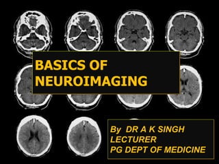
BASICS OF
NEUROIMAGING
What We Need to Know
• Air is very black (less than -300 HU)
• Water/CSF is black (near 0 HU)
• Bone is very dense/white (500-3000 HU)
• Blood is white (60-80 HU)
• Brain is gray 35-50 HU
Normal CT of brain
• Ventricles are normal sized,
the grey versus white distinction
is clear.
• Midline is straight.
• Sulci are symmetrical on
bothsides.
• Skull is intact with no
scalp edema.
Systemic Approach to Head CT
Interpretation
• Symmetry – Compare left and right side of the cranium
• Midline – Look for midline shift
• Cross-sectional anatomy – Review anatomical landmark for
each slide
– Brain tissue : gray matter, white matter , intracerebral lesions
– CSF space : ventricle, basal cistern, cortical sulci, fissure
– Skull and soft tissue : scalp swelling, fractures, sinuses, orbit
• Subdural windows : Look for blood collection adjacent to the
skull
• Bone windows : Skull, orbit and sinuses, intracranial air
Lateral View of Brain
Cross-sectional Anatomy
• Grey/White interface, Subcortical white
matter
Cross-sectional Anatomy
• Paired of crescent-shape = Twin bananas
Cross-sectional Anatomy
Basal ganglia
Cross-sectional Anatomy
• Third ventricle, Basal ganglia, Superior
cerebellar cistern
Physiologic Calcification
Cross-sectional Anatomy
• Third ventricle, Smiley face
Cross-sectional Anatomy
• Midbrain, Interpeduncular cistern
Cross-sectional Anatomy
• Star shape ~ Circle of Willis,
• Fourth ventricle, Temporal horn ~ slit
Cross-sectional Anatomy
• Base of skull, Midline bony prominence,
• Prepontine cistern, Pretrous bone, Frontal
Cross-sectional Anatomy
• Orbits, Ethmoid air cell
GREEN IS
TEMPORAL
LOBE
RED
DENOTES
FRONTAL
LOBE
GREEN
DENOTES
TEMPORAL
LOBE
RED
DENOTES
FRONTAL
LOBE
GREEN-
TEMPORAL
LOBE
RED-
FRONTAL
LOBE
GREEN-
TEMPORAL
LOBE
RED-
FRONTAL
LOBE
GREEN-
TEMPORAL
LOBE
YELLOW-
OCCIPITAL
LOBE
RED-
FRONTAL
LOBE
GREEN-
TEMPORAL
LOBE
YELLOW-
OCCIPITAL
LOBE
RED-
FRONTAL
LOBE
GREEN-
TEMPORAL
LOBE
YELLOW-
OCCIPITAL
LOBE
RED –FRONTAL
LOBE
BLUE-PARIETAL
LOBE
GREEN-
TEMPORAL
LOBE
YELLOW-
OCCIPITAL
LOBE
RED-
FRONTAL
LOBE
BLUE-
PARIETAL
LOBE
YELLOW-
OCCIPITAL
LOBE
RED –
FRONTAL
LOBE
BLUE-
PARIETAL
LOBE
YELLOW-
OCCIPITAL
LOBE
RED –
FRONTAL
LOBE
BLUE-
PARIETAL
LOBE
YELLOW-
OCCIPITAL
LOBE
FRONTAL
LOBE –RED
PARIETAL
LOBE - BLUE
RED –
FRONTAL
LOBE
BLUE-
TEMPORAL
LOBE
RED –
FRONTAL
LOBE
BLUE –
TEMPORAL
LOBE
Thalmus
Aqueduct of sylviusS
Cerebellum
Fourth
ventricle
Corpus
callosum
Midbrain
Pons
Medulla
Corpus callosum Thalamus Aqueduct of Sylvius
Fourth Ven. Mid-brain Pons Cerebellum Medulla oblongata
SECTION AT MID-SAGITTAL PLANE
MRI
• Based on the absorption and emission of radiofrequency
energy – so there is NO ionizing radiation.
• Uses magnets ranging in strength from 0.3 to 1.5 Tesla to
create a magnetic field around the patient.
• Magnetic field causes protons in the body to align and then
pulsed radiowaves are directed at the patient causing a
disturbance of the proton alignment.
• Atoms then realign and in doing so, emit the absorbed
radiofrequency
• The time it takes the protons to regain their equilibrium state =
• RELAXATION TIME.
„
• 2 types of relaxation time: T1 – Longitudinal (parallel to the magnetic
field) and T2 –transverse (perpendicular to the mag field).
„
• Relaxation Time and Proton Density are the main determinants of
signal strength.
„
• The main determinants of contrast or the weighting are:
‹
1)Repetition Time (TR) – the time between successive RF pulses
2)Echo Time (TE) – time between the arrival of the RF pulse that
excites
and the arrival of the return signal at the detector.
Short TR + Short TE = T1 weighted
•Dark
– CSF
– Increased Water –
edema,
– tumor, infarct,
inflammation,
– infection, hemorrhage
(hyperacute or
chronic)
– Low proton density,
calcification
– Flow Void
•Bright
– Fat
– Subacute hemorrhage
– Melanin
– Protein-rich Fluid
– Slowly flowing blood
– Gadolinium
– Laminar necrosis of
an infarct
Long TR + Long TE= T2 weighted
• Dark
– Low Proton Density,
– calcification, fibrous
tissue
– Paramagnetic
substances -
• deoxyhemoglobin,
• methemoglobin
(intracellular),
• iron, hemosiderin,
melanin
– Protein-rich fluid
– Flow Void
• Bright
– CSF
– Increased Water –
edema,
– tumor, infarct,
inflammation,
– infection, subdural
collection
– Methemoglobin
– (extracellular) in
subacute
– hemorrhage
Fluid-Attenuated Inversion Recovery
FLAIR
• Basically T2 without CSF brightness
• TE>80 and TR>10,000
• Edema and Gliosis are hyperintense
T1W / T2W / FLAIR
T1W T2W FLAIR
Fig. 1.1 Post Contrast Axial
MR Image of the brain
1
2
3
4
5
Post Contrast sagittal
T1 Weighted M.R.I.
Section at the level of
Foramen MagnumAnswers
1. Cisterna Magna
2. Cervical Cord
3. Nasopharynx
4. Mandible
5. Maxillary Sinus
Fig. 1.2 Post Contrast Axial
MR Image of the brain
7
6
Post Contrast sagittal
T1 Wtd M.R.I.
Section at the level of
medulla
Answers
6. Medulla
7. Sigmoid Sinus
Fig. 1.3 Post Contrast Axial
MR Image of the brain
15
8
9
10
11
12
13
14
16
17
Post Contrast sagittal
T1 Wtd M.R.I.
Section at the level of
PonsAnswers
8. Cerebellar
Hemisphere
9. Vermis
10. IV Ventricle
11. Pons
12. Basilar
Artery
13. Internal Carotid
Artery
14. Cavernous Sinus
15. Middle Cerebellar
Peduncle
16. Internal Auditory
Canal
17. Temporal Lobe
Fig. 1.4 Post Contrast Axial
MR Image of the brain
18
19
20
21
22
Post Contrast sagittal T1
Wtd M.R.I.
Section at the level of Mid
Brain
Answers
18. Aqueduct of Sylvius
19. Midbrain
20. Orbits
21. Posterior Cerebral
Artery
22. Middle Cerebral Artery
Fig. 1.5 Post Contrast Axial
MR Image of the brain
23
24
25
26
27
Post Contrast sagital T1 Wtd
M.R.I.
Section at the level of the
III Ventricle
Answers
23. Occipital Lobe
24. III Ventricle
25. Frontal Lobe
26. Temporal Lobe
27. Sylvian Fissure
Fig. 1.6 Post Contrast Axial
MR Image of the brain
28
29
30
31
32
38
33
34
36
35
37
Post Contrast sagittal
T1 Wtd M.R.I.
Section at the level of
Thalamus
Answers
28. Superior Sagittal
Sinus
29. Occipital Lobe
30. Choroid Plexus
within the occipital
horn
31. Internal Cerebral
Vein
32. Frontal Horn
33. Thalamus
34. Temporal Lobe
35. Internal Capsule
36. Putamen
37. Caudate Nucleus
38. Frontal Lobe
Fig. 1.7 Post Contrast Axial
MR Image of the brain
39
40
41
Post Contrast sagittal
T1 Wtd M.R.I.
Section at the level of
Corpus Callosum
Answers
39. Splenium of corpus callosum
40. Choroid plexus within the
body of lateral ventricle
41. Genu of corpus callosum
Fig. 1.8 Post Contrast Axial
MR Image of the brain
42
43
44
Post Contrast sagittal
T1 Wtd M.R.I.
Section at the level of
Body of Corpus
Callosum
Answers
42. Parietal Lobe
43. Body of the Corpus Callosum
44. Frontal Lobe
T1W T2W FLAIR
acute/subacute hemorrhage
acu
te
SUBACUTE T2WI T1WI
Strokes show up faster on MRI
than CT
MRI and CAT views of the same
whole R. hemispherical infarct
Some very big strokes settle down and don’t require surgical
decompression. This man opens his eyes to verbal on nasal
cannula and follows on the right side 10 days post stroke.
MR:44396
MRI appearances of acute
cerebral infarction
T2WI T1WI Flair
The End…

Más contenido relacionado

La actualidad más candente

Neuroradiology slideshare
Neuroradiology slideshareNeuroradiology slideshare
Neuroradiology slideshareREKHAKHARE
 
Basic approach to brain CT Dr. Muhammad Bin Zulfiqar
Basic approach to brain CT Dr. Muhammad Bin ZulfiqarBasic approach to brain CT Dr. Muhammad Bin Zulfiqar
Basic approach to brain CT Dr. Muhammad Bin ZulfiqarDr. Muhammad Bin Zulfiqar
 
Clinical correlation and interpretation of Brain MRI by dr.Sagor
Clinical correlation and interpretation of Brain MRI by dr.SagorClinical correlation and interpretation of Brain MRI by dr.Sagor
Clinical correlation and interpretation of Brain MRI by dr.SagorChittagong Medical College Hospital
 
MRI Procedure of Brain
MRI Procedure of BrainMRI Procedure of Brain
MRI Procedure of BrainSudil Paudyal
 
Imaging in stroke
Imaging in strokeImaging in stroke
Imaging in strokeikramdr01
 
Mri imaging finding in wilson disease
Mri imaging finding in wilson diseaseMri imaging finding in wilson disease
Mri imaging finding in wilson diseasegulabsoni
 
BASICS IN NEUROIMAGING: CT, MRI AND PET
BASICS IN NEUROIMAGING: CT, MRI AND PETBASICS IN NEUROIMAGING: CT, MRI AND PET
BASICS IN NEUROIMAGING: CT, MRI AND PETPramod Krishnan
 
Neuroimaging Lecture
Neuroimaging LectureNeuroimaging Lecture
Neuroimaging Lecturetest
 
CT imaging of Brain in Clinical Practice by Dr. Vaibhav Yawalkar
CT imaging of Brain in Clinical Practice by Dr. Vaibhav YawalkarCT imaging of Brain in Clinical Practice by Dr. Vaibhav Yawalkar
CT imaging of Brain in Clinical Practice by Dr. Vaibhav Yawalkarvaibhavyawalkar
 
Craniovertebral junction
Craniovertebral junctionCraniovertebral junction
Craniovertebral junctionNeurologyKota
 
Corpus Callosum Anomalies
Corpus Callosum  AnomaliesCorpus Callosum  Anomalies
Corpus Callosum AnomaliesMarwan Alhalabi
 
Sulcal and gyral neuroanatomy
Sulcal and gyral neuroanatomySulcal and gyral neuroanatomy
Sulcal and gyral neuroanatomyNavni Garg
 
Normal & abnormal radiology of brain part ii
Normal & abnormal radiology of brain part iiNormal & abnormal radiology of brain part ii
Normal & abnormal radiology of brain part iiMohammed Fathy
 
Diffusion tensor imaging in Neurology
Diffusion tensor imaging in NeurologyDiffusion tensor imaging in Neurology
Diffusion tensor imaging in NeurologyOsama Ragab
 
Presentation1.pptx, radiological imaging of cerebello pontine angle mass lesi...
Presentation1.pptx, radiological imaging of cerebello pontine angle mass lesi...Presentation1.pptx, radiological imaging of cerebello pontine angle mass lesi...
Presentation1.pptx, radiological imaging of cerebello pontine angle mass lesi...Abdellah Nazeer
 

La actualidad más candente (20)

Brain anatomy on ct & amp; mri
Brain anatomy on ct & amp; mriBrain anatomy on ct & amp; mri
Brain anatomy on ct & amp; mri
 
Radiological anatomy of the brain
Radiological anatomy of the brainRadiological anatomy of the brain
Radiological anatomy of the brain
 
Neuroradiology slideshare
Neuroradiology slideshareNeuroradiology slideshare
Neuroradiology slideshare
 
Basic approach to brain CT Dr. Muhammad Bin Zulfiqar
Basic approach to brain CT Dr. Muhammad Bin ZulfiqarBasic approach to brain CT Dr. Muhammad Bin Zulfiqar
Basic approach to brain CT Dr. Muhammad Bin Zulfiqar
 
Clinical correlation and interpretation of Brain MRI by dr.Sagor
Clinical correlation and interpretation of Brain MRI by dr.SagorClinical correlation and interpretation of Brain MRI by dr.Sagor
Clinical correlation and interpretation of Brain MRI by dr.Sagor
 
MRI Procedure of Brain
MRI Procedure of BrainMRI Procedure of Brain
MRI Procedure of Brain
 
Imaging in stroke
Imaging in strokeImaging in stroke
Imaging in stroke
 
Mri imaging finding in wilson disease
Mri imaging finding in wilson diseaseMri imaging finding in wilson disease
Mri imaging finding in wilson disease
 
BASICS IN NEUROIMAGING: CT, MRI AND PET
BASICS IN NEUROIMAGING: CT, MRI AND PETBASICS IN NEUROIMAGING: CT, MRI AND PET
BASICS IN NEUROIMAGING: CT, MRI AND PET
 
Neuroimaging Lecture
Neuroimaging LectureNeuroimaging Lecture
Neuroimaging Lecture
 
CT imaging of Brain in Clinical Practice by Dr. Vaibhav Yawalkar
CT imaging of Brain in Clinical Practice by Dr. Vaibhav YawalkarCT imaging of Brain in Clinical Practice by Dr. Vaibhav Yawalkar
CT imaging of Brain in Clinical Practice by Dr. Vaibhav Yawalkar
 
MR Spectroscopy
MR SpectroscopyMR Spectroscopy
MR Spectroscopy
 
Craniovertebral junction
Craniovertebral junctionCraniovertebral junction
Craniovertebral junction
 
Corpus Callosum Anomalies
Corpus Callosum  AnomaliesCorpus Callosum  Anomalies
Corpus Callosum Anomalies
 
Sulcal and gyral neuroanatomy
Sulcal and gyral neuroanatomySulcal and gyral neuroanatomy
Sulcal and gyral neuroanatomy
 
Normal & abnormal radiology of brain part ii
Normal & abnormal radiology of brain part iiNormal & abnormal radiology of brain part ii
Normal & abnormal radiology of brain part ii
 
Mr imaging of brain
Mr imaging of brainMr imaging of brain
Mr imaging of brain
 
Diffusion tensor imaging in Neurology
Diffusion tensor imaging in NeurologyDiffusion tensor imaging in Neurology
Diffusion tensor imaging in Neurology
 
Presentation1.pptx, radiological imaging of cerebello pontine angle mass lesi...
Presentation1.pptx, radiological imaging of cerebello pontine angle mass lesi...Presentation1.pptx, radiological imaging of cerebello pontine angle mass lesi...
Presentation1.pptx, radiological imaging of cerebello pontine angle mass lesi...
 
CSF cisterns
CSF cisternsCSF cisterns
CSF cisterns
 

Destacado

Brain imaging.pdf
Brain imaging.pdf Brain imaging.pdf
Brain imaging.pdf coburgpsych
 
Introduction to Neuroimaging
Introduction to NeuroimagingIntroduction to Neuroimaging
Introduction to NeuroimagingSunghyon Kyeong
 
fMRI preprocessing steps (in SPM8)
fMRI preprocessing steps (in SPM8)fMRI preprocessing steps (in SPM8)
fMRI preprocessing steps (in SPM8)Sunghyon Kyeong
 
fMRI Presentation
fMRI PresentationfMRI Presentation
fMRI Presentationricksw78
 
Miniatlas of Human Cross-Sectional Anatomy
Miniatlas of Human Cross-Sectional AnatomyMiniatlas of Human Cross-Sectional Anatomy
Miniatlas of Human Cross-Sectional AnatomyPhilip Tate
 
101 ct neuroimaging
101 ct neuroimaging101 ct neuroimaging
101 ct neuroimagingAhmad Shahir
 
Advances in neuroimaging techniques
Advances in neuroimaging techniquesAdvances in neuroimaging techniques
Advances in neuroimaging techniquesSreenivasa Raju
 
fMRI Brain Imaging
fMRI Brain ImagingfMRI Brain Imaging
fMRI Brain ImagingMerrett
 
Diagnostic Imaging of Brain Tumors
Diagnostic Imaging of Brain TumorsDiagnostic Imaging of Brain Tumors
Diagnostic Imaging of Brain TumorsMohamed M.A. Zaitoun
 
the brain ,parts and functions
 the brain ,parts and functions the brain ,parts and functions
the brain ,parts and functionsEduc_Louie Tanaka
 
Brain ppt
Brain pptBrain ppt
Brain pptcnps
 
The human brain presentation
The human brain presentationThe human brain presentation
The human brain presentationSilvia Borba
 
10 Brain.ppt
10 Brain.ppt10 Brain.ppt
10 Brain.pptShama
 

Destacado (20)

Neuroimaging
NeuroimagingNeuroimaging
Neuroimaging
 
Brain imaging.pdf
Brain imaging.pdf Brain imaging.pdf
Brain imaging.pdf
 
Neuroimaging methods
Neuroimaging methodsNeuroimaging methods
Neuroimaging methods
 
Introduction to Neuroimaging
Introduction to NeuroimagingIntroduction to Neuroimaging
Introduction to Neuroimaging
 
fMRI preprocessing steps (in SPM8)
fMRI preprocessing steps (in SPM8)fMRI preprocessing steps (in SPM8)
fMRI preprocessing steps (in SPM8)
 
fMRI Presentation
fMRI PresentationfMRI Presentation
fMRI Presentation
 
Miniatlas of Human Cross-Sectional Anatomy
Miniatlas of Human Cross-Sectional AnatomyMiniatlas of Human Cross-Sectional Anatomy
Miniatlas of Human Cross-Sectional Anatomy
 
101 ct neuroimaging
101 ct neuroimaging101 ct neuroimaging
101 ct neuroimaging
 
Advances in neuroimaging techniques
Advances in neuroimaging techniquesAdvances in neuroimaging techniques
Advances in neuroimaging techniques
 
Fmri overview
Fmri overviewFmri overview
Fmri overview
 
Introduction to fMRI
Introduction to fMRIIntroduction to fMRI
Introduction to fMRI
 
fMRI Brain Imaging
fMRI Brain ImagingfMRI Brain Imaging
fMRI Brain Imaging
 
Diagnostic Imaging of Brain Tumors
Diagnostic Imaging of Brain TumorsDiagnostic Imaging of Brain Tumors
Diagnostic Imaging of Brain Tumors
 
Functional MRI
Functional MRI Functional MRI
Functional MRI
 
fMRI Study Design
fMRI Study DesignfMRI Study Design
fMRI Study Design
 
Functions of the Brain
Functions of the BrainFunctions of the Brain
Functions of the Brain
 
the brain ,parts and functions
 the brain ,parts and functions the brain ,parts and functions
the brain ,parts and functions
 
Brain ppt
Brain pptBrain ppt
Brain ppt
 
The human brain presentation
The human brain presentationThe human brain presentation
The human brain presentation
 
10 Brain.ppt
10 Brain.ppt10 Brain.ppt
10 Brain.ppt
 

Similar a Basics of neuroimaging

Neuroimaging in Psychiatry
Neuroimaging in PsychiatryNeuroimaging in Psychiatry
Neuroimaging in PsychiatryEnoch R G
 
PRICIPLES OF MRI BRAIN FINAL COPY.pptx
PRICIPLES OF MRI BRAIN  FINAL COPY.pptxPRICIPLES OF MRI BRAIN  FINAL COPY.pptx
PRICIPLES OF MRI BRAIN FINAL COPY.pptxDRMANOJSEERVI
 
ctandmriinterpretation-150127105431-conversion-gate01.pdf
ctandmriinterpretation-150127105431-conversion-gate01.pdfctandmriinterpretation-150127105431-conversion-gate01.pdf
ctandmriinterpretation-150127105431-conversion-gate01.pdfrahul amte
 
Ctandmriinterpretation 150127105431-conversion-gate01
Ctandmriinterpretation 150127105431-conversion-gate01Ctandmriinterpretation 150127105431-conversion-gate01
Ctandmriinterpretation 150127105431-conversion-gate01Thamiil Selvan
 
pediatric neuroradiology essentials
pediatric neuroradiology essentialspediatric neuroradiology essentials
pediatric neuroradiology essentialsdr jyoti prajapati
 
MRI BRAIN(AIIMS DELHI).pptx
MRI BRAIN(AIIMS DELHI).pptxMRI BRAIN(AIIMS DELHI).pptx
MRI BRAIN(AIIMS DELHI).pptxGyandeep28
 
Sectional anatomy of brain part 1
Sectional  anatomy  of  brain  part 1Sectional  anatomy  of  brain  part 1
Sectional anatomy of brain part 1Manideep Malaka
 
Neurosurgery 1.pptx
Neurosurgery 1.pptxNeurosurgery 1.pptx
Neurosurgery 1.pptxjoendesh
 
ESSENTIALS_OF_CT_BRAIN_For_Undergraduate.pptx
ESSENTIALS_OF_CT_BRAIN_For_Undergraduate.pptxESSENTIALS_OF_CT_BRAIN_For_Undergraduate.pptx
ESSENTIALS_OF_CT_BRAIN_For_Undergraduate.pptxssuser39be96
 
Normal anatomy of brain on CT and MRI
Normal anatomy of brain on CT and MRINormal anatomy of brain on CT and MRI
Normal anatomy of brain on CT and MRIDr Anuj Aggarwal
 
Heart attacks & detection
Heart attacks & detectionHeart attacks & detection
Heart attacks & detectionAbhisek Anand
 
Radiology of central nervous system
Radiology of central nervous systemRadiology of central nervous system
Radiology of central nervous systemWEEKLYMEDIC
 

Similar a Basics of neuroimaging (20)

Neuroimaging in Psychiatry
Neuroimaging in PsychiatryNeuroimaging in Psychiatry
Neuroimaging in Psychiatry
 
PRICIPLES OF MRI BRAIN FINAL COPY.pptx
PRICIPLES OF MRI BRAIN  FINAL COPY.pptxPRICIPLES OF MRI BRAIN  FINAL COPY.pptx
PRICIPLES OF MRI BRAIN FINAL COPY.pptx
 
ctandmriinterpretation-150127105431-conversion-gate01.pdf
ctandmriinterpretation-150127105431-conversion-gate01.pdfctandmriinterpretation-150127105431-conversion-gate01.pdf
ctandmriinterpretation-150127105431-conversion-gate01.pdf
 
Ct and mri interpretation
Ct and mri interpretationCt and mri interpretation
Ct and mri interpretation
 
Ctandmriinterpretation 150127105431-conversion-gate01
Ctandmriinterpretation 150127105431-conversion-gate01Ctandmriinterpretation 150127105431-conversion-gate01
Ctandmriinterpretation 150127105431-conversion-gate01
 
pediatric neuroradiology essentials
pediatric neuroradiology essentialspediatric neuroradiology essentials
pediatric neuroradiology essentials
 
MRI BRAIN(AIIMS DELHI).pptx
MRI BRAIN(AIIMS DELHI).pptxMRI BRAIN(AIIMS DELHI).pptx
MRI BRAIN(AIIMS DELHI).pptx
 
Sectional anatomy of brain part 1
Sectional  anatomy  of  brain  part 1Sectional  anatomy  of  brain  part 1
Sectional anatomy of brain part 1
 
Neurosurgery 1.pptx
Neurosurgery 1.pptxNeurosurgery 1.pptx
Neurosurgery 1.pptx
 
ESSENTIALS_OF_CT_BRAIN_For_Undergraduate.pptx
ESSENTIALS_OF_CT_BRAIN_For_Undergraduate.pptxESSENTIALS_OF_CT_BRAIN_For_Undergraduate.pptx
ESSENTIALS_OF_CT_BRAIN_For_Undergraduate.pptx
 
Essentials of CT brain (For Undergraduates)
Essentials of CT brain (For Undergraduates)Essentials of CT brain (For Undergraduates)
Essentials of CT brain (For Undergraduates)
 
Normal anatomy of brain on CT and MRI
Normal anatomy of brain on CT and MRINormal anatomy of brain on CT and MRI
Normal anatomy of brain on CT and MRI
 
Brain stem
Brain stemBrain stem
Brain stem
 
Neonatal MRI Brain
Neonatal MRI BrainNeonatal MRI Brain
Neonatal MRI Brain
 
Introduction to cranial ct
Introduction to cranial ctIntroduction to cranial ct
Introduction to cranial ct
 
RAH Med 4 MHU - Brain CT 1
RAH Med 4 MHU - Brain CT 1RAH Med 4 MHU - Brain CT 1
RAH Med 4 MHU - Brain CT 1
 
Heart attacks & detection
Heart attacks & detectionHeart attacks & detection
Heart attacks & detection
 
CT BRAIN ANATOMY.pptx
CT BRAIN ANATOMY.pptxCT BRAIN ANATOMY.pptx
CT BRAIN ANATOMY.pptx
 
Radiology of central nervous system
Radiology of central nervous systemRadiology of central nervous system
Radiology of central nervous system
 
How To Read A Head CT Scan
How To Read A Head CT Scan How To Read A Head CT Scan
How To Read A Head CT Scan
 

Basics of neuroimaging