
Recomendados
Más contenido relacionado
Similar a p1185.pdf
Similar a p1185.pdf (20)
Último
Último (20)
p1185.pdf
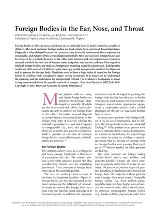
- 1. Foreign Bodies in the Ear, Nose, and Throat STEVEN W. HEIM, MD, MSPH, and KAREN L. MAUGHAN, MD University of Virginia School of Medicine, Charlottesville, Virginia M ost patients with ear, nose, and throat foreign bodies are children; intellectually chal- lenged or mentally ill adults are also at increased risk. Often, family phy- sicians are able to remove the foreign body in the office. Successful removal depends on several factors, including location of the foreign body, type of material, whether the material is graspable (i.e., soft and irregular) or nongraspable (i.e., hard and spherical), physician dexterity, and patient cooperation. Table 1 provides an overview of common foreign bodies, removal techniques, and indi- cations for referral.1-18 Ear Foreign Bodies The external auditory canal is a cartilaginous and bony passage lined with a thin layer of periosteum and skin. The osseous por- tion is extremely sensitive because the skin provides little cushion over the underlying periosteum. Thus, attempts at foreign body removal can be extremely painful. The external auditory canal narrows at the bony cartilaginous junction (Figure 1). Foreign bodies can become impacted at this point, increasing the difficulty of removal. Attempts to remove the foreign body may push it further into the canal and lodge it at this narrow point. In addition, the tympanic membrane can be damaged by pushing the foreign body further into the canal or by the instruments used during removal attempts. Adequate visualization, appropriate equip- ment, a cooperative patient, and a skilled physician are the keys to successful foreign body removal. In many cases, patients with foreign bod- ies in the ear are asymptomatic, and in chil- dren the foreign body is often an incidental finding.1,19 Other patients may present with pain, symptoms of otitis media, hearing loss, or a sense of ear fullness. In several large case series focusing on children, research- ers found that 75 percent of patients with ear foreign bodies were younger than eight years.1,2,19 Similar studies on adult patients are lacking. The most common ear foreign bodies include beads, plastic toys, pebbles, and popcorn kernels.2 Insects are more com- mon in patients older than 10 years. In one series, 30 percent of patients required gen- eral anesthesia to facilitate removal of an ear foreign body; the majority of those patients were younger than seven years.2 Graspable foreign bodies (e.g., foam rubber, paper, vegetable material) have higher rates of suc- cess for removal under direct visualization. In contrast, nongraspable foreign bodies (e.g., beads, pebbles, popcorn kernels) have Foreign bodies in the ear, nose, and throat are occasionally seen in family medicine, usually in children. The most common foreign bodies are food, plastic toys, and small household items. Diagnosis is often delayed because the causative event is usually unobserved, the symptoms are nonspecific, and patients often are misdiagnosed initially. Most ear and nose foreign bodies can be removed by a skilled physician in the office with minimal risk of complications. Common removal methods include use of forceps, water irrigation, and suction catheter. Pharyngeal or tracheal foreign bodies are medical emergencies requiring surgical consultation. Radiography results are often normal. Flexible or rigid endoscopy usually is required to confirm the diagnosis and to remove the foreign body. Physicians need to have a high index of suspicion for foreign bodies in children with unexplained upper airway symptoms. It is important to understand the anatomy and the indications for subspecialist referral. The evidence is inadequate to make strong recommendations for specific removal techniques. (Am Fam Physician 2007;76:1185-9. Copyright © 2007 American Academy of Family Physicians.) Downloaded from the American Family Physician Web site at www.aafp.org/afp. Copyright © 2007 American Academy of Family Physicians. For the private, noncommercial use of one individual user of the Web site. All other rights reserved. Contact copyrights@aafp.org for copyright questions and/or permission requests.
- 2. 1186 American Family Physician www.aafp.org/afp Volume 76, Number 8 ◆ October 15, 2007 lower rates of successful removal and are associated with more complications, par- ticularly canal lacerations.3 Many techniques to remove ear foreign bodies are available, and the choice depends on the clinical situation, the type of foreign body suspected, and the experience of the physician. Options include water irrigation, forceps removal (e.g., alligator forceps), cerumen loops, right-angle ball hooks, and suction catheters. Live insects can be killed rapidly by instilling alcohol, 2% lidocaine (Xylocaine), or mineral oil into the ear canal. This should be done before removal is attempted but should not be used when the tympanic membrane is perforated.20 Irrigation should be avoided in patients with button batteries in the ear because the electrical current and/or battery contents can cause a liquefaction tissue necrosis.21 SORT: KEY RECOMMENDATIONS FOR PRACTICE Clinical recommendation Evidence rating References Directly visible, “graspable” foreign bodies in the ear or nose can often be removed without subspecialist referral. C 1 Removal of hard, nongraspable, spherical foreign bodies in the ear canal against the tympanic membrane will most likely require subspecialist referral. C 1, 23 If a foreign body in the ear, nose, or throat cannot be directly visualized or if attempts at removal have been unsuccessful, the patient should be referred to a subspecialist. C 1, 3, 12 Patients with a suspected foreign body in the throat should be referred to a subspecialist unless the item is easily visible and graspable. C 14, 18 A = consistent, good-quality patient-oriented evidence; B = inconsistent or limited-quality patient-oriented evidence; C = consensus, disease-oriented evidence, usual practice, expert opinion, or case series. For information about the SORT evidence rating system, see page 1095 or http://www.aafp.org/afpsort.xml. Table 1. Management of Common Foreign Bodies in the Ear, Nose, and Throat Location Common foreign bodies Removal technique Indications for referral Ear Beads, plastic toys, pebbles, popcorn kernels2 Irrigation with water Grasping foreign body with forceps, cerumen loop, right-angle ball hook, or suction catheter Acetone to dissolve Styrofoam foreign body4 Need for sedation Canal or tympanic membrane trauma Foreign body is nongraspable, tightly wedged, or touching tympanic membrane Sharp foreign body Removal attempts unsuccessful1-3,12 Nose Beads, buttons, toy parts, pebbles, candle wax, food, paper, cloth, button batteries5,6 Grasping with forceps, curved hook, cerumen loop, or suction catheter Thin, lubricated, balloon-tip catheter7 Patient “blows nose” with opposite nostril obstructed PPV delivered to patient’s mouth with opposite nostril obstructed8,9,11 ; PPV may also be delivered by bag mask10 Tumor or mass suspected Removal attempts unsuccessful Edema, bony destruction, granulation tissue from chronic foreign body12 Throat (pharynx)* Plastic, metal pin, seeds, nuts, bones, coins, dental appliances14-18 Often need to be removed endoscopically, requiring sedation14,18 and, thus, referral Inadequate visualization Need for sedation Signs of airway compromise13,14 PPV = positive pressure ventilation. *—Most foreign bodies in the throat require consultation with a subspecialist. Information from references 1 through 18.
- 3. October 15, 2007 ◆ Volume 76, Number 8 www.aafp.org/afp American Family Physician 1187 Acetone may be used to dissolve Styrofoam foreign bodies4 or to loosen cyanoacrylate (i.e., superglue).22 The first attempt at removal is critical because success rates markedly decrease after the first failed attempt. Accordingly, com- plications increase as the number of failed removal attempts increases.23,24 Removal attempts are often painful, can cause bleed- ing that limits visualization, and can further wedge the foreign body into the canal. An otolaryngology referral should be obtained for patients requiring sedation or anesthe- sia. Other indications for referral include patients with trauma to the canal or tym- panic membrane; a nongraspable foreign body that is tightly wedged in the medial two thirds of the canal or is suspected of touching the tympanic membrane; foreign bodies with sharp edges (e.g., pieces of glass); or unsuccessful removal attempts.1-3 Multiple foreign bodies are not uncom- mon, especially in small children. Thus, all other orifices of the head should be inspected after removal of a foreign body from the external auditory canal.3 Otic anti- biotic drops are needed in patients with concurrent otitis externa and should be con- sidered when canal lacerations or trauma is present. Audiography should be considered if tympanic membrane trauma or hearing loss is suspected. Nasal Foreign Bodies The nose consists of two nasal fossae sepa- rated by a vertical septum and subdivided into three passages by the nasal turbinates. Nasal foreign bodies tend to be located on the floor of the nasal passage, just below the inferior turbinate, or in the upper nasal fossa anterior to the middle turbinate5 (Figure 2). Most nasal foreign bodies can be easily removed in the office or emergency department. Patients often present with uni- lateral, foul-smelling nasal discharge.25 Com- mon nasal foreign bodies include beads, buttons, toy parts, pebbles, candle wax, food, paper, cloth, and button batteries.5,6 Before foreign body removal, 0.5% phenyl ephrine (Neo-Synephrine) can be used to reduce mucosal edema, and topical lidocaine may be applied to provide analgesia. Tech- niques include removal with direct visualiza- tion using forceps, curved hooks, cerumen loops, or suction catheters. Additionally, suc- cessful removal has been achieved by passing a thin, lubricated, balloon-tip catheter (5 or 6 French Foley) past the foreign body, inflat- ing the balloon, and pulling the inflated Figure 1. The external auditory canal. Foreign bodies may become lodged in the narrowing at the bony cartilaginous junction. Figure 2. Nasopharyngeal and tracheal anatomy. Highlighted areas indicate points at which nasal foreign bodies may become lodged. Bony cartilaginous junction Osseous portion of ear canal Tympanic membrane Foreign body Foreign body Soft palate Epiglottis Esophagus Trachea Cricoid cartilage Vocal cords Upper, middle, and lower turbinates ILLUSTRATION BY christy krames ILLUSTRATION BY christy krames Foreign Bodies
- 4. 1188 American Family Physician www.aafp.org/afp Volume 76, Number 8 ◆ October 15, 2007 Foreign Bodies catheter balloon forward, thus moving the foreign body into the anterior nares where removal can be completed.7 Patients may be able to expel the nasal foreign body simply by “blowing their nose” while blocking the opposite nostril. If this fails, or if a nasal foreign body is present in a small child unable to cooper- ate, positive pressure ventilation can be delivered through the patient’s mouth. In this technique, the parent covers the child’s mouth with his or her mouth, plugs the unobstructed nostril with a finger, and gives a rapid, soft puff of air.8,9 Although the child will reflexively close the glottis to protect his or her lungs from the pressure, it is impor- tant that the parent not use a large-volume or high-pressure breath. Barotrauma to the ear is a theoretical risk of positive pressure ventilation, but this complication has not been reported. Appropriate infection-control precautions must be taken because the foreign body will likely be expelled against the parent’s cheek and will be covered with mucus and possibly blood. Positive pressure can also be deliv- ered through the mouth using a bag mask (Ambu Bag)10 or through the nose using oxygen tubing.11 Button batteries must be removed from the nose immediately because of the danger of liquefaction necrosis of the surrounding tissue.26 Attemptsatremovalmaypushthenasalfor- eign body into the pharynx, creating an air- way hazard. Sedation is discouraged because it can increase complications by reducing the gag and cough reflexes.12 Consultation should be obtained when the foreign body cannot be removed or adequately visualized, or when a tumor or mass is suspected. Throat Foreign Bodies The throat (pharynx) is bound superiorly by the base of the skull (nasopharynx) and infe- riorly by the cricoid cartilage/inferior border of the C6 vertebra (hypopharynx; Figure 2). The hypopharynx contains the larynx and the upper openings of the trachea and the esophagus. All pharyngeal foreign bodies are medical emergencies that require airway protection. Because complete airway obstruction usu- ally occurs at the time of aspiration and results in immediate respiratory distress, emergency intervention is essential. Com- mon obstructing foreign bodies in children include balloons, pieces of soft deform- able plastic, and food boluses.15 Patients with nonobstructing or partially obstruct- ing foreign bodies in the throat often pres- ent with a history of choking, dysphagia, odynophagia, or dysphonia.13 Pharyngeal foreign bodies should also be suspected in patients with undiagnosed coughing, stri- dor, or hoarseness.14 Parents and caregivers of children with symptoms of partial airway obstruction should be asked whether choking and aspi- ration have occurred. Diagnosis is often complicated by delayed presentation. Case reports describe foreign bodies in the throat that were misdiagnosed and treated as croup.15,27 Thus, physicians must have a high degree of suspicion in patients with unex- plained upper airway symptoms, especially in children who have a history of choking. The most common foreign bodies in the throat are pieces of plastic, metal pins, seeds, nuts, bones, coins, and dental appliances.14-17 Radiography can be helpful in localizing coins, button batteries, and other radiopaque objects, but most laryngeal foreign bodies, including many fish bones, are radiolu- cent.13,28 Therefore, the decision to pursue surgical intervention should be based on the patient’s history and a physical examination that suggests the presence of a foreign body rather than based on radiography alone.29 Early consultation is advisable because pharyngeal foreign bodies are difficult to visualize without the use of flexible or rigid endoscopy. Furthermore, removal attempts are difficult and are complicated by the gag reflex. Because the airway must be protected, most foreign bodies in the throat require otolaryngology intervention with sedation and endoscopic removal.14,18 Complications include airway obstruction, laryngeal edema, and pushing the foreign body into the sub- glottic space, esophagus, or trachea.14,18 Pharyngeal foreign bodies should be suspected in patients with undiag- nosed coughing, stridor, or hoarseness.
- 5. October 15, 2007 ◆ Volume 76, Number 8 www.aafp.org/afp American Family Physician 1189 Foreign Bodies The Authors STEVEN W. HEIM, MD, MSPH, is an associate professor of family medicine at the University of Virginia School of Medicine, Charlottesville. Dr. Heim received his medical degree from the University of Virginia School of Medicine and completed a family medicine residency and fellowship training at the University of Missouri–Columbia School of Medicine. KAREN L. MAUGHAN, MD, is an associate professor of family medicine at the University of Virginia School of Medicine. Dr. Maughan received her medical degree from McGill University Medical School, Montreal, Quebec, Canada, and completed a family medicine residency at the University of Ottawa, Ontario, Canada, and a fellowship at the University of Virginia School of Medicine. Address correspondence to Steven W. Heim, MD, MSPH, Dept. of Family Medicine, University of Virginia Health System, Box 800729, Charlottesville, VA 22908. Reprints are not available from the authors. Author disclosure: Nothing to disclose. REFERENCES 1. DiMuzio J Jr, Deschler DG. Emergency department management of foreign bodies of the external ear canal in children. Otol Neurotol 2002;23:473-5. 2. Ansley JF, Cunningham MJ. Treatment of aural foreign bodies in children. Pediatrics 1998;101(4 pt 1):638-41. 3. Thompson SK, Wein RO, Dutcher PO. External auditory canal foreign body removal: management practices and outcomes. Laryngoscope 2003;113:1912-5. 4. White SJ, Broner S. The use of acetone to dissolve a Styrofoam impaction of the ear. Ann Emerg Med 1994; 23:580-2. 5. Chan TC, Ufberg J, Harrigan RA, Vilke GM. Nasal for- eign body removal. J Emerg Med 2004;26:441-5. 6. Kalan A, Tariq M. Foreign bodies in the nasal cavities: a comprehensive review of the aetiology, diagnostic pointers, and therapeutic measures. Postgrad Med J 2000;76:484-7. 7. Fox JR. Fogarty catheter removal of nasal foreign bod- ies. Ann Emerg Med 1980;9:37-8. 8. Backlin SA. Positive-pressure technique for nasal for- eign body removal in children. Ann Emerg Med 1995; 25:554-5. 9. Botma M, Bader R, Kubba H. ‘A parent’s kiss’: evaluat- ing an unusual method for removing nasal foreign bod- ies in children. J Laryngol Otol 2000;114:598-600. 10. Finkelstein JA. Oral Ambu-bag insufflation to remove unilateral nasal foreign bodies. Am J Emerg Med 1996; 14:57-8. 11. Navitsky RC, Beamsley A, McLaughlin S. Nasal positive- pressure technique for nasal foreign body removal in children. Am J Emerg Med 2002;20:103-4. 12. Kadish H. Ear and nose foreign bodies: “It is all about the tools”. Clin Pediatr (Phila) 2005;44:665-70. 13. Ngo A, Ng KC, Sim TP. Otorhinolaryngeal foreign bodies in children presenting to the emergency department. Singapore Med J 2005;46:172-8. 14. Esclamado RM, Richardson MA. Laryngotracheal for- eign bodies in children. A comparison with bronchial foreign bodies. Am J Dis Child 1987;141:259-62. 15. Bloom DC, Christenson TE, Manning SC, Eksteen EC, Perkins JA, Inglis AF, et al. Plastic laryngeal foreign bodies in children: a diagnostic challenge. Int J Pediatr Otorhinolaryngol 2005;69:657-62. 16. Berkowitz RG, Lim WK. Laryngeal foreign bodies in children revisited. Ann Otol Rhinol Laryngol 2003;112: 866-8. 17. Gautam V, Phillips J, Bowmer H, Reichl M. Foreign body in the throat. J Accid Emerg Med 1994;11:113-5. 18. Puhakka H, Svedstrom E, Kero P, Valli P, Iisalo E. Tra- cheobronchial foreign bodies. A persistent problem in pediatric patients. Am J Dis Child 1989;143:543-5. 19. Brown L, Denmark TK, Wittlake WA, Vargas EJ, Watson T, Crabb JW. Procedural sedation use in the ED: man- agement of pediatric ear and nose foreign bodies. Am J Emerg Med 2004;22:310-4. 20. Antonelli PJ, Ahmadi A, Prevatt A. Insecticidal activity of common reagents for insect foreign bodies of the ear. Laryngoscope 2001;111:15-20. 21. McRae D, Premachandra DJ, Gatland DJ. Button batter- ies in the ear, nose and cervical esophagus: a destruc- tive foreign body. J Otolaryngol 1989;18:317-9. 22. Abadir WF, Nakhla V, Chong P. Removal of superglue from the external ear using acetone: case report and literature review. J Laryngol Otol 1995;109:1219-21. 23. Schulze SL, Kerschner J, Beste D. Pediatric external auditory canal foreign bodies: a review of 698 cases. Otolaryngol Head Neck Surg 2002;127:73-8. 24. Balbani AP, Sanchez TG, Butugan O, Kii MA, Angelico FV Jr, Ikino CM, et al. Ear and nose foreign body removal in children. Int J Pediatr Otorhinolaryngol 1998;46:37-42. 25. Kadish HA, Corneli HM. Removal of nasal foreign bod- ies in the pediatric population. Am J Emerg Med 1997; 15:54-6. 26. Loh WS, Leong JL, Tan HK. Hazardous foreign bodies: complications and management of button batteries in nose. Ann Otol Rhinol Laryngol 2003;112:379-83. 27. Robinson PJ. Laryngeal foreign bodies in children: first stop before the right main bronchus. J Paediatr Child Health 2003;39:477-9. 28. Kumar M, Joseph G, Kumar S, Clayton M. Fish bone as a foreign body. J Laryngol Otol 2003;117:568-9. 29. Silva AB, Muntz HR, Clary R. Utility of conventional radiography in the diagnosis and management of pedi- atric airway foreign bodies. Ann Otol Rhinol Laryngol 1998;107(10 pt 1):834-8.