
Recomendados
Más contenido relacionado
La actualidad más candente
La actualidad más candente (20)
Destacado
Destacado (9)
Similar a Circulatory system
Similar a Circulatory system (20)
Más de Muhammad Fahad Saleh
Más de Muhammad Fahad Saleh (20)
Último
Último (20)
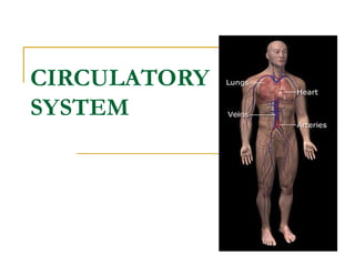
Circulatory system
- 2. The Heart 4 chambers Located between the lungs 2/3 of heart left of midline Apex points downward & contacts the diaphragm It lies in the pericardial cavity
- 3. The Heart It is separated from the other organs by a double- layered membrane = Pericardium The Pericardium is composed of a Fibrous Pericardium & a Serous Pericardium. The serous pericardium has 2 parts: 1. Parietal layer - attached to the back of the fibrous pericardium 2. Visceral layer (epicardium) - attached to the heart muscle These two are separated by a fluid filled space = pericardial cavity.
- 4. The Heart Wall A. Epicardium - outermost, = Visceral layer of the serous pericardium
- 5. Heart Wall B. Myocardium - middle, = Cardiac muscle cells (very thick)
- 6. The Heart Wall C. Endocardium - innermost, forms valves, & is continuous with the endothelium of the blood vessels that enter & leave the heart
- 7. Chambers 4 chambers 2 upper: Lt. & Rt. Atrium 2 lower: Lt. & Rt. Ventricle
- 8. Valves 4 valves 2 Atrioventricular (AV) Valves Rt. AV valve = tricuspid Lt. AV valve = bicuspid, mitral 2 semilunar valves: found at the base of 2 large vessels leaving the heart = Pulmonary & Aortic valves
- 9. Blood flow
- 10. Blood Flow 1. Rt. Atrium: receives deoxygenated (venous) blood from 3 vessels; A. Superior vena cava - blood from above the heart B. Inferior vena cava - blood from below the heart C. Coronary sinus - blood from the heart muscle
- 11. Blood Flow 2. Blood flows through Rt. AV valve into Rt. Ventricle (the flaps of AV valves are held in place by Chordae Tendineae & Papillary Muscles to prevent back flow)
- 12. Blood Flow 3. Rt. Ventricle contracts & blood exits through the Pulmonary Semilunar valve. It enters the Pulmonary trunk which divides into Lt. & Rt. Pulmonary arteries. Blood goes to lungs (carbon dioxide out, oxygen in)
- 13. Blood Flow cont. 4. Oxygenated blood returns from the lungs through the Pulmonary veins to the Lt. Atrium
- 14. Blood Flow 5. Blood flows through the Lt. AV valve (bicuspid, mitral) to the Lt. Ventricle
- 15. Blood Flow 6. Lt. Ventricle contracts & blood exits through the Aortic Semilunar valve & enters Ascending Aorta.
- 16. Coronary circulation (Blood flow to Heart Muscle) First vessels off of the Ascending Aorta = Lt. & Rt. Coronary Arteries
- 17. Coronary Circulation cont. The blood returns from the heart muscle via 2 major veins 1. Great Cardiac vein: brings deoxygenated blood back from the anterior heart wall 2. Middle Cardiac vein: brings deoxygenated blood back from the posterior heart wall. Both vessels empty into the Coronary Sinus (a large vein on back of heart). It empties into Rt. Atrium
- 18. Conduction system An electrical system. It determines the rate & rhythm of the heartbeat 1. Sinoatrial node (SA node, pacemaker) - Neurons fire at 70/80 beats per minute, causes atria to contract 2. Atrioventricular node (AV node) - neurons fire at 40-50 beats per minute; typically the SA node overrides it, but if SA node is not functioning it will ultimately cause ventricles to contract at a slower rate.
- 19. Conduction System 3. Atrioventricular Bundle (Bundle of His) - conducts impulses between ventricles 4. The AV Bundle divides into lt & rt Bundle Branches which go to the ventricles. 5. Purkinje fibers - deliver impulses directly to the myocardium of the ventricles.
- 20. Blood – connective tissue with fluid matrix A. Fluid = plasma B. Blood cells = formed elements 1. Red blood cells (RBC's) = ERYTHROCYTES a. Flattened, biconcave, anucleated discs b. Life span - 120 days c. Function: transport oxygen & carbon dioxide bound pigmented protein = hemoglobin
- 21. Blood cont. 2. White blood cells (WBC's) = LEUKOCYTES a.granulocytes i. eosinophils ii. Basophils iii. Neutrophils
- 22. Blood cont. 2. White blood cells (WBC's) = LEUKOCYTES b. Agranulocytes i. Monocytes ii. Lymphocytes
- 23. Blood cont. 3. Thrombocytes = PLATELETS; not cells. Cytoplasmic fragments of megakaryocytes. Assists in blood clot formation.
- 24. Hemopoiesis = Blood Cell formation. Occurs in red bone marrow. A. Erythropoiesis = RBC formation B. Leukopoiesis = WBC formation C. Thrombopoiesis = platelet formation
- 25. Blood vessels: blood flow Blood flows from the heart through progressively narrowing vessels; artery ->arteriole -> capillary And returns through progressively enlarging vessels; venules -> vein-> heart
- 26. Blood vessels Structure: arteries and veins have 3 tunics 1. Tunica Externa (adventitia) - Outermost, loose connective tissue, this is the thickest layer in veins 2. Tunica Media - middle, smooth muscle layer, this is the thickest layer in arteries 3. Tunica Intima - innermost a. Endothelium - simple squamous + c.t. b. Subendothelial layer - c.t.
- 27. Arteries (carry blood away from heart) Elastic - large amount of elastin expandable Muscular - tunica media is predominantly smooth muscle. There is an elastic lamina on each face of the tunica media
- 28. Arterioles - Smallest, tunica media very thin (<10 layers)
- 29. Capillaries "Functional units" of circulatory system, very thin-walled, allows for exchange of gases, nutrients, & waste products. Composed of the Tunica Intima only
- 30. Venules Usually lack a tunica media. They have the other two tunics
- 31. Veins Carry blood to the heart) All 3 tunics present. Veins have a very Low pressure, The blood flow through them is dependent on: A. Contraction of surrounding musculature = Skeletal muscle "pump" B. One-way valves