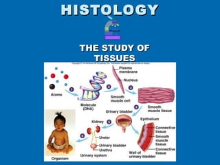
2. TISSUES
A tissue is a
functional collection of
cells and associated
intercellular material
that is specialized to
carry out a specific
role.
3. Technique of
Tissue
Sectioning
1. Tissue is preserved in a fixative
(chemical)
2. Cut into very thin slices by a special
machine
- slices are called histological
sections
3. Sections are mounted on microscope
slides
4. Sections on slides are stained
4.
5.
6.
7. Classification of the FOUR basic
tissues
1- Epithelial
2- Connective (CT)
3- Muscular
4- Nervous
8. Connective vs Epithelial
Lots of extra-cellular Little extra cellular
material, few cells material, lots of cells
Lots of blood vessels No blood vessels
Covered by other Usually form surface
tissues layers and are NOT
covered by another
tissue (*except blood
vessels)
Epithelial tissue
Connective tissue
9. Epithelial Tissues
Flat sheet of cells with upper surface exposed to
environment or internal space. (Barriers!
Protection!)
Covers body surface & lines body cavities
Forms external & internal lining of organs
Constitutes most gland tissues (Secretion!)
Little extra cellular material
No room for blood vessels (avascular) so
depends on blood vessels in CT for food and
waste movement
10. Epithelial Tissue Specifics
Basement membrane:
membrane
Anchors epith to CT
Apical Surface- faces surface, may have
Made of proteins, cilia, microtubules
acellular
Basal surface - epith
surface that sits on Connect cells
basement membrane to basement
membrane
Apical surface - epith Basal
surface that faces away Surface-
Adheres to
from basement. “Free
Basement
Surface” membrane
A thickening of the basement
membrane is a contributing cause
of blindness and kidney in diabetes.
15. Endothelium is the simple squamous
epithelium lining blood vessels
Two blood vessels seen in cross section
16. Quick Review
13. Identify the tissue.
14. Identify the structure.
15. What tissue makes up
this (#14) structure?
16. Identify the cellular
structure.
Click for Answers
13. Simple Cuboidal Epithelium
14. Blood Vessel
15. Simple Squamous epithelium
16. Nucleus
17. Stratified Squamous Epithelial
Characteristics:
Many layers becoming
flatter from basal to
Stratified layers
apical surfaces
of epithelial cells
Basal cells are mitotic
Apical cells are dead
Basal cells
LOCATIONS: Mitotic area
Cells are rounder
Keritanized
• skin
Stratified layers
Nonkeritanized
• Mouth, esophagus,
Apical cells will be sloughed off and
pharynx, vagina replaced by cells in layer below.
FUNCTION:
protection
18.
19. Simple Cuboidal Epithelium
Characteristics: single layer, square or
roundish
Location: Ducts of many glands, lines
kidney tubules, surface of ovary
Function: secretion and absorption