SlideShare una empresa de Scribd logo
1 de 52
Presenter
Praveen Gupta
Unit
Dr Santhosh Satheesh
JIPEMER
Pondicherry
India
11/02/2017
1
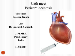
Cath meet
Pericardiocentesis
History
 22/F came to JIPMER with C/O
 Facial puffiness
 Cough with expectoration
 Dyspnoea since one month
 Atypical chest pain since last one month
2
O/E
 P=116/min
 BP-120/70 mmhg
 RS-Right side decreased air entry
 CVS-S1S2 Present, muffled heart sound
3
Chest X-ray
4
 Massive Right sided pleural
effusion ? Mild cardiomegaly
??Pericardial effusion
 ICD insertion was done for both
therapeutic and diagnostic
pleurocentesis
Chest X-ray showing Massive right sided pleural
effusion
JIPMER, Pulmonary medicine Department, 01/02/2017
JIPMER, Radiology Department, 01/02/2017
Chest X-ray(Post pleurocentesis)
5
JIPMER, Pulmonary medicine Department, 01/02/2017
JIPMER, Radiology Department, 01/02/2017
PA chest x-ray of the patient showing
right side mild pleural effusion with
ICD insitu
Lateral chest x-ray of the patient
showing right side mild pleural
effusion with ICD insitu
CECT Chest
6
CECT Chest of the patient showing right side mild pleural effusion with ICD
insitu with massive pericardial effusion
JIPMER, Pulmonary medicine Department, 02/02/2017
JIPMER, Radiology Department, 02/02/2017
CECT chest
7
CECT Chest of the patient showing right side mild pleural effusion with ICD
insitu with massive pericardial effusion
JIPMER, Pulmonary medicine Department, 03/02/2017
JIPMER, Radiology Department, 03/02/2017
CT chest
 SVC syndrome
 Right hilar lymphadenopathy
 Right pleural effusion
 Massive pericardial effusion
 Pericardial tamponade
8
ECHO showing large Pericardial effuison measureing 4 cm anteriorly, 3 cm laterally, 2 cms
posteriorly, RA/RV collpse was present suggestive of pericardial tamponade
9
JIPMER, Cardiology Department, SSB SIEMEN Cath
lab, 06/02/2017
Apical 4 chamber view showing Massive
pericardial effusion with RA/RV collapse during
diastole suggestive of tamponade physiology
Parasternal short axis view showing Massive
circumferential pericardial effusion
ECHO guided pericardial neddle inetion done…….
10
 18 No. gauge needle was
inserted under both
fluoroscopic and ECHO
guidance.
 Position of the needle was
conformed by injecting
agitated saline into the
pericardium
Apical 4 Chamber showing needle into the pericardium. Position of the needle conformed by injecting
agitated saline into the pericardial space. Presence of bubble into the pericardium conform our needle
position into the pericardium
JIPMER, Cardiology Department, SSB SIEMEN Cath lab, 06/02/2017
ECHO Apical 4 Chamber showing Presence of bubble into the pericardium…….
11 JIPMER, Cardiology Department, SSB SIEMEN Cath lab, 06/02/2017
ECHO guidance pericardiocentesis started…..
12
Apical 4 chamber view: Patient tachycardia started to subsided as soon as
pericardiocentesis was started
JIPMER, Cardiology Department, SSB SIEMEN Cath lab, 06/02/2017
Pericardiocentesis…..
13 JIPMER, Cardiology Department, SSB SIEMEN Cath lab, 06/02/2017
Pericardiocentesis…..
14 JIPMER, Cardiology Department, SSB SIEMEN Cath lab, 06/02/2017
ECHO suggestive of resolution of pericardial effusion with pericardial tamponade suggestive of successful
pericardiocentesis. Procedure was stoped after removing 590 ml of pericardial fluid. Piagtail catheter was kept insitu
for 72 hours
15 JIPMER, Cardiology Department, SSB SIEMEN Cath lab, 06/02/2017
Final diagnosis
 SVC syndrome
 Right hilar lymphadenopathy
 Right pleural effusion
 Massive pericardial effusion
 Pericardial tamponade
 S/P Successful Pericardiocentesis
 Presently patient is admitted in Infection and disease ward and under investigation for the
etiology of the pericardial effusion
16
JIPMER Data (SSB Cath lab)
(1 st Jan 2016-31 Dec 2016)
Pericardiocentesis(All patient had Pericardial effusion with tamponade)
17
Patient
NO
Age/Sex WD Diagnosis
1 40 yr/M CTVS WD Successful
2 50 yr/M SSBCCC
U
VT/S/P
AICD
insertion
Successful
3 53 yr/M SSB
CCCU
Post Left
atrial
appendag
e device
occlusion
Successful
4 40 yr/F Nephro CKD/PE Successful
5 40 yr/F OG CA cervix Successful
6 38 yr/F Medicine
WD
??Diagnos
is
Successful
JIPMER Data (EMS Cath lab)
(1 st Jan 2016-31 Dec 2016)
Pericardiocentesis(All patient had Pericardial effusion with tamponade)
18
Patien
t NO
Age/Sex WD Diagnosis
1 55 yr/M EMS CCCU AWMI/SVD Of LAD Successful
2 44 yr/M Medicine
WD
Pericardial effusion
?etio
Successful
3 60 yr/M Immunology RA/Pericardial
effusion
Successful
Approximate incidence of Pericardial tamponade after
Pacemaker/ICD/CRT at JIPMER
(Jan 2016-Dec 2016)
19
Total no of Device
inserted (Including
both
ICD/CRT/Pacemaker
(Approximate)
Total no of patient
who develop
Pericardial
effusion with
tamponade
Indicence of
PE/PT after
device
implantation at
JIPMER(2016)
300 1 0.33%
Management of pericardial effusion
20
Imazio M, Spodick DH, Brucato A, et al. Controversial issues in the management of
pericardial disease. Circulation 2010;121:916-928.)
Pericardiocentesis: Technique
21
 Performed in the cardiac
catheterization laboratory using a
combination of echocardiographic
and fluoroscopic guidance
Pericardiocentesis: Technique
22
 Two-dimensional echocardiogram just
prior to the procedure
 Document the presence, location, and size
of the effusion
 Determine the presence of loculation or
significant stranding
 Determine location on body surface where
effusion lies closest to the surface
 At which the fluid depth overlying the
heart is maximal
 Optimal direction for needle passage
 Approximate depth of needle insertion
Pericardiocentesis: Technique
23
 Access to pressure measurement,
continuous ECG and vital sign
monitoring, and fluoroscopy
with the ability to inject
radiographic contrast in the
cardiac catheterization
laboratory to be preferable,
particularly in difficult or
challenging cases, in patients
with small or localized effusions
or when complications ensue
Pericardiocentesis: Technique
24
 Access to adequate ancillary
support in hemodynamically
unstable patients
 Right heart pressure measurement
required if effusive-constrictive
pericarditis is suspected, the
effusion is small or loculated, or if
the patient is hemodynamically
unstable
Pericardiocentesis: Technique
25
 Instrument required
 10 ml/50 ml Syringe
 18 No Needle
 Pigtail catheter
 Guidewire
 Dilator
 Scalpal with blade
Pericardiocentesis: Technique
26
 Patient torso is propped up to a
level of about 45°
 Subxiphoid approach
 Skin nick made 1 to 2 cm below
costal margin just left of the
xiphoid process
Pericardiocentesis: Technique
27
 Needle path toward the
posterior aspect of the left
shoulder, passing anterior to
or through the anterior
capsule of the liver, and
entering the pericardial
space overlying the right
ventricle
Pericardiocentesis: Technique
28
 Echocardiography from subxiphoid
window confirm the optimal
direction and depth below the skin
 Posterior effusions or patients with
large body habitus, apical or low
parasternal intercostal puncture
sites are alternatives

Pericardiocentesis: Technique
 Since echocardiography does not image through air (and to avoid
pneumothorax), sites with significant intervening lung should be excluded; care
should be taken in the parasternal approach to avoid the internal mammary
artery that runs 3 to 5 cm from the parasternal border, and also the
neurovascular bundle at the lower margin of each rib
29
Pericardiocentesis: Technique
30
 Skin and subcutaneous tissues a infiltrated
with lidocaine with a small-gauge needle
along the proposed path of entry.
 Use a 5- to 8-cm, 18-gauge needle attached to
a 10-mL syringe filled with saline or
lidocaine and inserted following the echo-
determined trajectory
 As the needle is advanced, the syringe is
alternately aspirated to determine pericardial
space entry and injected to deliver more local
anesthesia along the route
Pericardiocentesis: Technique
31
 Three-way stopcock can be used to
connect to a pressure manifold via
a fluid-filled extension tube.
Pericardiocentesis: Technique
32
 Classically, electrocardiographic
monitoring of the needle (by
attaching its shaft to the V lead of
the ECG system using a sterile
alligator clip) was used to provide
an additional measure of safety
Pericardiocentesis: Technique
33
 ST segment recorded from the
needle should be isoelectric during
advancement, but dramatic
elevation of the ST segment
appears if the needle contacts the
right ventricular epicardium
 Needle withdrawn slightly until ST
elevation resolves to minimize the
chance of right ventricular puncture
or laceration.
Pericardiocentesis: Technique
34
 When the needle enters the pericardial
space, a distinct pop is usually felt and
it is possible to aspirate fluid
Pericardiocentesis: Technique
35
 Entry of the pericardial space
can be confirmed by injection of
radiographic contrast, agitated
saline echo contrast,
JIPMER, Cardiology Department, SSB SIEMEN Cath
lab, 06/02/2017
Apical 4 chamber shows agitated saline
bubble into the pericardium
Guide wire into the pericardium
36
 Entry of the pericardial
space can be confirmed by
advancement of an 0.035-
inch J wire in the
characteristic path
wrapping around the heart
Pericardiocentesis: Technique
37
 Turn the interposed stopcock to display
intrapericardial pressure, which should
be superimposable on the
simultaneously displayed right atrial
pressure from the right heart catheter
Femoral arterial (FA), right atrial (RA), and pericardial pressure before (A) and after (B) pericardiocentesis
in a patient with cardiac tamponade. Both RA and pericardial pressure are approximately 15 mm Hg before
pericardiocentesis. In this case there was a negligible paradoxical pulse. Note the presence of the x descent
but absence of the y descent before pericardiocentesis
Pericardiocentesis: Technique
38
 The waveform should
emphatically not resemble that
of right ventricular pressure
 If the pericardial needle tip
displays a right ventricular
waveform, the tip is quickly
but smoothly withdrawn under
continuous hemodynamic
monitoring until the overlying
pericardial space is entered
Pericardiocentesis: Technique
39
 An 8F dilator is then introduced over the
guidewire, followed by a drainage catheter
(straight or pigtail shaped, with multiple
side holes)
 Attach a 50-mL syringe and three-way
stopcock to the drainage catheter,
connecting an extension tube from the
other port of the three-way stopcock to a
drainage bag or vacuum bottle
 This allows fluid to be aspirated into the
syringe and transferred to the bottle.
Pericardiocentesis: Technique
40
 Removal of 50 mL fluid sufficient
to relieve tamponade and improve
hemodynamics.
 After removal of 100 to 200 mL of
fluid, it is informative to remeasure
the pericardial and right atrial
pressure before resuming aspiration
Pericardiocentesis: Technique
41
 When fluid can no longer be aspirated,
fluoroscopy should show that the
previously immobile cardiac silhouette
now exhibits a normal pulsation pattern,
and a repeat echocardiogram should show
only minimal posterior effusion
 . Occasional patients will experience
pericardial pain when the effusion is
tapped dry. Parenteral narcotic analgesics
and benzodiazepines can be administered,
and if the pain is severe, 50 mL of
pericardial fluid, sterile saline, or 10 to 20
mL of 1% Xylocaine can be reintroduced
to help ease the pain
Pericardiocentesis: Technique
42
Cardiac tamponade relieved if
(a) Pericardial pressure falls to a level 0 mm Hg
and separates from the right atrial pressure
(b) Right atrial pressure itself falls to the normal
range and exhibits return of the normal
diastolic y descent
c) Pulsus paradoxus is relieved
Pericardiocentesis: Technique
43
 Systemic arterial pressure rises in association with an increase in mixed venous
oxygen content, indicative of an increase in cardiac output
 Failure of pericardial pressure to fall close to 0 indicates reference height of
transducers incorrect or free or loculated pericardial fluid is still under pressure
Pericardiocentesis: Technique
44
 If the pericardial pressure falls
appropriately but the right atrial
pressures remain elevated with
prominent x and y descents, the
diagnosis of effusive-constrictive
pericarditis must be entertained,
with an ongoing element of
constriction after the tamponade
physiology has been relieved Pericardiocentesis results in a marked increase in FA pressure and
a marked decrease in RA pressure. During inspiration, pericardial
pressure becomes negative, there is clear separation between RA
and pericardial pressure, and the y descent is now prominent, thus
suggesting the possibility of an effusive-constrictive picture.
Pericardiocentesis: Technique
 We then sew the drainage catheter in place attached to a sterile fluid path (stopcock,
syringe, and drainage bag) to allow the postprocedure nursing staff to periodically
attempt additional aspiration.
 Sterility must be tightly maintained with this technique
 Catheter is removed when the drainage has decreased to <25 to 50 mL per 24 hours and
there is no echocardiographic evidence of reaccumulation of fluid
 Periodic echo reassessment for fluid reaccumulation should be performed.
 Larger effusions may benefit from slightly more prolonged drainage, but >48-hour dwell
time should be avoided to reduce the risk of infection
45
Pericardiocentesis: Complications
 The safety and success related to choice of entry site/ size of effusion
 Ùncomplicated if both anterior and posterior echo-free spaces 10 mm
 Avoided in minimally symptomatic patients with small incidental
effusions
 Risk increased in patients who are anticoagulated with warfarin
 Deferred if possible until INR within normal range
46
Pericardiocentesis: Complications
 If hemodynamic status demands urgent pericardiocentesis in the patient with
elevated INR, fresh frozen plasma should be administered in the catheterization
suite immediately after catheter access to the pericardium is achieved by an
expert operator and drainage is initiated (to avoid conversion of a free
hemorrhagic effusion into mixture of fluid and gelatinous clot)
47
Pericardiocentesis: Complications
 Laceration of a chamber wall or laceration of a coronary artery or vein
 Perforation of the ventricular myocardium with just the needle usually
does not result in significant bleeding and is usually well tolerated, but
reflex hypotension occur
 Ventricular and atrial arrhythmia,transient and not life threatening
48
Pericardiocentesis: Complications
 Pneumothorax
 Laceration of the liver or penetration of the stomach, colon, or spleen
 Right and left ventricular failure
 Pulmonary edema
 Exacerbation of bleeding from an ascending aortic dissection
49
Take home message
50
 Performed in the cardiac catheterization laboratory using a combination
of echocardiographic and fluoroscopic guidance
 Access to adequate ancillary support in hemodynamically unstable
patients is important
 Avoided in minimally symptomatic patients with small incidental
effusions
Reference
51
 Thank to Department of cardiology, Department of Pulmonary
medicine, Department of Radiodiagnosis JIPMER, Pondicherrry, India
 JIPMER SSB Cath lab staff
 Fang JC, Borlaug BA. Grossman & Baim's Cardiac Catheterization,
Angiography, and Intervention. InWolters Kluwer Health Adis (ESP)
2013 Oct 5.
 Goggle Image
52

Más contenido relacionado

La actualidad más candente

Coronary angiography
Coronary angiographyCoronary angiography
Coronary angiographyRaja Lahiri
 
Intra Aortic Balloon Pump by Rubina Shehzadi RN
Intra Aortic Balloon Pump by Rubina Shehzadi RNIntra Aortic Balloon Pump by Rubina Shehzadi RN
Intra Aortic Balloon Pump by Rubina Shehzadi RNRubina Shehzadi
 
Transesophageal echocardiography
Transesophageal echocardiographyTransesophageal echocardiography
Transesophageal echocardiographyAmit Gulati
 
Intra aortic balloon pump and ECMO
Intra aortic balloon pump and ECMOIntra aortic balloon pump and ECMO
Intra aortic balloon pump and ECMORachel Jeevakirubai
 
Aortic dissection nikku ppt
Aortic dissection nikku pptAortic dissection nikku ppt
Aortic dissection nikku pptNikhil Vaishnav
 
Defibrillation & Cardioversion
Defibrillation & CardioversionDefibrillation & Cardioversion
Defibrillation & CardioversionHIRANGER
 
Temporary cardiac pacing
Temporary cardiac pacingTemporary cardiac pacing
Temporary cardiac pacingMashiul Alam
 
Temporary Pacemaker Slides
Temporary Pacemaker SlidesTemporary Pacemaker Slides
Temporary Pacemaker Slidesmaltiaziz24
 
Cardiac cath complications
Cardiac cath complicationsCardiac cath complications
Cardiac cath complicationsFuad Farooq
 
Defibrillation & cardioversion by DJ
Defibrillation & cardioversion by DJDefibrillation & cardioversion by DJ
Defibrillation & cardioversion by DJDharmendra Joshi
 
Electrophysiology study basics
Electrophysiology study basicsElectrophysiology study basics
Electrophysiology study basicsSatyam Rajvanshi
 
BALLOON AORTIC VALVULOPLASTY
BALLOON AORTIC VALVULOPLASTYBALLOON AORTIC VALVULOPLASTY
BALLOON AORTIC VALVULOPLASTYPraveen Nagula
 

La actualidad más candente (20)

Coronary angiography
Coronary angiographyCoronary angiography
Coronary angiography
 
Intra Aortic Balloon Pump by Rubina Shehzadi RN
Intra Aortic Balloon Pump by Rubina Shehzadi RNIntra Aortic Balloon Pump by Rubina Shehzadi RN
Intra Aortic Balloon Pump by Rubina Shehzadi RN
 
Transesophageal echocardiography
Transesophageal echocardiographyTransesophageal echocardiography
Transesophageal echocardiography
 
Cardioversion
Cardioversion Cardioversion
Cardioversion
 
Intra aortic balloon pump and ECMO
Intra aortic balloon pump and ECMOIntra aortic balloon pump and ECMO
Intra aortic balloon pump and ECMO
 
Aortic dissection nikku ppt
Aortic dissection nikku pptAortic dissection nikku ppt
Aortic dissection nikku ppt
 
Defibrillation & Cardioversion
Defibrillation & CardioversionDefibrillation & Cardioversion
Defibrillation & Cardioversion
 
Temporary cardiac pacing
Temporary cardiac pacingTemporary cardiac pacing
Temporary cardiac pacing
 
Temporary Pacemaker Slides
Temporary Pacemaker SlidesTemporary Pacemaker Slides
Temporary Pacemaker Slides
 
Coronary angiogram
Coronary angiogramCoronary angiogram
Coronary angiogram
 
Cardioversion
CardioversionCardioversion
Cardioversion
 
Treadmill test (TMT)
Treadmill test (TMT)Treadmill test (TMT)
Treadmill test (TMT)
 
Transesophageal echocardiography
Transesophageal echocardiographyTransesophageal echocardiography
Transesophageal echocardiography
 
Cardiac cath complications
Cardiac cath complicationsCardiac cath complications
Cardiac cath complications
 
Defibrillation & cardioversion by DJ
Defibrillation & cardioversion by DJDefibrillation & cardioversion by DJ
Defibrillation & cardioversion by DJ
 
Stress echocardiography
Stress echocardiographyStress echocardiography
Stress echocardiography
 
Cath hemodynamics vir
Cath hemodynamics virCath hemodynamics vir
Cath hemodynamics vir
 
2D ECHO Basics
2D ECHO Basics2D ECHO Basics
2D ECHO Basics
 
Electrophysiology study basics
Electrophysiology study basicsElectrophysiology study basics
Electrophysiology study basics
 
BALLOON AORTIC VALVULOPLASTY
BALLOON AORTIC VALVULOPLASTYBALLOON AORTIC VALVULOPLASTY
BALLOON AORTIC VALVULOPLASTY
 

Destacado

DM cardiology Exam Spotter
DM cardiology Exam SpotterDM cardiology Exam Spotter
DM cardiology Exam SpotterPRAVEEN GUPTA
 
Chest xray for evaluation of cardiovascular system
Chest xray for evaluation of cardiovascular systemChest xray for evaluation of cardiovascular system
Chest xray for evaluation of cardiovascular systemPRAVEEN GUPTA
 
Cardiology window Blog
Cardiology window BlogCardiology window Blog
Cardiology window BlogPRAVEEN GUPTA
 
Cardiac catheteriztion, Oximetery study in a patient with VSD
Cardiac catheteriztion, Oximetery study in a patient with VSDCardiac catheteriztion, Oximetery study in a patient with VSD
Cardiac catheteriztion, Oximetery study in a patient with VSDPRAVEEN GUPTA
 
2016 acc focused update on new pharmacological therapy for heart failure
2016 acc focused update on new pharmacological therapy for heart failure2016 acc focused update on new pharmacological therapy for heart failure
2016 acc focused update on new pharmacological therapy for heart failurePRAVEEN GUPTA
 
Análisis comparativo entre las organizaciones actuales y actuales
Análisis comparativo entre las organizaciones actuales y actualesAnálisis comparativo entre las organizaciones actuales y actuales
Análisis comparativo entre las organizaciones actuales y actualesjessica michelle martinez dueñas
 
Going Beyond Google Analytics
Going Beyond Google AnalyticsGoing Beyond Google Analytics
Going Beyond Google Analyticsbrentjacobsen
 
Peter's Pain - US Presentation - 2013 2014
Peter's Pain - US Presentation - 2013 2014Peter's Pain - US Presentation - 2013 2014
Peter's Pain - US Presentation - 2013 2014Peter's Pain
 
Xamarin security talk slideshare
Xamarin security talk slideshareXamarin security talk slideshare
Xamarin security talk slideshareMarcus de Wilde
 
Pericardiocentesis - Just in Time Training
Pericardiocentesis - Just in Time TrainingPericardiocentesis - Just in Time Training
Pericardiocentesis - Just in Time TrainingNikita Joshi
 
Introduction to Electrocardiography
Introduction to ElectrocardiographyIntroduction to Electrocardiography
Introduction to ElectrocardiographyRanjeet Dalvi
 
Dialysis
Dialysis Dialysis
Dialysis Sai Ram
 

Destacado (20)

DM cardiology Exam Spotter
DM cardiology Exam SpotterDM cardiology Exam Spotter
DM cardiology Exam Spotter
 
Cardiology exam MCQ
Cardiology exam MCQCardiology exam MCQ
Cardiology exam MCQ
 
Chest xray for evaluation of cardiovascular system
Chest xray for evaluation of cardiovascular systemChest xray for evaluation of cardiovascular system
Chest xray for evaluation of cardiovascular system
 
Cardiology window Blog
Cardiology window BlogCardiology window Blog
Cardiology window Blog
 
Cardiac catheteriztion, Oximetery study in a patient with VSD
Cardiac catheteriztion, Oximetery study in a patient with VSDCardiac catheteriztion, Oximetery study in a patient with VSD
Cardiac catheteriztion, Oximetery study in a patient with VSD
 
PERICARDIOCENTESIS
PERICARDIOCENTESISPERICARDIOCENTESIS
PERICARDIOCENTESIS
 
2016 acc focused update on new pharmacological therapy for heart failure
2016 acc focused update on new pharmacological therapy for heart failure2016 acc focused update on new pharmacological therapy for heart failure
2016 acc focused update on new pharmacological therapy for heart failure
 
SUSTAINED LOW EFFICIENCY DAILY DIALYSIS (SLEDD)
SUSTAINED LOW EFFICIENCY DAILY DIALYSIS (SLEDD)SUSTAINED LOW EFFICIENCY DAILY DIALYSIS (SLEDD)
SUSTAINED LOW EFFICIENCY DAILY DIALYSIS (SLEDD)
 
Normativa deportiva
Normativa deportivaNormativa deportiva
Normativa deportiva
 
Análisis comparativo entre las organizaciones actuales y actuales
Análisis comparativo entre las organizaciones actuales y actualesAnálisis comparativo entre las organizaciones actuales y actuales
Análisis comparativo entre las organizaciones actuales y actuales
 
Car care
Car careCar care
Car care
 
Going Beyond Google Analytics
Going Beyond Google AnalyticsGoing Beyond Google Analytics
Going Beyond Google Analytics
 
Presentation1
Presentation1Presentation1
Presentation1
 
Peter's Pain - US Presentation - 2013 2014
Peter's Pain - US Presentation - 2013 2014Peter's Pain - US Presentation - 2013 2014
Peter's Pain - US Presentation - 2013 2014
 
Shagnal
ShagnalShagnal
Shagnal
 
Xamarin security talk slideshare
Xamarin security talk slideshareXamarin security talk slideshare
Xamarin security talk slideshare
 
Human Iris Biometry
Human Iris BiometryHuman Iris Biometry
Human Iris Biometry
 
Pericardiocentesis - Just in Time Training
Pericardiocentesis - Just in Time TrainingPericardiocentesis - Just in Time Training
Pericardiocentesis - Just in Time Training
 
Introduction to Electrocardiography
Introduction to ElectrocardiographyIntroduction to Electrocardiography
Introduction to Electrocardiography
 
Dialysis
Dialysis Dialysis
Dialysis
 

Similar a Pericardiocentesis

CT Coronary Angiography.pptx, eart muscle) Acute angina (type of chest pain) ...
CT Coronary Angiography.pptx, eart muscle) Acute angina (type of chest pain) ...CT Coronary Angiography.pptx, eart muscle) Acute angina (type of chest pain) ...
CT Coronary Angiography.pptx, eart muscle) Acute angina (type of chest pain) ...RukamaneeYadav
 
Severe Aortic Stenosis with Ischemic Heart Disease. (Management of TAVI) - Pa...
Severe Aortic Stenosis with Ischemic Heart Disease. (Management of TAVI) - Pa...Severe Aortic Stenosis with Ischemic Heart Disease. (Management of TAVI) - Pa...
Severe Aortic Stenosis with Ischemic Heart Disease. (Management of TAVI) - Pa...Yukta Wankhede
 
Austin Journal of Anesthesia and Analgesia
Austin Journal of Anesthesia and AnalgesiaAustin Journal of Anesthesia and Analgesia
Austin Journal of Anesthesia and AnalgesiaAustin Publishing Group
 
Combined Spinal Epidural Anesthesia
Combined Spinal Epidural AnesthesiaCombined Spinal Epidural Anesthesia
Combined Spinal Epidural AnesthesiaBilal Baig
 
Desun Hospital Health Insights : Modern Approach on Angioplasty
Desun Hospital Health Insights : Modern Approach on AngioplastyDesun Hospital Health Insights : Modern Approach on Angioplasty
Desun Hospital Health Insights : Modern Approach on AngioplastyDESUN Hospital
 
17. minor surgical procedures copy
17. minor surgical procedures   copy17. minor surgical procedures   copy
17. minor surgical procedures copyMD Specialclass
 
minor surgical procedures
minor surgical proceduresminor surgical procedures
minor surgical proceduresMD Specialclass
 
17 Minor Surgical Procedures
17  Minor Surgical Procedures17  Minor Surgical Procedures
17 Minor Surgical ProceduresMD Specialclass
 
Introduction to echocardiography
Introduction to echocardiography Introduction to echocardiography
Introduction to echocardiography Bibini Bab
 
Percutaneous coronary intervention
Percutaneous coronary interventionPercutaneous coronary intervention
Percutaneous coronary interventionKhairul Bashar
 
Managment for dvr surgery
Managment for dvr surgeryManagment for dvr surgery
Managment for dvr surgeryNilesh Parmar
 
Managment DVR Surgery
Managment DVR SurgeryManagment DVR Surgery
Managment DVR SurgeryNilesh Parmar
 
Dr. Escobar’s CMC X-Ray Mastery Project: December Cases
Dr. Escobar’s CMC X-Ray Mastery Project: December CasesDr. Escobar’s CMC X-Ray Mastery Project: December Cases
Dr. Escobar’s CMC X-Ray Mastery Project: December CasesSean M. Fox
 
Ct pulmonary angiogram
Ct pulmonary angiogramCt pulmonary angiogram
Ct pulmonary angiogrammuhammed Yasar
 
Adult Chest X-Rays Of The Month - #46
Adult Chest X-Rays Of The Month - #46Adult Chest X-Rays Of The Month - #46
Adult Chest X-Rays Of The Month - #46Sean M. Fox
 

Similar a Pericardiocentesis (20)

CT Coronary Angiography.pptx, eart muscle) Acute angina (type of chest pain) ...
CT Coronary Angiography.pptx, eart muscle) Acute angina (type of chest pain) ...CT Coronary Angiography.pptx, eart muscle) Acute angina (type of chest pain) ...
CT Coronary Angiography.pptx, eart muscle) Acute angina (type of chest pain) ...
 
Severe Aortic Stenosis with Ischemic Heart Disease. (Management of TAVI) - Pa...
Severe Aortic Stenosis with Ischemic Heart Disease. (Management of TAVI) - Pa...Severe Aortic Stenosis with Ischemic Heart Disease. (Management of TAVI) - Pa...
Severe Aortic Stenosis with Ischemic Heart Disease. (Management of TAVI) - Pa...
 
tamponade.pptx
tamponade.pptxtamponade.pptx
tamponade.pptx
 
Austin Journal of Anesthesia and Analgesia
Austin Journal of Anesthesia and AnalgesiaAustin Journal of Anesthesia and Analgesia
Austin Journal of Anesthesia and Analgesia
 
Combined Spinal Epidural Anesthesia
Combined Spinal Epidural AnesthesiaCombined Spinal Epidural Anesthesia
Combined Spinal Epidural Anesthesia
 
Inahrs 2018 abstracts
Inahrs 2018 abstractsInahrs 2018 abstracts
Inahrs 2018 abstracts
 
Desun Hospital Health Insights : Modern Approach on Angioplasty
Desun Hospital Health Insights : Modern Approach on AngioplastyDesun Hospital Health Insights : Modern Approach on Angioplasty
Desun Hospital Health Insights : Modern Approach on Angioplasty
 
Echocardiography basics
Echocardiography basicsEchocardiography basics
Echocardiography basics
 
17. minor surgical procedures copy
17. minor surgical procedures   copy17. minor surgical procedures   copy
17. minor surgical procedures copy
 
minor surgical procedures
minor surgical proceduresminor surgical procedures
minor surgical procedures
 
17 Minor Surgical Procedures
17  Minor Surgical Procedures17  Minor Surgical Procedures
17 Minor Surgical Procedures
 
Introduction to echocardiography
Introduction to echocardiography Introduction to echocardiography
Introduction to echocardiography
 
Diary of Practical Training
Diary of Practical Training Diary of Practical Training
Diary of Practical Training
 
Percutaneous coronary intervention
Percutaneous coronary interventionPercutaneous coronary intervention
Percutaneous coronary intervention
 
Coronary Artery Bypass Graft
Coronary Artery Bypass GraftCoronary Artery Bypass Graft
Coronary Artery Bypass Graft
 
Managment for dvr surgery
Managment for dvr surgeryManagment for dvr surgery
Managment for dvr surgery
 
Managment DVR Surgery
Managment DVR SurgeryManagment DVR Surgery
Managment DVR Surgery
 
Dr. Escobar’s CMC X-Ray Mastery Project: December Cases
Dr. Escobar’s CMC X-Ray Mastery Project: December CasesDr. Escobar’s CMC X-Ray Mastery Project: December Cases
Dr. Escobar’s CMC X-Ray Mastery Project: December Cases
 
Ct pulmonary angiogram
Ct pulmonary angiogramCt pulmonary angiogram
Ct pulmonary angiogram
 
Adult Chest X-Rays Of The Month - #46
Adult Chest X-Rays Of The Month - #46Adult Chest X-Rays Of The Month - #46
Adult Chest X-Rays Of The Month - #46
 

Más de PRAVEEN GUPTA

Praveen's Indian cardiology board review and self-assessment
Praveen's Indian cardiology board review and self-assessmentPraveen's Indian cardiology board review and self-assessment
Praveen's Indian cardiology board review and self-assessmentPRAVEEN GUPTA
 
Evaluation and management of Pacemaker malfunction
Evaluation and management of Pacemaker malfunctionEvaluation and management of Pacemaker malfunction
Evaluation and management of Pacemaker malfunctionPRAVEEN GUPTA
 
Air pollution and cardiovascular disease
Air pollution and cardiovascular diseaseAir pollution and cardiovascular disease
Air pollution and cardiovascular diseasePRAVEEN GUPTA
 
Chronic thromboembolic pulmonary hypertension (CTEPH)
Chronic thromboembolic pulmonary hypertension (CTEPH)Chronic thromboembolic pulmonary hypertension (CTEPH)
Chronic thromboembolic pulmonary hypertension (CTEPH)PRAVEEN GUPTA
 
ICD in Non-ischemic cardiomyopathy
ICD in Non-ischemic cardiomyopathyICD in Non-ischemic cardiomyopathy
ICD in Non-ischemic cardiomyopathyPRAVEEN GUPTA
 
2015 Jones criteria for the diagnosis of rheumatic fever
2015 Jones criteria for the diagnosis of rheumatic fever2015 Jones criteria for the diagnosis of rheumatic fever
2015 Jones criteria for the diagnosis of rheumatic feverPRAVEEN GUPTA
 
Non-Invasive physiological assessment of coronary circulatory function
Non-Invasive physiological assessment of coronary circulatory functionNon-Invasive physiological assessment of coronary circulatory function
Non-Invasive physiological assessment of coronary circulatory functionPRAVEEN GUPTA
 
Pulseless electrical activity, Ventricular flutter, Ventricular fibrillation
Pulseless electrical activity, Ventricular flutter, Ventricular fibrillationPulseless electrical activity, Ventricular flutter, Ventricular fibrillation
Pulseless electrical activity, Ventricular flutter, Ventricular fibrillationPRAVEEN GUPTA
 
Hybrid atrial fibrillation ablation
Hybrid atrial fibrillation ablationHybrid atrial fibrillation ablation
Hybrid atrial fibrillation ablationPRAVEEN GUPTA
 
Cardiology Journal club
Cardiology Journal clubCardiology Journal club
Cardiology Journal clubPRAVEEN GUPTA
 
Cardiovascular complication of cancer chemotherapy
Cardiovascular complication of cancer chemotherapyCardiovascular complication of cancer chemotherapy
Cardiovascular complication of cancer chemotherapyPRAVEEN GUPTA
 
Cardiology Journal scan
Cardiology Journal scanCardiology Journal scan
Cardiology Journal scanPRAVEEN GUPTA
 
SELECT-LV study (2017)
SELECT-LV study (2017)SELECT-LV study (2017)
SELECT-LV study (2017)PRAVEEN GUPTA
 
Wearable defibrillator
Wearable defibrillatorWearable defibrillator
Wearable defibrillatorPRAVEEN GUPTA
 
Diabetic cardiomyopathy (Pathophysiology)
Diabetic cardiomyopathy (Pathophysiology)Diabetic cardiomyopathy (Pathophysiology)
Diabetic cardiomyopathy (Pathophysiology)PRAVEEN GUPTA
 

Más de PRAVEEN GUPTA (20)

Praveen's Indian cardiology board review and self-assessment
Praveen's Indian cardiology board review and self-assessmentPraveen's Indian cardiology board review and self-assessment
Praveen's Indian cardiology board review and self-assessment
 
Evaluation and management of Pacemaker malfunction
Evaluation and management of Pacemaker malfunctionEvaluation and management of Pacemaker malfunction
Evaluation and management of Pacemaker malfunction
 
Air pollution and cardiovascular disease
Air pollution and cardiovascular diseaseAir pollution and cardiovascular disease
Air pollution and cardiovascular disease
 
Chronic thromboembolic pulmonary hypertension (CTEPH)
Chronic thromboembolic pulmonary hypertension (CTEPH)Chronic thromboembolic pulmonary hypertension (CTEPH)
Chronic thromboembolic pulmonary hypertension (CTEPH)
 
ICD in Non-ischemic cardiomyopathy
ICD in Non-ischemic cardiomyopathyICD in Non-ischemic cardiomyopathy
ICD in Non-ischemic cardiomyopathy
 
2015 Jones criteria for the diagnosis of rheumatic fever
2015 Jones criteria for the diagnosis of rheumatic fever2015 Jones criteria for the diagnosis of rheumatic fever
2015 Jones criteria for the diagnosis of rheumatic fever
 
Non-Invasive physiological assessment of coronary circulatory function
Non-Invasive physiological assessment of coronary circulatory functionNon-Invasive physiological assessment of coronary circulatory function
Non-Invasive physiological assessment of coronary circulatory function
 
Cardiology mcq
Cardiology mcq Cardiology mcq
Cardiology mcq
 
Heart block
Heart blockHeart block
Heart block
 
Pulseless electrical activity, Ventricular flutter, Ventricular fibrillation
Pulseless electrical activity, Ventricular flutter, Ventricular fibrillationPulseless electrical activity, Ventricular flutter, Ventricular fibrillation
Pulseless electrical activity, Ventricular flutter, Ventricular fibrillation
 
Kawasaki diseases
Kawasaki diseasesKawasaki diseases
Kawasaki diseases
 
Hybrid atrial fibrillation ablation
Hybrid atrial fibrillation ablationHybrid atrial fibrillation ablation
Hybrid atrial fibrillation ablation
 
Cardiology Journal club
Cardiology Journal clubCardiology Journal club
Cardiology Journal club
 
AICD programming
AICD programmingAICD programming
AICD programming
 
Cardiovascular complication of cancer chemotherapy
Cardiovascular complication of cancer chemotherapyCardiovascular complication of cancer chemotherapy
Cardiovascular complication of cancer chemotherapy
 
Cardiology Journal scan
Cardiology Journal scanCardiology Journal scan
Cardiology Journal scan
 
SELECT-LV study (2017)
SELECT-LV study (2017)SELECT-LV study (2017)
SELECT-LV study (2017)
 
Wearable defibrillator
Wearable defibrillatorWearable defibrillator
Wearable defibrillator
 
Diabetic cardiomyopathy (Pathophysiology)
Diabetic cardiomyopathy (Pathophysiology)Diabetic cardiomyopathy (Pathophysiology)
Diabetic cardiomyopathy (Pathophysiology)
 
Heart sounds
Heart soundsHeart sounds
Heart sounds
 

Último

4.16.24 Poverty and Precarity--Desmond.pptx
4.16.24 Poverty and Precarity--Desmond.pptx4.16.24 Poverty and Precarity--Desmond.pptx
4.16.24 Poverty and Precarity--Desmond.pptxmary850239
 
Full Stack Web Development Course for Beginners
Full Stack Web Development Course  for BeginnersFull Stack Web Development Course  for Beginners
Full Stack Web Development Course for BeginnersSabitha Banu
 
USPS® Forced Meter Migration - How to Know if Your Postage Meter Will Soon be...
USPS® Forced Meter Migration - How to Know if Your Postage Meter Will Soon be...USPS® Forced Meter Migration - How to Know if Your Postage Meter Will Soon be...
USPS® Forced Meter Migration - How to Know if Your Postage Meter Will Soon be...Postal Advocate Inc.
 
Music 9 - 4th quarter - Vocal Music of the Romantic Period.pptx
Music 9 - 4th quarter - Vocal Music of the Romantic Period.pptxMusic 9 - 4th quarter - Vocal Music of the Romantic Period.pptx
Music 9 - 4th quarter - Vocal Music of the Romantic Period.pptxleah joy valeriano
 
ICS2208 Lecture6 Notes for SL spaces.pdf
ICS2208 Lecture6 Notes for SL spaces.pdfICS2208 Lecture6 Notes for SL spaces.pdf
ICS2208 Lecture6 Notes for SL spaces.pdfVanessa Camilleri
 
Student Profile Sample - We help schools to connect the data they have, with ...
Student Profile Sample - We help schools to connect the data they have, with ...Student Profile Sample - We help schools to connect the data they have, with ...
Student Profile Sample - We help schools to connect the data they have, with ...Seán Kennedy
 
Global Lehigh Strategic Initiatives (without descriptions)
Global Lehigh Strategic Initiatives (without descriptions)Global Lehigh Strategic Initiatives (without descriptions)
Global Lehigh Strategic Initiatives (without descriptions)cama23
 
Keynote by Prof. Wurzer at Nordex about IP-design
Keynote by Prof. Wurzer at Nordex about IP-designKeynote by Prof. Wurzer at Nordex about IP-design
Keynote by Prof. Wurzer at Nordex about IP-designMIPLM
 
Virtual-Orientation-on-the-Administration-of-NATG12-NATG6-and-ELLNA.pdf
Virtual-Orientation-on-the-Administration-of-NATG12-NATG6-and-ELLNA.pdfVirtual-Orientation-on-the-Administration-of-NATG12-NATG6-and-ELLNA.pdf
Virtual-Orientation-on-the-Administration-of-NATG12-NATG6-and-ELLNA.pdfErwinPantujan2
 
Food processing presentation for bsc agriculture hons
Food processing presentation for bsc agriculture honsFood processing presentation for bsc agriculture hons
Food processing presentation for bsc agriculture honsManeerUddin
 
ENGLISH 7_Q4_LESSON 2_ Employing a Variety of Strategies for Effective Interp...
ENGLISH 7_Q4_LESSON 2_ Employing a Variety of Strategies for Effective Interp...ENGLISH 7_Q4_LESSON 2_ Employing a Variety of Strategies for Effective Interp...
ENGLISH 7_Q4_LESSON 2_ Employing a Variety of Strategies for Effective Interp...JhezDiaz1
 
How to do quick user assign in kanban in Odoo 17 ERP
How to do quick user assign in kanban in Odoo 17 ERPHow to do quick user assign in kanban in Odoo 17 ERP
How to do quick user assign in kanban in Odoo 17 ERPCeline George
 
ISYU TUNGKOL SA SEKSWLADIDA (ISSUE ABOUT SEXUALITY
ISYU TUNGKOL SA SEKSWLADIDA (ISSUE ABOUT SEXUALITYISYU TUNGKOL SA SEKSWLADIDA (ISSUE ABOUT SEXUALITY
ISYU TUNGKOL SA SEKSWLADIDA (ISSUE ABOUT SEXUALITYKayeClaireEstoconing
 
ANG SEKTOR NG agrikultura.pptx QUARTER 4
ANG SEKTOR NG agrikultura.pptx QUARTER 4ANG SEKTOR NG agrikultura.pptx QUARTER 4
ANG SEKTOR NG agrikultura.pptx QUARTER 4MiaBumagat1
 
Daily Lesson Plan in Mathematics Quarter 4
Daily Lesson Plan in Mathematics Quarter 4Daily Lesson Plan in Mathematics Quarter 4
Daily Lesson Plan in Mathematics Quarter 4JOYLYNSAMANIEGO
 
Grade 9 Quarter 4 Dll Grade 9 Quarter 4 DLL.pdf
Grade 9 Quarter 4 Dll Grade 9 Quarter 4 DLL.pdfGrade 9 Quarter 4 Dll Grade 9 Quarter 4 DLL.pdf
Grade 9 Quarter 4 Dll Grade 9 Quarter 4 DLL.pdfJemuel Francisco
 
Q4-PPT-Music9_Lesson-1-Romantic-Opera.pptx
Q4-PPT-Music9_Lesson-1-Romantic-Opera.pptxQ4-PPT-Music9_Lesson-1-Romantic-Opera.pptx
Q4-PPT-Music9_Lesson-1-Romantic-Opera.pptxlancelewisportillo
 
Barangay Council for the Protection of Children (BCPC) Orientation.pptx
Barangay Council for the Protection of Children (BCPC) Orientation.pptxBarangay Council for the Protection of Children (BCPC) Orientation.pptx
Barangay Council for the Protection of Children (BCPC) Orientation.pptxCarlos105
 
AUDIENCE THEORY -CULTIVATION THEORY - GERBNER.pptx
AUDIENCE THEORY -CULTIVATION THEORY -  GERBNER.pptxAUDIENCE THEORY -CULTIVATION THEORY -  GERBNER.pptx
AUDIENCE THEORY -CULTIVATION THEORY - GERBNER.pptxiammrhaywood
 

Último (20)

4.16.24 Poverty and Precarity--Desmond.pptx
4.16.24 Poverty and Precarity--Desmond.pptx4.16.24 Poverty and Precarity--Desmond.pptx
4.16.24 Poverty and Precarity--Desmond.pptx
 
Full Stack Web Development Course for Beginners
Full Stack Web Development Course  for BeginnersFull Stack Web Development Course  for Beginners
Full Stack Web Development Course for Beginners
 
USPS® Forced Meter Migration - How to Know if Your Postage Meter Will Soon be...
USPS® Forced Meter Migration - How to Know if Your Postage Meter Will Soon be...USPS® Forced Meter Migration - How to Know if Your Postage Meter Will Soon be...
USPS® Forced Meter Migration - How to Know if Your Postage Meter Will Soon be...
 
Music 9 - 4th quarter - Vocal Music of the Romantic Period.pptx
Music 9 - 4th quarter - Vocal Music of the Romantic Period.pptxMusic 9 - 4th quarter - Vocal Music of the Romantic Period.pptx
Music 9 - 4th quarter - Vocal Music of the Romantic Period.pptx
 
ICS2208 Lecture6 Notes for SL spaces.pdf
ICS2208 Lecture6 Notes for SL spaces.pdfICS2208 Lecture6 Notes for SL spaces.pdf
ICS2208 Lecture6 Notes for SL spaces.pdf
 
YOUVE_GOT_EMAIL_PRELIMS_EL_DORADO_2024.pptx
YOUVE_GOT_EMAIL_PRELIMS_EL_DORADO_2024.pptxYOUVE_GOT_EMAIL_PRELIMS_EL_DORADO_2024.pptx
YOUVE_GOT_EMAIL_PRELIMS_EL_DORADO_2024.pptx
 
Student Profile Sample - We help schools to connect the data they have, with ...
Student Profile Sample - We help schools to connect the data they have, with ...Student Profile Sample - We help schools to connect the data they have, with ...
Student Profile Sample - We help schools to connect the data they have, with ...
 
Global Lehigh Strategic Initiatives (without descriptions)
Global Lehigh Strategic Initiatives (without descriptions)Global Lehigh Strategic Initiatives (without descriptions)
Global Lehigh Strategic Initiatives (without descriptions)
 
Keynote by Prof. Wurzer at Nordex about IP-design
Keynote by Prof. Wurzer at Nordex about IP-designKeynote by Prof. Wurzer at Nordex about IP-design
Keynote by Prof. Wurzer at Nordex about IP-design
 
Virtual-Orientation-on-the-Administration-of-NATG12-NATG6-and-ELLNA.pdf
Virtual-Orientation-on-the-Administration-of-NATG12-NATG6-and-ELLNA.pdfVirtual-Orientation-on-the-Administration-of-NATG12-NATG6-and-ELLNA.pdf
Virtual-Orientation-on-the-Administration-of-NATG12-NATG6-and-ELLNA.pdf
 
Food processing presentation for bsc agriculture hons
Food processing presentation for bsc agriculture honsFood processing presentation for bsc agriculture hons
Food processing presentation for bsc agriculture hons
 
ENGLISH 7_Q4_LESSON 2_ Employing a Variety of Strategies for Effective Interp...
ENGLISH 7_Q4_LESSON 2_ Employing a Variety of Strategies for Effective Interp...ENGLISH 7_Q4_LESSON 2_ Employing a Variety of Strategies for Effective Interp...
ENGLISH 7_Q4_LESSON 2_ Employing a Variety of Strategies for Effective Interp...
 
How to do quick user assign in kanban in Odoo 17 ERP
How to do quick user assign in kanban in Odoo 17 ERPHow to do quick user assign in kanban in Odoo 17 ERP
How to do quick user assign in kanban in Odoo 17 ERP
 
ISYU TUNGKOL SA SEKSWLADIDA (ISSUE ABOUT SEXUALITY
ISYU TUNGKOL SA SEKSWLADIDA (ISSUE ABOUT SEXUALITYISYU TUNGKOL SA SEKSWLADIDA (ISSUE ABOUT SEXUALITY
ISYU TUNGKOL SA SEKSWLADIDA (ISSUE ABOUT SEXUALITY
 
ANG SEKTOR NG agrikultura.pptx QUARTER 4
ANG SEKTOR NG agrikultura.pptx QUARTER 4ANG SEKTOR NG agrikultura.pptx QUARTER 4
ANG SEKTOR NG agrikultura.pptx QUARTER 4
 
Daily Lesson Plan in Mathematics Quarter 4
Daily Lesson Plan in Mathematics Quarter 4Daily Lesson Plan in Mathematics Quarter 4
Daily Lesson Plan in Mathematics Quarter 4
 
Grade 9 Quarter 4 Dll Grade 9 Quarter 4 DLL.pdf
Grade 9 Quarter 4 Dll Grade 9 Quarter 4 DLL.pdfGrade 9 Quarter 4 Dll Grade 9 Quarter 4 DLL.pdf
Grade 9 Quarter 4 Dll Grade 9 Quarter 4 DLL.pdf
 
Q4-PPT-Music9_Lesson-1-Romantic-Opera.pptx
Q4-PPT-Music9_Lesson-1-Romantic-Opera.pptxQ4-PPT-Music9_Lesson-1-Romantic-Opera.pptx
Q4-PPT-Music9_Lesson-1-Romantic-Opera.pptx
 
Barangay Council for the Protection of Children (BCPC) Orientation.pptx
Barangay Council for the Protection of Children (BCPC) Orientation.pptxBarangay Council for the Protection of Children (BCPC) Orientation.pptx
Barangay Council for the Protection of Children (BCPC) Orientation.pptx
 
AUDIENCE THEORY -CULTIVATION THEORY - GERBNER.pptx
AUDIENCE THEORY -CULTIVATION THEORY -  GERBNER.pptxAUDIENCE THEORY -CULTIVATION THEORY -  GERBNER.pptx
AUDIENCE THEORY -CULTIVATION THEORY - GERBNER.pptx
 

Pericardiocentesis

  • 1. Presenter Praveen Gupta Unit Dr Santhosh Satheesh JIPEMER Pondicherry India 11/02/2017 1 Cath meet Pericardiocentesis
  • 2. History  22/F came to JIPMER with C/O  Facial puffiness  Cough with expectoration  Dyspnoea since one month  Atypical chest pain since last one month 2
  • 3. O/E  P=116/min  BP-120/70 mmhg  RS-Right side decreased air entry  CVS-S1S2 Present, muffled heart sound 3
  • 4. Chest X-ray 4  Massive Right sided pleural effusion ? Mild cardiomegaly ??Pericardial effusion  ICD insertion was done for both therapeutic and diagnostic pleurocentesis Chest X-ray showing Massive right sided pleural effusion JIPMER, Pulmonary medicine Department, 01/02/2017 JIPMER, Radiology Department, 01/02/2017
  • 5. Chest X-ray(Post pleurocentesis) 5 JIPMER, Pulmonary medicine Department, 01/02/2017 JIPMER, Radiology Department, 01/02/2017 PA chest x-ray of the patient showing right side mild pleural effusion with ICD insitu Lateral chest x-ray of the patient showing right side mild pleural effusion with ICD insitu
  • 6. CECT Chest 6 CECT Chest of the patient showing right side mild pleural effusion with ICD insitu with massive pericardial effusion JIPMER, Pulmonary medicine Department, 02/02/2017 JIPMER, Radiology Department, 02/02/2017
  • 7. CECT chest 7 CECT Chest of the patient showing right side mild pleural effusion with ICD insitu with massive pericardial effusion JIPMER, Pulmonary medicine Department, 03/02/2017 JIPMER, Radiology Department, 03/02/2017
  • 8. CT chest  SVC syndrome  Right hilar lymphadenopathy  Right pleural effusion  Massive pericardial effusion  Pericardial tamponade 8
  • 9. ECHO showing large Pericardial effuison measureing 4 cm anteriorly, 3 cm laterally, 2 cms posteriorly, RA/RV collpse was present suggestive of pericardial tamponade 9 JIPMER, Cardiology Department, SSB SIEMEN Cath lab, 06/02/2017 Apical 4 chamber view showing Massive pericardial effusion with RA/RV collapse during diastole suggestive of tamponade physiology Parasternal short axis view showing Massive circumferential pericardial effusion
  • 10. ECHO guided pericardial neddle inetion done……. 10  18 No. gauge needle was inserted under both fluoroscopic and ECHO guidance.  Position of the needle was conformed by injecting agitated saline into the pericardium Apical 4 Chamber showing needle into the pericardium. Position of the needle conformed by injecting agitated saline into the pericardial space. Presence of bubble into the pericardium conform our needle position into the pericardium JIPMER, Cardiology Department, SSB SIEMEN Cath lab, 06/02/2017
  • 11. ECHO Apical 4 Chamber showing Presence of bubble into the pericardium……. 11 JIPMER, Cardiology Department, SSB SIEMEN Cath lab, 06/02/2017
  • 12. ECHO guidance pericardiocentesis started….. 12 Apical 4 chamber view: Patient tachycardia started to subsided as soon as pericardiocentesis was started JIPMER, Cardiology Department, SSB SIEMEN Cath lab, 06/02/2017
  • 13. Pericardiocentesis….. 13 JIPMER, Cardiology Department, SSB SIEMEN Cath lab, 06/02/2017
  • 14. Pericardiocentesis….. 14 JIPMER, Cardiology Department, SSB SIEMEN Cath lab, 06/02/2017
  • 15. ECHO suggestive of resolution of pericardial effusion with pericardial tamponade suggestive of successful pericardiocentesis. Procedure was stoped after removing 590 ml of pericardial fluid. Piagtail catheter was kept insitu for 72 hours 15 JIPMER, Cardiology Department, SSB SIEMEN Cath lab, 06/02/2017
  • 16. Final diagnosis  SVC syndrome  Right hilar lymphadenopathy  Right pleural effusion  Massive pericardial effusion  Pericardial tamponade  S/P Successful Pericardiocentesis  Presently patient is admitted in Infection and disease ward and under investigation for the etiology of the pericardial effusion 16
  • 17. JIPMER Data (SSB Cath lab) (1 st Jan 2016-31 Dec 2016) Pericardiocentesis(All patient had Pericardial effusion with tamponade) 17 Patient NO Age/Sex WD Diagnosis 1 40 yr/M CTVS WD Successful 2 50 yr/M SSBCCC U VT/S/P AICD insertion Successful 3 53 yr/M SSB CCCU Post Left atrial appendag e device occlusion Successful 4 40 yr/F Nephro CKD/PE Successful 5 40 yr/F OG CA cervix Successful 6 38 yr/F Medicine WD ??Diagnos is Successful
  • 18. JIPMER Data (EMS Cath lab) (1 st Jan 2016-31 Dec 2016) Pericardiocentesis(All patient had Pericardial effusion with tamponade) 18 Patien t NO Age/Sex WD Diagnosis 1 55 yr/M EMS CCCU AWMI/SVD Of LAD Successful 2 44 yr/M Medicine WD Pericardial effusion ?etio Successful 3 60 yr/M Immunology RA/Pericardial effusion Successful
  • 19. Approximate incidence of Pericardial tamponade after Pacemaker/ICD/CRT at JIPMER (Jan 2016-Dec 2016) 19 Total no of Device inserted (Including both ICD/CRT/Pacemaker (Approximate) Total no of patient who develop Pericardial effusion with tamponade Indicence of PE/PT after device implantation at JIPMER(2016) 300 1 0.33%
  • 20. Management of pericardial effusion 20 Imazio M, Spodick DH, Brucato A, et al. Controversial issues in the management of pericardial disease. Circulation 2010;121:916-928.)
  • 21. Pericardiocentesis: Technique 21  Performed in the cardiac catheterization laboratory using a combination of echocardiographic and fluoroscopic guidance
  • 22. Pericardiocentesis: Technique 22  Two-dimensional echocardiogram just prior to the procedure  Document the presence, location, and size of the effusion  Determine the presence of loculation or significant stranding  Determine location on body surface where effusion lies closest to the surface  At which the fluid depth overlying the heart is maximal  Optimal direction for needle passage  Approximate depth of needle insertion
  • 23. Pericardiocentesis: Technique 23  Access to pressure measurement, continuous ECG and vital sign monitoring, and fluoroscopy with the ability to inject radiographic contrast in the cardiac catheterization laboratory to be preferable, particularly in difficult or challenging cases, in patients with small or localized effusions or when complications ensue
  • 24. Pericardiocentesis: Technique 24  Access to adequate ancillary support in hemodynamically unstable patients  Right heart pressure measurement required if effusive-constrictive pericarditis is suspected, the effusion is small or loculated, or if the patient is hemodynamically unstable
  • 25. Pericardiocentesis: Technique 25  Instrument required  10 ml/50 ml Syringe  18 No Needle  Pigtail catheter  Guidewire  Dilator  Scalpal with blade
  • 26. Pericardiocentesis: Technique 26  Patient torso is propped up to a level of about 45°  Subxiphoid approach  Skin nick made 1 to 2 cm below costal margin just left of the xiphoid process
  • 27. Pericardiocentesis: Technique 27  Needle path toward the posterior aspect of the left shoulder, passing anterior to or through the anterior capsule of the liver, and entering the pericardial space overlying the right ventricle
  • 28. Pericardiocentesis: Technique 28  Echocardiography from subxiphoid window confirm the optimal direction and depth below the skin  Posterior effusions or patients with large body habitus, apical or low parasternal intercostal puncture sites are alternatives 
  • 29. Pericardiocentesis: Technique  Since echocardiography does not image through air (and to avoid pneumothorax), sites with significant intervening lung should be excluded; care should be taken in the parasternal approach to avoid the internal mammary artery that runs 3 to 5 cm from the parasternal border, and also the neurovascular bundle at the lower margin of each rib 29
  • 30. Pericardiocentesis: Technique 30  Skin and subcutaneous tissues a infiltrated with lidocaine with a small-gauge needle along the proposed path of entry.  Use a 5- to 8-cm, 18-gauge needle attached to a 10-mL syringe filled with saline or lidocaine and inserted following the echo- determined trajectory  As the needle is advanced, the syringe is alternately aspirated to determine pericardial space entry and injected to deliver more local anesthesia along the route
  • 31. Pericardiocentesis: Technique 31  Three-way stopcock can be used to connect to a pressure manifold via a fluid-filled extension tube.
  • 32. Pericardiocentesis: Technique 32  Classically, electrocardiographic monitoring of the needle (by attaching its shaft to the V lead of the ECG system using a sterile alligator clip) was used to provide an additional measure of safety
  • 33. Pericardiocentesis: Technique 33  ST segment recorded from the needle should be isoelectric during advancement, but dramatic elevation of the ST segment appears if the needle contacts the right ventricular epicardium  Needle withdrawn slightly until ST elevation resolves to minimize the chance of right ventricular puncture or laceration.
  • 34. Pericardiocentesis: Technique 34  When the needle enters the pericardial space, a distinct pop is usually felt and it is possible to aspirate fluid
  • 35. Pericardiocentesis: Technique 35  Entry of the pericardial space can be confirmed by injection of radiographic contrast, agitated saline echo contrast, JIPMER, Cardiology Department, SSB SIEMEN Cath lab, 06/02/2017 Apical 4 chamber shows agitated saline bubble into the pericardium
  • 36. Guide wire into the pericardium 36  Entry of the pericardial space can be confirmed by advancement of an 0.035- inch J wire in the characteristic path wrapping around the heart
  • 37. Pericardiocentesis: Technique 37  Turn the interposed stopcock to display intrapericardial pressure, which should be superimposable on the simultaneously displayed right atrial pressure from the right heart catheter Femoral arterial (FA), right atrial (RA), and pericardial pressure before (A) and after (B) pericardiocentesis in a patient with cardiac tamponade. Both RA and pericardial pressure are approximately 15 mm Hg before pericardiocentesis. In this case there was a negligible paradoxical pulse. Note the presence of the x descent but absence of the y descent before pericardiocentesis
  • 38. Pericardiocentesis: Technique 38  The waveform should emphatically not resemble that of right ventricular pressure  If the pericardial needle tip displays a right ventricular waveform, the tip is quickly but smoothly withdrawn under continuous hemodynamic monitoring until the overlying pericardial space is entered
  • 39. Pericardiocentesis: Technique 39  An 8F dilator is then introduced over the guidewire, followed by a drainage catheter (straight or pigtail shaped, with multiple side holes)  Attach a 50-mL syringe and three-way stopcock to the drainage catheter, connecting an extension tube from the other port of the three-way stopcock to a drainage bag or vacuum bottle  This allows fluid to be aspirated into the syringe and transferred to the bottle.
  • 40. Pericardiocentesis: Technique 40  Removal of 50 mL fluid sufficient to relieve tamponade and improve hemodynamics.  After removal of 100 to 200 mL of fluid, it is informative to remeasure the pericardial and right atrial pressure before resuming aspiration
  • 41. Pericardiocentesis: Technique 41  When fluid can no longer be aspirated, fluoroscopy should show that the previously immobile cardiac silhouette now exhibits a normal pulsation pattern, and a repeat echocardiogram should show only minimal posterior effusion  . Occasional patients will experience pericardial pain when the effusion is tapped dry. Parenteral narcotic analgesics and benzodiazepines can be administered, and if the pain is severe, 50 mL of pericardial fluid, sterile saline, or 10 to 20 mL of 1% Xylocaine can be reintroduced to help ease the pain
  • 42. Pericardiocentesis: Technique 42 Cardiac tamponade relieved if (a) Pericardial pressure falls to a level 0 mm Hg and separates from the right atrial pressure (b) Right atrial pressure itself falls to the normal range and exhibits return of the normal diastolic y descent c) Pulsus paradoxus is relieved
  • 43. Pericardiocentesis: Technique 43  Systemic arterial pressure rises in association with an increase in mixed venous oxygen content, indicative of an increase in cardiac output  Failure of pericardial pressure to fall close to 0 indicates reference height of transducers incorrect or free or loculated pericardial fluid is still under pressure
  • 44. Pericardiocentesis: Technique 44  If the pericardial pressure falls appropriately but the right atrial pressures remain elevated with prominent x and y descents, the diagnosis of effusive-constrictive pericarditis must be entertained, with an ongoing element of constriction after the tamponade physiology has been relieved Pericardiocentesis results in a marked increase in FA pressure and a marked decrease in RA pressure. During inspiration, pericardial pressure becomes negative, there is clear separation between RA and pericardial pressure, and the y descent is now prominent, thus suggesting the possibility of an effusive-constrictive picture.
  • 45. Pericardiocentesis: Technique  We then sew the drainage catheter in place attached to a sterile fluid path (stopcock, syringe, and drainage bag) to allow the postprocedure nursing staff to periodically attempt additional aspiration.  Sterility must be tightly maintained with this technique  Catheter is removed when the drainage has decreased to <25 to 50 mL per 24 hours and there is no echocardiographic evidence of reaccumulation of fluid  Periodic echo reassessment for fluid reaccumulation should be performed.  Larger effusions may benefit from slightly more prolonged drainage, but >48-hour dwell time should be avoided to reduce the risk of infection 45
  • 46. Pericardiocentesis: Complications  The safety and success related to choice of entry site/ size of effusion  Ùncomplicated if both anterior and posterior echo-free spaces 10 mm  Avoided in minimally symptomatic patients with small incidental effusions  Risk increased in patients who are anticoagulated with warfarin  Deferred if possible until INR within normal range 46
  • 47. Pericardiocentesis: Complications  If hemodynamic status demands urgent pericardiocentesis in the patient with elevated INR, fresh frozen plasma should be administered in the catheterization suite immediately after catheter access to the pericardium is achieved by an expert operator and drainage is initiated (to avoid conversion of a free hemorrhagic effusion into mixture of fluid and gelatinous clot) 47
  • 48. Pericardiocentesis: Complications  Laceration of a chamber wall or laceration of a coronary artery or vein  Perforation of the ventricular myocardium with just the needle usually does not result in significant bleeding and is usually well tolerated, but reflex hypotension occur  Ventricular and atrial arrhythmia,transient and not life threatening 48
  • 49. Pericardiocentesis: Complications  Pneumothorax  Laceration of the liver or penetration of the stomach, colon, or spleen  Right and left ventricular failure  Pulmonary edema  Exacerbation of bleeding from an ascending aortic dissection 49
  • 50. Take home message 50  Performed in the cardiac catheterization laboratory using a combination of echocardiographic and fluoroscopic guidance  Access to adequate ancillary support in hemodynamically unstable patients is important  Avoided in minimally symptomatic patients with small incidental effusions
  • 51. Reference 51  Thank to Department of cardiology, Department of Pulmonary medicine, Department of Radiodiagnosis JIPMER, Pondicherrry, India  JIPMER SSB Cath lab staff  Fang JC, Borlaug BA. Grossman & Baim's Cardiac Catheterization, Angiography, and Intervention. InWolters Kluwer Health Adis (ESP) 2013 Oct 5.  Goggle Image
  • 52. 52