Femoral Triangle Anatomy Guide
•
1 recomendación•247 vistas

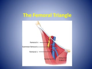
The femoral triangle is located in the upper thigh. It is bounded superiorly by the inguinal ligament, laterally by the sartorius muscle, and medially by the adductor longus muscle. The floor of the femoral triangle consists of three muscles - the iliacus, psoas major, and pectineus/adductor longus - forming a gutter shape. It contains the femoral nerve, femoral artery, and femoral vein from lateral to medial.

Recomendados
Más contenido relacionado
La actualidad más candente
La actualidad más candente (20)
Similar a Femoral Triangle Anatomy Guide
Similar a Femoral Triangle Anatomy Guide (20)
Más de Pathshallacom
Último
Último (20)
Femoral Triangle Anatomy Guide
- 2. The Femoral Triangle is a region found at the medial aspect of the upper thigh
- 3. The Femoral Triangle is a region found at the medial aspect of the upper thigh The floor of the triangle is gutter shaped
- 4. Femoral Triangle Boundaries Superior Border •Inguinal ligament Inguinal ligament Right Femoral Triangle
- 5. Femoral Triangle Boundaries Superior Border •Inguinal ligament Lateral Border •Sartorius – medial edge Inguinal ligament Right Femoral Triangle
- 6. Femoral Triangle Boundaries Superior Border •Inguinal ligament Lateral Border •Sartorius – medial edge Inguinal ligament Right Femoral Triangle
- 7. Femoral Triangle Boundaries Superior Border •Inguinal ligament Lateral Border •Sartorius – medial edge Medial Border •Adductor Longus - medial edge Inguinal ligament Right Femoral Triangle
- 8. Femoral Triangle Boundaries Superior Border •Inguinal ligament Lateral Border •Sartorius – medial edge Medial Border •Adductor Longus - medial edge Inguinal ligament Right Femoral Triangle
- 9. Femoral Triangle Boundaries Superior Border •Inguinal ligament Lateral Border •Sartorius – medial edge Medial Border •Adductor Longus - medial edge Inguinal ligament Right Femoral Triangle medial lateral
- 10. Floor of the Femoral Triangle Inguinal ligament Right Femoral Triangle medial lateral The floor of the Femoral Triangle consists of 3 muscles It is not flat but gutter shaped
- 11. Floor of the Femoral Triangle Inguinal ligament Right Femoral Triangle medial lateral From lateral to medial
- 12. Floor of the Femoral Triangle Inguinal ligament Right Femoral Triangle medial lateral From lateral to medial Iliacus
- 13. Floor of the Femoral Triangle Inguinal ligament Right Femoral Triangle medial lateral From lateral to medial Iliacus Psoas Major psoas major
- 14. Floor of the Femoral Triangle Inguinal ligament Right Femoral Triangle medial lateral From lateral to medial Iliacus Psoas Pectineus and Adductor Longus psoas pectineus
- 15. Femoral Triangle Contents From Lateral to Medial N - Femoral Nerve
- 16. Femoral Triangle Contents From Lateral to Medial N - Femoral Nerve A -Femoral Artery
- 17. Femoral Triangle Contents From Lateral to Medial N - Femoral Nerve A -Femoral Artery V - Femoral Vein
- 18. Femoral Triangle Contents From Lateral to Medial N - Femoral Nerve A -Femoral Artery V - Femoral Vein (femoral canal containing lymph nodes)
- 19. Femoral Triangle Contents From Lateral to Medial N - Femoral Nerve A -Femoral Artery Inside Femoral Sheath V - Femoral Vein (femoral canal containing lymph nodes)
- 20. Blood Supply
- 21. External Iliac Artery Arterial Supply
- 22. External Iliac Artery Common Femoral Artery Arterial Supply
- 23. Superficial Femoral Artery External Iliac Artery Common Femoral Artery Arterial Supply
- 24. Superficial Femoral Artery External Iliac Artery Common Femoral Artery Arterial Supply
- 25. Superficial Femoral Artery External Iliac Artery Common Femoral Artery Deep Femoral Artery or Profunda Femoris Arterial Supply
- 26. Superficial Femoral Artery Deep Femoral Artery or Profunda Femoris External Iliac Artery Common Femoral Artery Medial Circumflex Femoral Artery Arterial Supply
- 27. Deep Femoral Artery or Profunda Femoris External Iliac Artery Common Femoral Artery Medial Circumflex Femoral Artery Superficial Femoral Artery Lateral Circumflex Femoral Artery Arterial Supply
- 28. Femoral Vein Deep System Venous System
- 29. Great Saphenous Vein Superficial System Femoral Vein Deep System Venous System
- 30. Femoral Vein Deep System Sapheno-Femoral Junction Great Saphenous Vein Superficial System Venous System
- 31. Summary of Muscles in the Femoral Triangle
- 32. Sartorius Origin Anterior superior iliac spine Insertion Superior aspect medial surface of tibial shaft Action Flexes, adducts and laterally rotates the hip joint and flexes the knee Nerve Supply Anterior division of femoral nerve (L3, L4) Blood Supply Femoral artery Anterior Thigh
- 33. Hip Flexors Iliacus Origin Insertion Action Nerve Supply Blood Supply Upper 2/3 of iliac fossa of ilium, lateral aspect of sacrum Lesser trochanter inferior aspect Flexes and medially rotates hip Muscular branch of femoral nerve (L2, L3) Lumbar branch of iliopsoas branch of internal iliac artery
- 34. Hip Flexors Psoas Origin Insertion Action Nerve Supply Blood Supply Anterior surfaces and lower borders of transverse processes of L1 - L5 and bodies and discs of T12 - L5 Lesser trochanter Flexes and medially rotates the hip Anterior primary rami L1,2 Lumbar branch of iliopsoas branch of internal iliac artery
- 35. Anterior Thigh Pectineus Origin Pectineal surface of the pubis Insertion Pectineal line of femur below lesser trochanter Action Adducts the thigh, flexes hip joint Nerve Supply Femoral nerve & some innervation from the obturator nerve (L2, L3, L4) Blood Supply Medial circumflex femoral branch of femoral artery and obturator artery
- 36. Adductor Longus Origin Anterior surface of body of pubis, just lateral to pubic symphysis Insertion Middle third of linea aspera, between the more lateral adductor magnus and brevis insertions and the more medial origin of the vastus medialis Action Adducts, flexes and medially rotates the hip joint Nerve Supply Anterior division of obturator nerve (L2, L3, L4) Blood Supply Obturator artery and medial circumflex femoral artery Anterior Thigh