Smartphone-powered Ophthalmic Diagnostics
•
6 likes•1,381 views

Overview of the emerging field of smartphone-powered ophthalmic diagnostics that has the potential to bring down the cost and improve access to healthcare especially in developing countries. Alternative download link: https://www.dropbox.com/s/c3ef13nlw2mywa7/iphoneFundusCamera.pdf?dl=0

Recommended
Recommended
More Related Content
What's hot
What's hot (17)
Viewers also liked
Viewers also liked (19)
Similar to Smartphone-powered Ophthalmic Diagnostics
Similar to Smartphone-powered Ophthalmic Diagnostics (20)
More from PetteriTeikariPhD
More from PetteriTeikariPhD (15)
Recently uploaded
Recently uploaded (20)
Smartphone-powered Ophthalmic Diagnostics
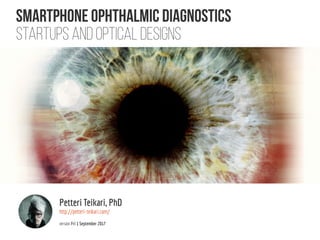
- 1. SMARTphone OPHTHALMIC DIAGNOSTICS Startups and optical designs Petteri Teikari, PhD http://petteri-teikari.com/ version Fri 1 September 2017
- 2. Smartphone fundus cameras #1 http://www.odocs-tech.com/ http://www.peekvision.org/ d-eyecare.com ophthalmicdocs.com OPEN-SOURCECOMMUNITY
- 3. Smartphone fundus cameras #2 volk.com medisave.co.uk
- 4. PORTABLE/SMARTPHONE fundus cameras #3 Why would you need someone else to take a picture of your own eye? http://web.media.mit.edu/~tswedish/projects/eyeSelfie.html http://www.optomed.com/
- 5. OPHTHALMOLOGICAL OPTICS A The human eye as an optical system (Sliney 2005) - estimates of human eye focal length f vary between 17-24 mm, see e.g. hypertextbook.com (f = 22.3 mm standardized result) B Photoreceptor optics (Warrant and Nilsson 1998)
- 6. SMARTPHONE SENSOR&OPTICS anandtech.com dpreview.com anandtech.com “Easiest to design to iPhones as the optomechanics are the same, whereas with different Android phones, the optics should be customized to each model”
- 7. OPTICAL DESIGNS #1 Giardini et al. (2014) [PDF] Fang et al. (2015)
- 8. OPTICAL DESIGNS #2 Exploded view of the D-Eye module (angles and distances between components are approximated). Retinal images are acquired using coaxial illumination and imaging paths thanks to a beam splitter (C). The blue arrow depicts the path of the light; red arrow depicts the path of fundus imaging. Device components are glass platelet (A) with imprinted negative lens (A ′), photo-absorbing wall (B), beam splitter (C), mirror (D), plastic case (E), diaphragm (F), polarized filters (G, H), flash and camera glass (J, I), and magnetic external ring (K). Russo et al. (2015)
- 9. OPTICAL DESIGNS #3 http://web.media.mit.edu/~tswedish/projects/eyeSelfie.html “The subject aligns his/her own for optimal image”
- 10. OPTICAL DESIGNS #4 Bergeles et al. (2015)
- 11. OPTICAL DESIGNS #5 3ders.org 3D print your own parts, or have them printed for you at Shapeways
- 12. OPTICAL DESIGNS #6 https://hackaday.io/project/11943-open-indirect-ophthalmoscope Including CAD files for 3D Printing g and Eagle files for the PCB design
- 13. Medical optics Small-scale micro-optics http://nanocomp.fi/what-we-do/applications/healthcare/ https://www.luxexcel.com/portfolio/microstructures-micro-optics/ 3D printed custom microstructures and micro optics, LUXeXceL http://dx.doi.org/10.1038/nphoton.2016.121 Two-photon direct laser writing of ultracompact multi-lens objectives
- 14. Mobile ERG Phone as DAQ essentially