SlideShare una empresa de Scribd logo
1 de 11
Descargar para leer sin conexión
CHEMOKINES, CYTOKINES, AND INTERLEUKINS
cAMP-induced PKC␨ activation increases functional CXCR4 expression
on human CD34ϩ hematopoietic progenitors
Polina Goichberg, Alexander Kalinkovich, Nataliya Borodovsky, Melania Tesio, Isabelle Petit, Arnon Nagler, Izhar Hardan,
and Tsvee Lapidot
Chemokines are key regulators of hema-
topoiesis and host defense. We report
here that functional expression of the
chemokine receptor CXCR4 on human
immature CD34؉ hematopoietic progeni-
tors was increased as a result of sus-
tained elevation in cellular cAMP by db-
cAMP and prostaglandin E2. This effect
of cAMP was specifically mediated by
PKC␨ activity. CXCR4 expression and
PKC␨ activation by cAMP were decreased
after the inhibition of cAMP effector-Rap1
by Spa1 overexpression. Interference with
the activation of Rac1, a downstream
target of Rap1, prevented the cAMP-
induced increase in PKC␨ activity and
CXCR4 levels. Functional manifestation
of the effects of cAMP-elevating agents
revealed an increased ability of human
CD34؉ cells to transmigrate the bone
marrow (BM) endothelial layer and adhere
to BM stroma in vitro, and it augmented
the homing potential to the BM and
spleens of immunodeficient mice in a
Rac1- and a PKC␨-dependent manner.
cAMP- and TNF␣-stimulated pathways
converged in PKC␨-activated CXCR4 ex-
pression and MMP-2/MMP-9 secretion.
cAMP treatment had a beneficial effect on
CD34؉ cell survival in a PKC␨-mediated
fashion. Taken together, our data reveal
major roles for cAMP-induced PKC␨ acti-
vation in signaling governing the motility
and development of CD34؉ cells. (Blood.
2006;107:870-879)
© 2006 by The American Society of Hematology
Introduction
Considerable effort has been invested in recent years toward
understanding the mechanisms that govern hematopoietic stem/
progenitor cell trafficking and development. Chemokine stromal
cell–derived factor-1 (SDF-1/CXCL12) is the only known power-
ful chemoattractant for defined populations of primitive human
(CD34ϩCD38Ϫ/low) and murine (Sca-1ϩThy-1lowc-kitϩLinϪ) hema-
topoietic progenitor cells.1-3 The primary receptor for SDF-1,
CXCR4, is expressed on a variety of immature and mature
hematopoietic cells and on neuronal, endothelial, and epithelial
cells.4-6 Multiple studies have demonstrated major roles for
SDF-1/CXCR4 interaction in human stem cell migration in
vivo. Mice lacking either SDF-1 or CXCR4 exhibit several
lethal defects, including impairment in hematopoiesis and stem
cell seeding of the fetal BM.7-9 We have previously shown that
homing and engraftment of human CD34ϩCD38Ϫ/low hematopoi-
etic progenitors transplanted into immunodeficient mice are
regulated by cell surface human CXCR4 expression and BM-
produced murine SDF-1.2,10-13 The in vitro migratory capacity of
enriched CD34ϩ progenitors, particularly the expression and
functionality of CXCR4 on these cells, directly correlates with
clinical hematopoietic recovery after autologous stem cell
transplantation.14-16 Thus, understanding the mechanisms and
molecular pathways that affect CXCR4 expression and cellular
signaling might have important implications for clinical stem/
progenitor cell transplantation.
Recently, we identified a specific isoform of the PKC family,
atypical PKC␨, as a key regulator of SDF-1/CXCR4–activated
signaling in human hematopoietic progenitors, demonstrating that
ectopic PKC␨ expression increases SDF-1–induced motility,
whereas the inhibition of PKC␨ activity impairs survival, prolifera-
tion, adhesion and, engraftment of immature CD34ϩ progenitors.17
Unlike conventional (␣, ␤, ␥) and novel (␦, ⑀, ␩, ␪) PKC
isoenzymes, atypical PKCs ␨ and ␭/␫ are not activated by calcium
and diacylglycerol.18,19 Atypical PKCs are key regulators of cell
polarity, motility, and gene expression.20-22 Others’ and our find-
ings17,23-25 have positioned PKC␨ in the center of chemoattractant-
and immunoregulator-induced responses, yet the molecular events
regulating PKC␨ activation and signaling in hematopoietic cells
have not been completely elucidated.
In general, cAMP has been well established as a secondary
messenger regulating a wide variety of cell functions, such as
motility, growth, and metabolism. The effects of cAMP are
primarily mediated by PKA- and Rap1-activated signaling path-
ways (for recent reviews, see Bos et al26 and Kopperud et al27).
Multiple stimuli, including immunomodulators such as prosta-
glandins, cause increases in cellular cAMP production.28 The
From the Immunology Department, The Weizmann Institute of Science,
Rehovot, Israel; and the Bone Marrow Transplantation Department, Chaim
Sheba Medical Center, Tel Hashomer, Israel.
Submitted March 8, 2005; accepted September 15, 2005. Prepublished online as
Blood First Edition Paper, October 4, 2005; DOI 10.1182/blood-2005-03-0941.
Supported by grants from the Ares-Serono Group, the Israel Science
Foundation, and the Charles and David Wolfson Charitable Trust, through the
Stem Cell Center in Establishment at the Weitzmann Institute of Science.
P.G. designed and performed the experiments, analyzed the data, and wrote
the manuscript. A.K. and N.B. performed the experiments. M.T. performed real-
time polymerase chain reaction analysis. I.P. assisted in designing the
experiments. A.N. and I.H. provided cord blood and mobilized peripheral blood.
T.L. supervised the work and prepared the manuscript.
The online version of this article contains a data supplement.
Reprints: Tsvee Lapidot, Immunology Department, The Weizmann Institute of
Science, Rehovot 76100, Israel; e-mail: tsvee.lapidot@weizmann.ac.il.
The publication costs of this article were defrayed in part by page charge
payment. Therefore, and solely to indicate this fact, this article is hereby
marked ‘‘advertisement’’ in accordance with 18 U.S.C. section 1734.
© 2006 by The American Society of Hematology
870 BLOOD, 1 FEBRUARY 2006 ⅐ VOLUME 107, NUMBER 3
For personal use only.on July 14, 2015.by guestwww.bloodjournal.orgFrom
fact that changes in cellular cAMP level have been implicated in
the activation of CXCR4 expression in T cells,29-32 maturing
dendritic cells,33,34 endothelium,35 and astroglia36 calls attention
to its importance in the regulation of hematopoietic progenitor
cell biology.
In the present work, we have examined the effect of cAMP-
activated signaling on primary normal human CD34-enriched
(CD34ϩ) hematopoietic progenitor cells that are commonly used
for clinical stem cell transplantation, obtained from both cord blood
(CB) and adult mobilized peripheral blood (MPBL). We found that
a sustained elevation in cellular cAMP level significantly increased
CXCR4 expression, adhesion, survival, and homing of CD34ϩ
progenitors, and we describe a novel role for PKC␨ in this pathway.
Materials and methods
Human cells and reagents
Human cord blood (CB) and adult G-CSF MPBL mononuclear cells were
obtained after informed consent in accordance with procedures approved by
the human ethics committee of The Weizmann Institute of Science. CD34ϩ
cell enrichment was performed using magnetic bead separation, as previ-
ously described.12 Cells were cultured in RPMI 1640 medium supple-
mented with 10% heat-inactivated FCS, antibiotics, and glutamine. The
pre–B-cell acute lymphocyte leukemia (ALL) cell lines G2 and B1 (kindly
provided by M. Freedman, Hospital for Sick Children, Toronto, Canada)
were cultured in 10% FCS-supplemented IMDM. For the stimulation of
cAMP-mediated signaling, cells were treated with 500 ␮M N,62Ј-O-
dibutyryl adenosine 3Ј,5Ј-cyclic monophosphate (dbcAMP), 10 nM prosta-
glandin E2 (PGE2), or 2 ␮M cholesterol sulfate, all purchased from
Sigma-Aldrich (Rehovot, Israel). Cell-permeable myristolated pseudosub-
strate (PS) inhibitor peptides for PKC␨, PKC␣/␤, and PKC⑀ were pur-
chased from BioMol Research Laboratories (Plymouth Meeting, PA), and
Rac1 inhibitor (NSC23766) was purchased from Calbiochem, Merck
Biosciences GmbH (Darmstadt, Germany).
Flow cytometry analysis of membranal and intracellular
CXCR4 expression
Membranal CXCR4 expression was determined by flow cytometry (FACS-
calibur; Becton Dickinson, San Jose, CA) using purified anti–human
CXCR4 mAb, clone 12G5 (R&D Systems, Minneapolis, MN), followed by
secondary Alexa 488–conjugated donkey anti–mouse IgG (Molecular
Probes, Eugene, OR). Isotype-matched IgG2a was used as control, showing
background labeling with no differences between the treatments. Ratios of
mean values of Ab-labeled cells to secondary IgG–only control samples
were calculated using CellQuest software (Becton Dickinson), and the
results were expressed in arbitrary units (AU). Flow cytometry analysis
with additional anti-CXCR4 mAbs, clones 6H8 and 1D9, demonstrated
increases similar to that of the 12G5 increase after dbcAMP treatment. For
the analysis of intracellular CXCR4 levels, after blocking of membranal
receptor by 1-hour incubation with 10 ␮g/mL nonconjugated 12G5 mAb,
G2 cells were fixed, permeabilized, and immunolabeled with anti–human
CXCR4-PE Ab (12G5; R&D Systems), as previously described.37 Mem-
branal CXCR4 internalization was assayed as described38: G2 cells were
preincubated for 30 minutes at 4°C with 10 ␮g/mL anti–human CXCR4-PE
Ab, incubated at 37°C for the indicated times, and washed for 2 minutes on
ice at normal (pH 7.4) or acidic (pH 2.2) buffer to remove the surface-bound
Ab. The relative proportion of internalized receptor (labeling after acid
wash) out of total fluorescence intensity was determined by flow cytometry.
For the analysis of CXCR4 membranal re-expression after SDF-1–induced
internalization, G2 cells were incubated for 2 hours at 37°C with 250 nM
SDF-1 (PeproTech, Rocky Hill, NJ), washed in excess growth medium, and
further incubated at 37°C for the indicated times. CXCR4 expression was
monitored by flow cytometry, as described.
Activated Rap1 pull-down assay
GTP-Rap1 pull-down was performed using Ral GDS-RBD agarose slurry
(Upstate Cell Signaling Solutions, Lake Placid, NY), according to the
manufacturer’s instruction, and was detected, after 15% sodium dodecyl
sulfate–polyacrylamide gel electrophoresis (SDS-PAGE) by anti-Rap1
mAb (BD Biosciences, Bedford, MA).
Spa1 overexpression
MPBL CD34ϩ cells were transiently transfected with Spa1-GFP or
GFP-only expressing vector,39 kindly provided by M. Hattori and N. Minato
(Kyoto University, Kyoto, Japan), using the Amaxa Nucleofector kit
(Amaxa, Berlin, Germany) according to the manufacturer’s protocol.
Immunoprecipitation and immunoblot
Immunoprecipitation of PKC␨ was performed as previously described17 by
2-hour incubation at 4°C of G2 cell lysates with protein A–coated beads
(Bio-Rad Laboratories, Hercules, CA) preconjugated to polyclonal anti-
PKC␨ Ab (Santa Cruz Biotechnology, Santa Cruz, CA). Samples were
resolved by 7.5% SDS-PAGE and immunoblotted with anti–phospho-PKC␨
or monoclonal anti–total neutrophil PKC␨ (both purchased from Santa
Cruz). For the detection of ERK1/2 phosphorylation level, 20 ␮g G2 cell
lysate was subjected to 10% SDS-PAGE followed by immunoblot with
either anti–phospho-ERK1/2 Ab or anti–total-ERK1/2 Ab (both obtained
from Sigma-Aldrich).
Membranal PKC␨ translocation
MPBL CD34ϩ cells were transiently transfected as described, fixed,
permeabilized, and indirectly immunolabeled with anti–PKC␨ Ab (C-20;
Santa Cruz Biotechnology), as described.40 CXCR4 was labeled with 10
␮g/mL anti–human CXCR4 mAb (12G5; R&D Systems). Cells were
mounted on poly-L-lysine (Sigma-Aldrich)–coated glass coverslips. Im-
ages were acquired by the DeltaVision system using Resolve 3D software
(Applied Precision, Issaquah, WA) with an Axiovert 100TV inverted
microscope (Zeiss, Oberkochen, Germany) and a 100ϫ/1.40 Plan-
Apochromat objective lens, and images were processed in Adobe Photo-
shop 7.0 ME software (Adobe Systems, Mountain View, CA).
Real-time RT-PCR (reverse transcriptase polymerase
chain reaction)
Total RNA was isolated from G2 cells (5 ϫ 106 cells per sample) using
Tri-reagent (Molecular Research Center, Cincinnati, OH) according to the
manufacturer’s protocol and was subjected to cDNAsynthesis as previously
described.13 CXCR4 expression was analyzed by real-time quantitative
PCR using a LightCycler instrument and LightCycler-Fast Start DNA
Master SyBr Green I reagents (Roche Molecular Biochemicals, Indianapo-
lis, IN). The following primers were used: CXCR4 sense, 5Ј-TCTAGGCAG-
GACCTGT-3Ј; CXCR4 antisense, 5Ј-CACTTTGGGCTTTGGTT-3Ј; ␤-ac-
tin sense, 5Ј-GCTCAGGAGGAGCAAT-3Ј; ␤-actin antisense, 5Ј-
GGCATCCACGAAACTAC-3Ј. Standard curve was calculated using 5
different cDNA concentrations. Reactions were performed in triplicate at
95°C for 10 seconds, followed by 45 cycles of 95°C for 15 seconds, 60°C
for 10 seconds, 72°C for 20 seconds, 65°C for 40 seconds, and a melt curve
analysis to confirm the purity of the reaction products. Raw data from
reactions using CXCR4 primers were normalized to the corresponding data
from reactions using ␤-actin primers.
Analysis of CREB phosphorylation
For flow cytometry analysis of phosphorylated CREB content (P-CREB),
cells were stimulated for 1 hour with 500 ␮M dbcAMP, washed in PBS, and
fixed in 70% ice-cold ethanol. After overnight incubation at 4°C, samples
were washed in PBS, permeabilized with 0.25% Triton X100 in PBS for 10
minutes on ice, washed, and indirectly immunolabeled with anti–P-CREB
Ab (Upstate Cell Signaling Solutions, Waltham, MA), followed by Alexa
488–goat anti–rabbit IgG (Molecular Probes).
cAMP ACTIVATES PKC␨ IN HEMATOPOIETIC PROGENITORS 871BLOOD, 1 FEBRUARY 2006 ⅐ VOLUME 107, NUMBER 3
For personal use only.on July 14, 2015.by guestwww.bloodjournal.orgFrom
Adhesion and transmigration assays
Adhesion to MS-5 mouse stroma cells (kindly provided by J.C. Gutierrez-
Ramos, Millennium Pharmaceuticals, Cambridge, MA) was assayed as previ-
ously described.17 Briefly, CB CD34ϩ cells were preincubated for 24 hours with
or without 500 ␮M dbcAMP, labeled with CFSE (Molecular Probes), and
allowed to adhere for 30 to 40 minutes to a confluent MS-5 cell layer. After
extensive washings, cells were collected in 5 mM EDTA containing buffer, and
the number of CFSEϩ cells in each sample was determined by flow cytometry.
Where indicated, before the assay, cells were pretreated for 20 minutes at room
temperature with 10 ␮g/mL anti-CD49d (VLA-4; Serotec, Oxford, United
Kingdom) mAb or IgG1 control. For transendothelial migration, cAMP-treated
CFSE-labeled CD34ϩ cells, treated as above, were allowed to migrate for 6 hours
through human bone marrow SV–transformed endothelial cells (BMECs, a kind
gift from S. Rafii, Weill Medical College of Cornell University, New York, NY)
that were plated (2 ϫ 105 cells/well) 24 hours in advance on Costar tran-
swells precoated for 1 hour at 37°C with 25 ␮g/cm2 fibronectin (Sigma-Aldrich).
The integrity of the BMEC layer was verified microscopically. The number of
cells transmigrated spontaneously or toward 15 nM SDF-1 was quantified by
flow cytometry.
Homing experiments
␤2-microglobulin knockout NOD/LtSz-PrKdcscid (NOD/SCIDB2mnull) mice
were bred and housed as previously described.12 All experiments were approved
by the animal care committee of The Weizmann Institute of Science. Enriched
human CD34ϩ cells were treated for 24 hours with 500 ␮M dbcAMP with or
without subsequent washing (leading to identical results) and were injected
(0.5 ϫ 106 cells/mouse) into the tail veins of mice that were sublethally irradiated
(350 cGy) 24 hours before transplantation. Four to 6 hours after transplantation,
cells were recovered from the BM and spleens of the recipient mice and were
analyzed by flow cytometry using human-specific CD45-FITC mAb (IQ
Products, Groningen, The Netherlands) acquiring 1.5 ϫ 106 cells/sample. Cells
obtained from mice that did not undergo transplantation, or cells labeled with
mouse IgG1-FITC only, were used as negative controls.
Gelatin zymography
Zymography of supernatants obtained from CD34ϩ cells cultured in serum-free
RPMI 1640 for 40 hours untreated or in the presence of 500 ␮M dbcAMP, 1.15
pM TNF␣ (PeproTech), or both was performed as previously described.41
Cell survival and cell cycle analysis
CD34ϩ cells were cultured for the indicated lengths of time in 10%
FCS-supplemented RPMI for cell proliferation or serum-free IMDM
supplemented with 2% BSA, 25 U/mL insulin, 0.1 mM 2-mercaptoethanol,
10 mM HEPES, 2 mM L-glutamine, and antibiotics for cell survival. For
determination of DNA content, cells were fixed in 70% ice-cold ethanol and
incubated for 30 minutes at 37°C with 100 ␮g/mL RNase A and 50 ␮g/mL
propidium iodide (PI) (both from Sigma-Aldrich), and PI intensity was
determined by flow cytometry. In some assays, cell viability was deter-
mined by PI exclusion after brief incubation with nonfixed cells. For Ki-67
staining, G2 cells were cultured in serum-supplemented IMDM, washed in
excess PBS, fixed as described, and indirectly immunolabeled with mouse
anti–human Ki-67 mAb (DakoCytomation, Glostrup, Denmark) followed
by Alexa 488–conjugated donkey anti–mouse IgG (Molecular Probes).
Statistical analysis
Significance levels of data were determined by Student t test for the
differences in mean values.
Results
Cellular cAMP elevation increases CXCR4 expression and
membranal presentation on human hematopoietic progenitors
To increase the cellular cAMP level, we applied a cell-permeable
cAMP analog, dbcAMP. We found that 24-hour treatment with
dbcAMP increased more than 5-fold the membranal CXCR4
expression on MPBL and CB CD34ϩ cells (Figure 1A). Of interest,
CB-derived mononuclear cells of different lineages and primary
cells from patients with acute myeloid leukemia and leukemia-
derived cell lines displayed heterogeneous responses to the dbcAMP
treatment in terms of CXCR4 expression (P.G., unpublished
observation, May 2005), suggesting that although the increase in
CXCR4 in response to cAMP was not unique to immature
hematopoietic progenitors, it was not necessarily a general phenom-
enon for all the hematopoietic cell types. Given that CD34ϩ
hematopoietic progenitors were shown to respond to prostaglandin
E2 (PGE2) stimulation42 and based on the fact that PGE2 is a potent
activator of cellular cAMP synthesis,28 we attempted to increase the
cellular cAMP level by PGE2 treatment to study early processes in
the activation of CD34ϩ cells. Accordingly, stimulation by PGE2
had also elevated significantly the CXCR4 level expressed on
CD34ϩ progenitors (Figure 1A).
To further elucidate this effect, we used the G2 pre-B ALL cell
line that was previously shown to recapitulate several features of
normal hematopoietic progenitors.17 Treatment of G2 cells with
dbcAMP resulted in Rap1 activation, as assayed by the GTP-bound
Rap1 content (Figure 1B). Rap1 is a member of the Ras subgroup
of small GTP-binding proteins. To interfere with the Rap1 activity,
we transiently overexpressed Spa1, the hematopoietic cell–specific
Rap1 GTPase activating protein.43 As depicted in Figure 1C, the
excess of Rap1 inhibitor decreased dbcAMP-induced and, to a
lesser extent, basal level CXCR4 expression in MPBL CD34ϩ
progenitors, indicating that Rap1 activity was required for the
cAMP-mediated effect on CXCR4 expression. Interestingly, mem-
branal CXCR4 elevation stimulated by dbcAMP was not prevented
when we inhibited PKA activity by H89, KT5720, or several other
serine/threonine kinases—including phosphatidyl inositol 3-kinase
(PI3K)—by wortmannin and LY294002 and Rho-kinase by Y27632
(Figure S1, available on the Blood website; see the Supplemental
Materials link at the top of the online article).
CXCR4 expression induced by cAMP is PKC␨ dependent
To investigate the mechanisms underlying the cAMP effect on
CXCR4 expression, we examined downstream constituents of the
cAMP-activated pathway in hematopoietic progenitors. We thus
focused on PKC␨, which has been recently implicated in cAMP-
induced signaling.44,45 We found that an increase in cellular cAMP
level had a major effect on PKC␨ activity. As shown in Figure 2A,
24-hour treatment of G2 cells with dbcAMP increased by 2-fold the
phosphorylation of PKC␨, a hallmark of its activation. In MPBL
CD34ϩ progenitors, interference with the cAMP signaling by the
overexpression of Spa1 resulted in the inhibition of dbcAMP-
induced PKC␨ membranal translocation (Figure 2B), which is
implicated in PKC molecule activation. Surprisingly, despite the
profound decrease in the total number of cells with membranal
PKC␨ in Spa1-overexpressing samples (Figure 2B, histogram), we
still detected Spa1GFP-positive cells displaying membranal PKC␨
(Figure 2B, lower row), suggesting that Rap1 inhibition might
affect signaling upstream to PKC␨ translocation. Members of the
Rac family of small GTPases have been implicated downstream of
cAMP-activated Rap1 in the signaling pathway, regulating the
expression of amyloid precursor protein,46 whereas functional
Rap1 was found to recruit Rac-activating guanine nucleotide
exchange factors.47 It is suggested that in epithelial cells, Rac
mediates the localization and activation of atypical PKCs.48
Therefore, we examined the involvement of RacGTPases in the
cAMP-induced PKC␨ activation in hematopoietic progenitors. Our
872 GOICHBERG et al BLOOD, 1 FEBRUARY 2006 ⅐ VOLUME 107, NUMBER 3
For personal use only.on July 14, 2015.by guestwww.bloodjournal.orgFrom
data showed that blocking Rac1 activation by NSC23766, a
pharmacologic compound preventing Rac1 interactions with gua-
nine nucleotide exchange factors Trio and Tiam1,49 resulted in
concentration-dependent inhibition of the cAMP-induced PKC␨
phosphorylation in G2 cells (Figure 2C), implying that functional
Rac1 is required for PKC␨ activation by cAMP.
We next examined PKC␨ involvement in cAMP-activated
CXCR4 expression. Adding the PKC␨ inhibitory pseudosubstrate
(PS) substantially affected basal and completely abrogated cAMP-
induced CXCR4 expression in CD34ϩ cells. Blocking PKC␨
activity by a similar approach also inhibited the CXCR4 expression
in dbcAMP-treated human peripheral blood CD3ϩ T cells (data not
shown). In contrast, inhibiting the other PKC isoenzymes, namely
PKC⑀ and PKC␣/␤, had no effect on CXCR4 expression (Figure
3A). In addition, cholesterol sulfate, a chemical activator of
PKC␨,50 increased the membranal CXCR4 level to one similar to
that for dbcAMP-treated cells (Figure 3A). To further confirm these
findings, we used a PKC␨-deficient B1 cell line.17 As shown in
Figure 3B, neither dbcAMP nor PKC␨ PS affected CXCR4
expression in these cells. Importantly, blocking Rac1 activation in
CD34ϩ cells caused a reduction comparable to that for PKC␨ PS in
membranal CXCR4 expression (Figure 3A) in a manner that was
neither synergistic nor additive in the presence of both inhibitors,
supporting our suggestion that Rac1 and PKC␨ constitute the same
pathway in cAMP-stimulated CXCR4 elevation.
The changes in membranal and intracellular CXCR4 levels
were then examined comparatively in G2 cells incubated for 24
hours with dbcAMP. Results revealed that whereas cell surface
(membranal) expression of the receptor was up-regulated 2.5 times,
the intracellular molecule content was increased by just 1.4-fold
(Figure 3C). Furthermore, time-course analysis of surface-bound
anti–CXCR4 mAb internalization showed that in cAMP-stimulated
cells, CXCR4 receptor endocytosis was reduced 2 times compared
with untreated counterparts (Figure 3D), suggesting that in the
absence of a ligand, cAMP stimulation prolonged the time of
CXCR4 presence on the cell membrane. In addition, cAMP-treated
cells displayed a more efficient recovery from SDF-1–induced
CXCR4 desensitization (Figure 3D, inset). These results are in line
with previous findings demonstrating that cAMP treatment reduces
the CXCR4 internalization rate and antagonizes SDF-1–induced
receptor endocytosis in peripheral blood mononuclear cells.29
In view of reports that cAMP-induced CXCR4 expression in T
cells relies on de novo protein synthesis,31,32 we examined whether
this was also the case in hematopoietic cells. Time-lapse analysis of
changes in membranal CXCR4 expression in dbcAMP-stimulated
G2 cells revealed that the increase in CXCR4 took place after at
least 6 hours of incubation, reaching maximal level at approxi-
mately 24 hours of treatment (Figure 1D). Accordingly, we found
that in G2 cells cycloheximide treatment antagonized the CXCR4
elevation by cAMP (data not shown). Real-time RT-PCR analysis
of G2 cells stimulated with dbcAMP for 6 hours showed a 4-fold
increase in CXCR4 mRNA level that was significantly attenuated
in the presence of the PKC␨ PS (Figure 3E). The transcription
factor CREB is a major cAMP target in the regulation of gene
expression.51 CREB-mediated transcription was implicated in the
cAMP-induced CXCR4 expression.31 We found that in CD34ϩ
progenitors treated for 1 hour with dbcAMP (the time period of
maximal CREB binding to the CXCR4 promoter in peripheral
blood T cells),31 the phosphorylated (activated) CREB content was
increased by almost 2-fold, whereas pretreatment with PKC␨ PS
attenuated the cAMP-induced CREB phosphorylation (Figure 3F).
Figure 1. cAMP-stimulated CXCR4 expression in CD34؉ hematopoietic progenitors
is Rap1 dependent. (A) Membranal CXCR4 expression on human MPBL or CB CD34ϩ
cells treated for 24 hours with 500 ␮M dbcAMP (cAMP) and 10 nM PGE2, untreated, or
vehicle EtOH-treated as control (CTRL), indirectly immunolabeled with anti–human
CXCR4 mAb. Results are shown as mean Ϯ SD. For each cell source, 20 cAMP and 5
PGE2 independent experiments were conducted. P values indicate statistically significant
differences compared with CTRL. Representative flow cytometry analysis is shown at the
bottom. IgG indicates secondary Ab–only labeled samples. (B) Representative analysis of
activated Rap1 (Rap1-GTP) status in G2 cells stimulated for 24 hours with 500 ␮M cAMP
or left untreated (CTRL). (C) Effect of Spa1GFP overexpression on membranal CXCR4
level in MPBL CD34ϩ cells incubated 18 hours after transfection and left untreated (CTRL)
or treated with 500 ␮M cAMP. Results are shown as fold change compared with the
CXCR4 intensity in GFP-only transfected samples taken as 1 (mean Ϯ SD of 4 indepen-
dent experiments). (D) Time-lapse analysis of membranal CXCR4 expression in G2 cells
treated with 500 ␮M cAMP for the indicated time periods. Results (mean Ϯ SD of 3
independent experiments) are expressed as fold change compared with CXCR4 intensity
of untreated counterparts.
cAMP ACTIVATES PKC␨ IN HEMATOPOIETIC PROGENITORS 873BLOOD, 1 FEBRUARY 2006 ⅐ VOLUME 107, NUMBER 3
For personal use only.on July 14, 2015.by guestwww.bloodjournal.orgFrom
Interestingly, cAMP-activated CREB phosphorylation and mem-
branal CXCR4 expression in these cells were not affected by the
inhibition of NF-␬B activity when we applied a cell-permeable
inhibitory peptide NF-␬BSN50 (Figures 3F and S1, respectively),
implying that NF-␬B is not involved in cAMP-induced CXCR4
transcription.
CXCR4 induced by cAMP is functional and depends on PKC␨
for its activity
The following experiments were designed to investigate the ability
of cAMP-treated cells to respond to SDF-1, the CXCR4 ligand.
The first set of experiments was based on the in vitro assays. As
depicted in Figure 4A, cAMP-stimulated progenitors migrated
more efficiently than controls through BMEC-coated transwell
filters. The results were further substantiated by the observation
that cAMP treatment increased by 40% the ability of CD34ϩ cells
to adhere to the SDF-1–expressing BM stroma cell line MS-552
(Figure 4B). In accordance with our previous findings showing that
CD34ϩ cell adhesion to MS-5 is CXCR4 and PKC␨ dependent,17
we found that pretreatment with PKC␨ PS abolished the cAMP-
induced adhesion (Figure 4B). SDF-1–activated adhesion of CB
CD34ϩ cells to MS-5 is partially dependent on VLA-4/VCAM-1
interactions.17 We observed that blocking VLA-4 activity by
neutralizing Abs had an effect similar to that of PKC␨ PS on the
cAMP-induced adhesion (Figure 4B), further supporting our
suggestion that cAMP-stimulated cells adhere to the SDF-1–
expressing stroma by the CXCR4/PKC␨-mediated pathway.
The function of the cAMP-induced CXCR4 receptor was then
tested in vivo with the homing assay. We found that human CD34ϩ
cells exposed for 24 hours to dbcAMP home more efficiently to the
BM and spleens of recipient NOD/SCIDB2mnull mice, as measured
4 to 6 hours after transplantation (Figure 4C). Similar results were
obtained with PGE2-treated progenitors (data not shown), yet there
was no increase in the long-term repopulation capacity of dbcAMP-
treated cells, and the positive effect of cAMP on homing declined
with time. In fact, it was already less significant 24 hours after
transplantation (Figure S2A) because cAMP-induced effects, includ-
ing CXCR4 elevation (Figure S2B), are rapidly reversed once
dbcAMP is removed from the immediate cell environment, as in
intravenous injection. Increases in short-term progenitor cell
homing were abrogated in cAMP-stimulated cells pretreated with
the PKC␨ inhibitor just before transplantation (Figure 4C,
cAMPϩPS␨). In agreement with our findings implicating Rac1
activity in cAMP-induced PKC␨ activation (Figures 2C, 3A), we
found that Rac1 inhibition opposed the effect of cAMP on BM
homing of CD34ϩ progenitors (Figure 4C, cAMPϩRac1 inh).
To further define the role of PKC␨ in cAMP-stimulated
processes, we looked into the effects of TNF␣ because PKC␨ has
been well established as a major downstream target of TNF␣.53 The
experiments were based on the reports that TNF␣ is a potent
activator of matrix metalloproteinase-2 (MMP-2) and MMP-9
expression in human CD34ϩ cells,41 in a PKC␨-dependent man-
ner,54 and that MMP-9 activity is implicated in the regulation of
progenitor cell trafficking in vivo.41,55-57 Surprisingly, we found
that dbcAMP treatment also increased MMP-2 and MMP-9 expres-
sion by CD34ϩ progenitors, though to a much lower extent than
that of TNF␣ (Figure 5A). Moreover, MMP-2 and MMP-9
secretion was apparently further elevated in cells costimulated
with dbcAMP and TNF␣ (Figure 5A). We have previously
shown that MMP-9 expression induced by SDF-1 in CD34ϩ
cells is PKC␨ dependent.17 We here found that PKC␨ inhibition
abrogated dbcAMP- and TNF␣-activated MMP-2 and MMP-9
secretion and, significantly, MMP expression in cells costimu-
lated with both agents (Figure 5A, PS␨). Furthermore, we
observed that dbcAMP and TNF␣ had an additive effect on
CXCR4 expression in immature MPBL CD34ϩ progenitors
(Figure 5B) but not on CB CD34ϩ cells (data not shown). These
findings indicate that cAMP- and TNF␣-activated pathways use
PKC␨ for MMP-2, MMP-9, and CXCR4 induction in hematopoi-
etic progenitors.
Figure 2. cAMP activates PKC␨ in hematopoietic progenitor cells. (A) PKC␨ phosphorylation and expression levels in G2 cells cultured for 24 hours either untreated
(CTRL) or in the presence of 500 ␮M dbcAMP (cAMP), as assayed by immunoblot. PKC␨-phosphorylated (P-PKC␨) level was determined by densitometry, normalized to the
PKC␨ protein level (Total), and expressed as fold change versus CTRL cells. Results are mean Ϯ SD of 5 independent experiments. (B) PKC␨ membranal translocation in
cAMP-treated cells. MPBL CD34ϩ cells were transfected with GFP (green fluorescent protein) only (top and middle rows) or with Spa1GFP (bottom row) expressing vector and
further incubated for 18 hours either untreated (CTRL) or in the presence of 500 ␮M cAMP and indirectly immunolabeled for PKC␨ (red). GFP vector transfected cells were
colabeled for CXCR4 (green) because no GFP signal was retained after the permeabilization procedure. Note the increased CXCR4 expression and colocalization with PKC␨
at the membrane of cAMP-stimulated cells (middle row). Spa1GFP overexpression decreased the proportion of cells displaying membranal PKC␨ labeling (histogram on the
right, representative of 2 independent experiments; 250 cells in each sample), yet PKC␨ translocation to the membrane could be detected in Spa1GFP-transfected cells
(bottom row, asterisks). Bar represents 10 ␮m. (C) Inhibition of the cAMP-induced PKC␨ phosphorylation (P-PKC␨) by the NSC23766 (Rac1 inh) in G2 cells treated and
assayed as in panel A. Results of the densitometry analysis, expressed in arbitrary units (AU), are represented as mean Ϯ SD of 3 independent experiments.
874 GOICHBERG et al BLOOD, 1 FEBRUARY 2006 ⅐ VOLUME 107, NUMBER 3
For personal use only.on July 14, 2015.by guestwww.bloodjournal.orgFrom
Cellular cAMP elevation has opposing effects on progenitor
cell survival and proliferation
Activation of cAMP signaling has been well documented as a way
to promote the survival of a variety of cell types, including
hematopoietic cells.58,59 In accordance with this, we found that
dbcAMP stimulation substantially increased the survival of CD34ϩ
progenitors under cytokine-deprived conditions, as determined
through monitoring the viable cell count by flow cytometry and the
number of dead cells with fragmented DNA (Figure 6A). This
effect of cAMP was abrogated in the presence of PKC␨ PS (Figure
6A, inset). Strikingly, DNA content analysis revealed that dbcAMP
treatment also resulted in a 2-fold decrease in the percentage of
dividing CD34ϩ cells (Figure 6B). We found that the expression of
proliferation-associated antigen Ki-67 was reduced by 5-fold in
cAMP-treated G2 cells (Figure 6C). To address this issue, we
examined the activity of extracellular signal–regulated protein
kinase (ERK1/2), a major regulator of cell-cycle progression. The
outcome of the cAMP elevation on ERK activity and proliferation
is cell type and signal specific.60 We found that 24-hour exposure to
dbcAMP almost completely decreased the ERK1/2 phosphoryla-
tion in G2 cells (Figure 6D), suggesting that the negative effect of
cAMP on cell proliferation might be exerted through the inhibition
of ERK1/2 activity.
Figure 3. cAMP-induced increase in CXCR4 expression depends on PKC␨
activity. Membranal CXCR4 expression in CB and MPBL CD34ϩ (A) or B1 (B) cells,
incubated for 24 hours with 500 ␮M dbcAMP (cAMP), 2 ␮M cholesterol sulfate
(Choles Sulfate), or left untreated (Ϫ). Where indicated, 10 ␮M PS of PKC␨ (PS␨),
PKC⑀ (PS⑀), PS␣/␤ (PS␣/␤), or 25 ␮M NSC23766 (Rac1 inh) were applied. Flow
cytometry analysis data are shown in arbitrary units (AU) as mean Ϯ SD; 10
independent experiments were conducted for PS␨, and 6 were conducted for Rac1
inh. (C) Increase in membranal and intracellular CXCR4 labeling in G2 cells
incubated for 24 hours in the presence of 500 ␮M cAMP compared with untreated
cells (CTRL) taken as 1. Flow cytometry analysis data (mean Ϯ SD of 3 independent
experiments) are shown. (D) Decreased internalization rate of anti–CXCR4-PE mAb
in G2 cells treated for 24 hours with cAMP compared with CTRL. Results are
mean Ϯ SD of 3 independent experiments. P Ͻ .02; statistically significant differ-
ences compared with CTRL at each time point. Increased recovery after SDF-1–
induced receptor internalization is shown in the inset. Mean fluorescence intensity
(MFI) is calculated as percentage of CXCR4 expression in CTRL or cAMP-treated
cells before the application of SDF-1 (original MFI). (E) Relative CXCR4 expression
in G2 cells either untreated (Ϫ) or stimulated for 6 hours with 500 ␮M cAMP in the
absence or presence of 10 ␮M PS␨, as analyzed by real-time RT-PCR. Data are
expressed in AU as a ratio of CXCR4 and ␤-actin mRNA level (mean Ϯ SD of 3
independent experiments). (F) Changes in CREB phosphorylation (P-CREB) in
MPBL CD34ϩ progenitors stimulated for 1 hour with 500 ␮M cAMP (mean Ϯ SD of 3
independent experiments). Where specified, cells were pretreated with either 10 ␮M
PS␨ or 18 ␮M NF-␬BSN50 (NF-␬B inh). Representative flow cytometry analysis is
shown. IgG indicates secondary Ab–labeled cells; CTRL, untreated samples.
Figure 4. CXCR4 overexpressed on cAMP-stimulated CD34؉ progenitors is
functional. (A) Transendothelial migration of dbcAMP (cAMP)–treated cells. CD34ϩ
progenitors were incubated for 24 hours with 500 ␮M cAMP or left untreated (Ϫ),
labeled with CFSE, and allowed to migrate either spontaneously (spont) or toward
SDF-1 (directional) through BMEC-coated transwells. Data represent the percent-
ages of migrated cells (mean Ϯ SD of 2 independent experiments). (B) Adhesion to
BM cells after cAMP treatment. Cells treated for 24 hours with 500 ␮M cAMP or left
untreated (CTRL) were labeled with CFSE and allowed to adhere for 40 minutes to
MS-5 cell layer (mean Ϯ SD of at least 2 independent experiments, each in triplicate
samples). Where indicated, cAMP-treated cells were preincubated with 10 ␮M PKC␨
PS (PS␨), 10 ␮g/mL VLA-4 blocking mAb (␣VLA-4), or IgG1 control. (C) Homing
efficiency of cAMP-treated cells. CB or MPBL CD34ϩ cells were treated for 24 hours
with 500 ␮M cAMP or left untreated (CTRL) and injected intravenously into the
NOD/SCIDB2mnull mice. Where indicated, cAMP-treated cells were incubated for 30
minutes before injection with 10 ␮M PS␨ or cultured in the presence of 25 ␮M
NSC23766 (Rac1 inh). BM and spleen of recipient mice were analyzed for the
presence of human cells (mean Ϯ SD of at least 3 independent experiments, 2 to 3
mice per treatment in each experiment; *P Ͻ .05 relative to other treatments).
Representative flow cytometry analyses are shown on the right. IgG indicates
samples labeled with the isotype control Ab. Number of CD45ϩ human cells per 106
acquired is indicated in each case.
cAMP ACTIVATES PKC␨ IN HEMATOPOIETIC PROGENITORS 875BLOOD, 1 FEBRUARY 2006 ⅐ VOLUME 107, NUMBER 3
For personal use only.on July 14, 2015.by guestwww.bloodjournal.orgFrom
Discussion
The present study addressed molecular aspects of the regulation of
CXCR4 in human hematopoietic progenitors, namely primary
normal CD34ϩ cells enriched by MPBL and CB and pre-B ALL G2
cells. Our results showed that CXCR4 expression was increased
after elevation of the cellular cAMP level and implicated PKC␨
activity in the cAMP-mediated effects. We showed that in progeni-
tor cells, PKC␨ was directly stimulated by cAMP-activated signal-
ing. We further found that cAMP-induced and, to a lesser extent,
basal CXCR4 expression are PKC␨ dependent because, in the
PKC␨-deficient cell line B1, the CXCR4 level was not affected by
cAMP stimulation or PKC␨ inhibition, whereas the blocking of
PKC␨ activity in CD34ϩ cells completely abrogated membranal
CXCR4 expression. Importantly, we found that the inhibition of
several other PKC isoenzymes had no effect on CXCR4 expression
in hematopoietic progenitors (present work) or on their adhesion,
polarization, and chemotaxis.17
Our attempts to elucidate the molecular mechanisms underlying
the processes of cAMP-induced CXCR4 elevation led us to Rap1, a
member of the Ras family of small GTPases, which is a key
regulator of cell polarity, motility, adhesion, and prolifera-
tion.26,61,62 Our data indicate that Rap1 activity is required for
cAMP-induced CXCR4 up-regulation because the overexpression
of Spa1, a principal Rap1GAP in hematopoietic cells, attenuated
CXCR4 expression by cAMP. We have also observed reduced
membranal PKC␨ translocation in Spa1-overexpressing CD34ϩ
progenitors, indicating that interference with Rap1 signaling af-
fects PKC␨ activity. During the establishment of cell polarity and
intercellular junction formation, atypical PKCs are activated by
binding to the Par6/Par3/Cdc42 molecular complex.20,63 Rap1 was
positioned upstream of Cdc42 in determining neuronal cell asym-
metry mediated by atypical PKC family members.64 It has been
shown that Rac1 binds Par6 and potentially activates PKC␨.65
Accordingly, we found that Rac1 inhibition decreased cAMP-
induced PKC␨ phosphorylation and had an inhibitory effect on
CXCR4 expression identical to that of PKC␨ PS. Therefore, as
summarized in the model depicted in Figure 7, our data suggested
that Rap1, Rac1, and PKC␨ constitute a signaling pathway of
cAMP-induced CXCR4 expression. Our current and previous17
studies also point to a positive feedback loop between PKC␨
activation by cAMP, which results in elevated CXCR4 expression
and thus might further enhance PKC␨ activity.
We found that the increase in CXCR4 expression in cAMP-
stimulated cells resulted from enhanced transcription and, primar-
ily, prolonged time of membranal receptor representation. Previous
studies29 and our present findings on anti-CXCR4 Ab internaliza-
tion suggest that cAMP treatment leads to a decrease in CXCR4
endocytosis. Our preliminary results indicated that PKC␨ inhibition
significantly increases membranal CXCR4 turnover; this is under
further investigation. Consistent with previous reports, we also
observed cAMP-activated CXCR4 mRNA expression attenuated in
the presence of PKC␨ inhibitor. We show that CREB phosphoryla-
tion was induced by cAMP treatment of CD34ϩ progenitors,
whereas PKC␨ inhibition reduced this effect. Given that in T cells
cAMP-mediated CXCR4 expression is partially regulated by
CREB,31 we suggest that PKC␨ activation increases the CXCR4
Figure 5. cAMP and TNF␣ signaling converge in MMP-2/MMP-9 secretion and
induction of CXCR4 expression by CD34؉ progenitors. (A) MMP-2 and MMP-9
levels in the conditioned media of CB and MPBL CD34ϩ cells cultured for 24 hours
with the indicated agents, as determined by gelatin zymography. Positive control
(contr) is the conditioned medium of HT1080 cells. Representative gel and densitom-
etry analysis data are shown as mean Ϯ SD of 3 independent experiments ex-
pressed in arbitrary units (AU). Note that the presence of PKC␨ PS (PS␨) abrogates
MMP-2/MMP-9 secretion by cAMP and TNF␣ costimulated cells. (B) Membranal
CXCR4 expression in MPBL CD34ϩ cells treated as in panel A. Data are shown as
mean Ϯ SD of 6 independent experiments. Representative flow cytometry analysis is
presented at the bottom. IgG indicates secondary IgG only; CTRL, untreated cells.
Figure 6. cAMP increases CD34؉ progenitor cell survival by PKC␨-activated pathway while inhibiting their proliferation. (A) Increased survival of CB CD34ϩ cells
cultured for 3 days under cytokine-deprived conditions in the presence of 500 ␮M dbcAMP (cAMP) as analyzed by flow cytometry. Data represented as fold change
(mean Ϯ SD of 6 independent experiments each in triplicate) in the number of PI-positive (Dead cells) in cAMP-treated compared with untreated (CTRL) samples. Inset
shows representative changes in the proportion of CD34ϩ cells with fragmented DNA (dead cells [%]) cultured for the indicated time periods untreated (CTRL), treated
with cAMP, or treated with cAMP and 10 ␮M PKC␨ PS (cAMPϩPS␨). (B) Comparison of the percentage of dividing CD34ϩ cells (mean Ϯ SD of 4 independent
experiments) cultured for 3 days in serum-supplemented medium with 500 ␮M cAMP or without (CTRL), fixed, and PI labeled for cell cycle analysis. (C) Ki-67
expression in G2 cells cultured in full growth medium for 3 days either untreated (CTRL) or with 500 ␮M cAMP. Data are shown as mean Ϯ SD of 3 independent
experiments. Representative flow cytometry analyses are shown on the right; numbers indicate percentages of Ki-67–positive cells. (D) Immunoblot analysis of ERK1/2
phosphorylation (P-ERK1/2) status compared with the total ERK1/2 level in G2 cells incubated for 24 hours with 500 ␮M cAMP or left untreated (CTRL). Representative
of 1 of 3 independent experiments with similar results.
876 GOICHBERG et al BLOOD, 1 FEBRUARY 2006 ⅐ VOLUME 107, NUMBER 3
For personal use only.on July 14, 2015.by guestwww.bloodjournal.orgFrom
mRNA level by CREB-mediated transcription. In several cellular
systems, CXCR4 expression required NF-␬B activity.66-68 Interest-
ingly, however, we found no significant effect of NF-␬B inhibition
on CREB phosphorylation or cAMP-induced membranal CXCR4
elevation. Similarly, NF-␬B–independent PKC␨-mediated gene
transcription in human epithelial and monocytic cells was demon-
strated in response to Toll-like receptor 2 activation.69 Our data also
suggest that cAMP-stimulated CXCR4 expression by PKC␨ prob-
ably did not require PI3K (Figure S1). Accordingly, it was recently
reported that PKC␨ activation by TC10 (a member of the Rho small
GTPase family), which mediates insulin-stimulated PKC␨ activa-
tion in adipocytes, is PI3K-independent and is, rather, regulated by
the guanyl nucleotide exchange factor C3G.70 C3G is also a
Rap1-activating protein.71 These findings imply that members of
the small GTP-binding protein superfamily might compose a
PI3K-independent pathway for PKC␨ activation (Figure 7).
Our data suggest that PKC␨ stimulation and CXCR4 expression
in cAMP-treated hematopoietic progenitors increased their
adhesion and migration in vitro. Indeed, Rap family proteins,
effectors of cAMP signaling, are critical regulators of chemokine-
induced integrin activation and motility in lymphocytes,61,72
including SDF-1/CXCR4–mediated cell polarization, adhesion,
and migration.73,74 It was also shown that along with RhoA,
PKC␨ stimulates CCL21-induced adhesion and in vivo homing
of lymphocytes.75 Human hematopoietic progenitor cell homing
in immunodeficient mice and adhesion to BM endothelial and
stromal cells are CXCR4-dependent processes (for a review, see
Lapidot and Kollet5). In accordance, we observed facilitated
short-term homing of cAMP-treated CD34ϩ cells. However,
because of the transient nature of the stimulation by cAMP,
reversed within a few hours, we did not detect improved
repopulation capacity by these cells. The positive effect of
cAMP treatment on homing was abrogated on the inhibition of
Rac1 or PKC␨ activity. Our findings imply that functional Rac1
is required for cAMP-induced PKC␨ activation. In murine
hematopoietic progenitors, lack of Rac1 results in significant
impairment in hematopoietic reconstitution, and the combined
Rac1/Rac2 deficiency causes progenitor mobilization.76 Recent
reports indicate that a defect in progenitor retention in the stem
cell niche at the endosteum occurs in the absence of Rac1 and is
more severe in cells lacking both Rac1 and Rac2.77 Reminiscent
of our findings on PKC␨ inhibition,17 the injection of the Rac1
inhibitory compound NSC23766 increases the number of circu-
lating murine hematopoietic progenitors,77 pointing to the direct
role of PKC␨ and RacGTPases in progenitor adhesion and
retention. At present, the hematopoietic progenitors of PKC␨
knockout mice have not been characterized. However, PKC␨-
deficient mice display profound defects in B-cell development
and function,25,78 the processes that are dependent on SDF-1/
CXCR4 signaling. Data from the present study and our previous
work17 indicate that a variety of clinical conditions might benefit
from approaches targeting PKC␨,79 a key signaling molecule in
cAMP- and SDF-1–activated pathways.
Numerous studies imply that an increase in cellular cAMP level
inhibits immune cell responses to chemotactic stimuli.80,81 In that
respect, we found that whereas sustained cAMP elevation activated
PKC␨, resulting in increased CXCR4-mediated responses, short
(30-minute) priming with dbcAMP antagonized PKC␨ activity and
largely inhibited SDF-1–induced PKC␨ phosphorylation, cell polar-
ization, and spreading, with no apparent effect on CXCR4 expres-
sion (Figure S3). The duality of cell responses to the changes in
cellular cAMP might reflect physiologic situations such as inflam-
mation, in which chemokines and cytokines act in concert with
noncytokine mediators, among them PGE2 and neuromediators, in
modulating the immune response.28,82 These ligand-induced changes
in cAMP concentration, which vary in duration and amplitude, are
integrated and converged on the signaling pathways activated by
cytokines, regulating cell trafficking. In accordance, we found that
sustained cAMP elevation in CD34ϩ progenitors had additive and
synergistic effects on PKC␨ activation by TNF␣, manifested by the
increased CXCR4 expression and MMP-2 and MMP-9 production.
We further show that cAMP treatment affected progenitor cell
survival and proliferation. Cellular cAMP elevation was demon-
strated to have an antiapoptotic/survival effect on various experi-
mental settings.83-86 Interestingly, enhanced CREB phosphoryla-
tion was recently correlated with improved SDF-1–induced survival
in the MO7e megakaryocytic cell line.87 In that respect, we
observed a significantly better survival of cAMP-stimulated CD34ϩ
cells under the cytokine-deprived conditions, and this effect of
cAMP was abrogated in the presence of PKC␨ PS or NF-␬B
inhibitor (data not shown). These findings, in line with our previous
results,17 indicate that cAMP increases hematopoietic progenitor
cell survival by activating the PKC␨ pathway. PKC␨ activity is
controlled by several adaptor proteins, including the negative
regulator Par-4,53 which is shown to antagonize the positive effect
of PKC␨ on cell survival.23,88-90 Thus, one may speculate that by
decreasing Par-4 expression/function, cAMP stimulates PKC␨
activity and cell survival. In addition, we have found that sustained
stimulation with cAMP significantly decreased CD34ϩ progenitor
cell proliferation, pointing to a variety of mechanisms affecting the
ultimate size of progenitor cell population. It was previously shown
that cAMP-elevating agents reduce the proliferation of murine
hematopoietic precursors.91 In contrast, deregulated Rap1 activity
in Spa-1–deficient mice causes enhanced expansion of the BM
hematopoietic progenitors.62 Our data suggest that the effect of
cAMP on the cell cycle is mediated by the inhibition of ERK1/2
Figure 7. Schematic representation of a model of the PKC␨ regulation by cAMP
and SDF-1/CXCR4. Cellular cAMP accumulation is induced by the ligands (eg,
PGE2) that activate G␣s-coupled receptors. Persistent (“prolonged” hours) stimula-
tion with the cAMP-elevating agents results in PKC␨ activation in a Rap1/Rac1-
dependent manner and a subsequent increase in membranal CXCR4 expression.
CXCR4 binding to SDF-1 further activates PKC␨, resulting in a positive feedback
loop. On the other hand, SDF-1/CXCR4 interactions might reduce the cellular cAMP
level as a result of G␣i activity, establishing a negative feedback loop on the
cAMP-induced PKC␨ activation. SDF-1 activates PKC␨ by a distinct from the cAMP,
PI3K-dependent pathway.17 PKC␨ activity is required for multiple cellular functions,
including MMP-2/MMP-9 secretion, motility, adhesion, and survival. cAMP reduces
cell proliferation in a PKC␨-independent manner because of a negative effect on
ERK1/2 activity. In contrast to “prolonged” cAMP activation, “short” (minutes) priming
with the cAMP-elevating agents antagonizes PKC␨-mediated SDF-1/CXCR4–
induced responses.
cAMP ACTIVATES PKC␨ IN HEMATOPOIETIC PROGENITORS 877BLOOD, 1 FEBRUARY 2006 ⅐ VOLUME 107, NUMBER 3
For personal use only.on July 14, 2015.by guestwww.bloodjournal.orgFrom
activity. Several studies have demonstrated the negative role of
Rap1 on cell division and ERK activation,92,93 suggesting that
cAMP-induced inhibition of CD34ϩ cell proliferation is Rap1
dependent. Finally, given that we have previously shown that a
positive effect of SDF-1 on cell proliferation and ERK activation
was mediated by PKC␨,17 we propose that in progenitor cells the
cAMP-induced inhibition of cell division and ERK phosphoryla-
tion is PKC␨ independent (Figure 7).
In conclusion, we present novel evidence of the role and
molecular mechanisms of cAMP-induced PKC␨ activation in
human immature hematopoietic progenitor cells that result in
increased CXCR4 expression and signaling and that affect cell
motility and growth.
Acknowledgments
We thank LoyaAbel andYaronVagima for their excellent technical help
and Amiela Globerson for her valuable contribution to manuscript
preparation. We also thank Masakazu Hattori and Nagahiro Minato for
providing us with the Spa1-GFP–expressing vector.Tsvee Lapidot holds
the professional Edith Stein Immunology Chair.
References
1. Aiuti A, Webb IJ, Bleul C, Springer T, Gutierrez-
Ramos JC. The chemokine SDF-1 is a chemoat-
tractant for human CD34ϩ hematopoietic pro-
genitor cells and provides a new mechanism to
explain the mobilization of CD34ϩ progenitors to
peripheral blood. J Exp Med. 1997;185:111-120.
2. Peled A, Petit I, Kollet O, et al. Dependence of
human stem cell engraftment and repopulation of
NOD/SCID mice on CXCR4. Science. 1999;283:
845-848.
3. Wright DE, Bowman EP, Wagers AJ, Butcher EC,
Weissman IL. Hematopoietic stem cells are
uniquely selective in their migratory response to
chemokines. J Exp Med. 2002;195:1145-1154.
4. Kucia M, Jankowski K, Reca R, et al. CXCR4-
SDF-1 signalling, locomotion, chemotaxis and
adhesion. J Mol Histol. 2004;35:233-245.
5. Lapidot T, Kollet O. The essential roles of the che-
mokine SDF-1 and its receptor CXCR4 in human
stem cell homing and repopulation of trans-
planted immune-deficient NOD/SCID and NOD/
SCID/B2m(null) mice. Leukemia. 2002;16:1992-
2003.
6. Lataillade JJ, Domenech J, Le Bousse-Kerdiles
MC. Stromal cell-derived factor-1 (SDF-1)/
CXCR4 couple plays multiple roles on haemato-
poietic progenitors at the border between the old
cytokine and new chemokine worlds: survival, cell
cycling and trafficking. Eur Cytokine Netw. 2004;
15:177-188.
7. Ara T, Tokoyoda K, Sugiyama T, Egawa T, Kawa-
bata K, Nagasawa T. Long-term hematopoietic
stem cells require stromal cell-derived factor-1 for
colonizing bone marrow during ontogeny. Immu-
nity. 2003;19:257-267.
8. Nagasawa T, Hirota S, Tachibana K, et al. Defects
of B-cell lymphopoiesis and bone-marrow myelo-
poiesis in mice lacking the CXC chemokine
PBSF/SDF-1. Nature. 1996;382:635-638.
9. Zou YR, Kottmann AH, Kuroda M, Taniuchi I, Litt-
man DR. Function of the chemokine receptor
CXCR4 in haematopoiesis and in cerebellar de-
velopment. Nature. 1998;393:595-599.
10. Kahn J, Byk T, Jansson-Sjostrand L, et al. Over-
expression of CXCR4 on human CD34ϩ progeni-
tors increases their proliferation, migration, and
NOD/SCID repopulation. Blood. 2004;103:2942-
2949.
11. Kollet O, Petit I, Kahn J, et al. Human CD34(ϩ)
CXCR4(Ϫ) sorted cells harbor intracellular
CXCR4, which can be functionally expressed and
provide NOD/SCID repopulation. Blood. 2002;
100:2778-2786.
12. Kollet O, Spiegel A, Peled A, et al. Rapid and effi-
cient homing of human CD34(ϩ)CD38(Ϫ/low)
CXCR4(ϩ) stem and progenitor cells to the bone
marrow and spleen of NOD/SCID and NOD/
SCID/B2m(null) mice. Blood. 2001;97:3283-
3291.
13. Ponomaryov T, Peled A, Petit I, et al. Induction of
the chemokine stromal-derived factor-1 following
DNA damage improves human stem cell function.
J Clin Invest. 2000;106:1331-1339.
14. Christopherson KW 2nd, Hangoc G, Mantel CR,
Broxmeyer HE. Modulation of hematopoietic
stem cell homing and engraftment by CD26. Sci-
ence. 2004;305:1000-1003.
15. Spencer A, Jackson J, Baulch-Brown C. Enu-
meration of bone marrow ’homing’ haemopoietic
stem cells from G-CSF-mobilised normal donors
and influence on engraftment following allogeneic
transplantation. Bone Marrow Transplant. 2001;
28:1019-1022.
16. Voermans C, Kooi ML, Rodenhuis S, van der Le-
lie H, van der Schoot CE, Gerritsen WR. In vitro
migratory capacity of CD34ϩ cells is related to
hematopoietic recovery after autologous stem
cell transplantation. Blood. 2001;97:799-804.
17. Petit I, Goichberg P, Spiegel A, et al. Atypical
PKC-␨ regulates SDF-1-mediated migration and
development of human CD34ϩ progenitor cells.
J Clin Invest. 2005;115:168-176.
18. Liu WS, Heckman CA. The sevenfold way of PKC
regulation. Cell Signal. 1998;10:529-542.
19. Toker A. Signaling through protein kinase C.
Front Biosci. 1998;3:D1134-D1147.
20. Henrique D, Schweisguth F. Cell polarity: the ups
and downs of the Par6/aPKC complex. Curr Opin
Genet Dev. 2003;13:341-350.
21. Hirai T, Chida K. Protein kinase C␨ (PKC␨): acti-
vation mechanisms and cellular functions. J Bio-
chem (Tokyo). 2003;133:1-7.
22. Laudanna C, Sorio C, Tecchio C, et al. Motility
analysis of pancreatic adenocarcinoma cells re-
veals a role for the atypical zeta isoform of protein
kinase C in cancer cell movement. Lab Invest.
2003;83:1155-1163.
23. Duran A, Rodriguez A, Martin P, et al. Crosstalk
between PKC␨ and the IL4/Stat6 pathway during
T-cell-mediated hepatitis. EMBO J. 2004;23:
4595-4605.
24. Laudanna C, Mochly-Rosen D, Liron T, Constan-
tin G, Butcher EC. Evidence of zeta protein ki-
nase C involvement in polymorphonuclear neu-
trophil integrin-dependent adhesion and
chemotaxis. J Biol Chem. 1998;273:30306-
30315.
25. Martin P, Duran A, Minguet S, et al. Role of zeta
PKC in B-cell signaling and function. EMBO J.
2002;21:4049-4057.
26. Bos JL, de Bruyn K, Enserink J, et al. The role of
Rap1 in integrin-mediated cell adhesion. Bio-
chem Soc Trans. 2003;31:83-86.
27. Kopperud R, Krakstad C, Selheim F, Doskeland
SO. cAMP effector mechanisms: novel twists for
an ’old’ signaling system. FEBS Lett. 2003;546:
121-126.
28. Van Miert AS. Present concepts on the inflamma-
tory modulators with special reference to cyto-
kines. Vet Res Commun. 2002;26:111-126.
29. Cole SW, Jamieson BD, Zack JA. cAMP up-regu-
lates cell surface expression of lymphocyte
CXCR4: implications for chemotaxis and HIV-1
infection. J Immunol. 1999;162:1392-1400.
30. Cristillo AD, Bierer BE. Regulation of CXCR4 ex-
pression in human T lymphocytes by calcium and
calcineurin. Mol Immunol. 2003;40:539-553.
31. Cristillo AD, Highbarger HC, Dewar RL, Dimitrov
DS, Golding H, Bierer BE. Up-regulation of HIV
coreceptor CXCR4 expression in human T lym-
phocytes is mediated in part by a cAMP-respon-
sive element. FASEB J. 2002;16:354-364.
32. Cristillo AD, Xiao X, Campbell-Malone R, Dimitrov
DS, Bierer BE. Differences between CEM and
human peripheral blood T lymphocytes in cAMP-
dependent HIV viral fusion and CXCR4 expres-
sion. Exp Mol Pathol. 2002;73:9-18.
33. Gagliardi MC, De Magistris MT. Maturation of hu-
man dendritic cells induced by the adjuvant chol-
era toxin: role of cAMP on chemokine receptor
expression. Vaccine. 2003;21:856-861.
34. Heystek HC, Thierry AC, Soulard P, Moulon C.
Phosphodiesterase 4 inhibitors reduce human
dendritic cell inflammatory cytokine production
and Th1-polarizing capacity. Int Immunol. 2003;
15:827-835.
35. Salcedo R, Zhang X, Young HA, et al. Angiogenic
effects of prostaglandin E2 are mediated by up-
regulation of CXCR4 on human microvascular
endothelial cells. Blood. 2003;102:1966-1977.
36. Odemis V, Moepps B, Gierschik P, Engele J. In-
terleukin-6 and cAMP induce stromal cell-derived
factor-1 chemotaxis in astroglia by up-regulating
CXCR4 cell surface expression: implications for
brain inflammation. J Biol Chem. 2002;277:
39801-39808.
37. Spiegel A, Kollet O, Peled A, et al. Unique SDF-1-
induced activation of human precursor-B ALL
cells as a result of altered CXCR4 expression and
signaling. Blood. 2004;103:2900-2907.
38. Signoret N, Oldridge J, Pelchen-Matthews A, et
al. Phorbol esters and SDF-1 induce rapid endo-
cytosis and down modulation of the chemokine
receptor CXCR4. J Cell Biol. 1997;139:651-664.
39. Harazaki M, Kawai Y, Su L, et al. Specific recruit-
ment of SPA-1 to the immunological synapse:
involvement of actin-bundling protein actinin. Im-
munol Lett. 2004;92:221-226.
40. Nguyen BT, Dessauer CW. Relaxin stimulates
protein kinase C Zeta translocation: requirement
for cyclic adenosine 3’,5’-monophosphate pro-
duction. Mol Endocrinol. 2005;19:1012-1023.
41. Janowska-Wieczorek A, Marquez LA, Nabholtz
JM, et al. Growth factors and cytokines upregu-
late gelatinase expression in bone marrow
CD34(ϩ) cells and their transmigration through
reconstituted basement membrane. Blood. 1999;
93:3379-3390.
42. Fossiez F, Banchereau J, Murray R, Van Kooten
C, Garrone P, Lebecque S. Interleukin-17. Int Rev
Immunol. 1998;16:541-551.
43. Kurachi H, Wada Y, Tsukamoto N, et al. Human
SPA-1 gene product selectively expressed in lym-
phoid tissues is a specific GTPase-activating pro-
tein for Rap1 and Rap2: segregate expression
profiles from a rap1GAP gene product. J Biol
Chem. 1997;272:28081-28088.
878 GOICHBERG et al BLOOD, 1 FEBRUARY 2006 ⅐ VOLUME 107, NUMBER 3
For personal use only.on July 14, 2015.by guestwww.bloodjournal.orgFrom
44. Foey AD, Brennan FM. Conventional protein ki-
nase C and atypical protein kinase C␨ differen-
tially regulate macrophage production of tumour
necrosis factor-alpha and interleukin-10. Immu-
nology. 2004;112:44-53.
45. McConkey M, Gillin H, Webster CR, Anwer MS.
Cross-talk between protein kinases C␨ and B in
cyclic AMP-mediated sodium taurocholate co-
transporting polypeptide translocation in hepato-
cytes. J Biol Chem. 2004;279:20882-20888.
46. Maillet M, Robert SJ, Cacquevel M, et al.
Crosstalk between Rap1 and Rac regulates se-
cretion of sAPPalpha. Nat Cell Biol. 2003;5:633-
639.
47. Arthur WT, Quilliam LA, Cooper JA. Rap1 pro-
motes cell spreading by localizing Rac guanine
nucleotide exchange factors. J Cell Biol. 2004;
167:111-122.
48. Tunggal JA, Helfrich I, Schmitz A, et al. E-cad-
herin is essential for in vivo epidermal barrier
function by regulating tight junctions. EMBO J.
2005;24:1146-1156.
49. Gao Y, Dickerson JB, Guo F, Zheng J, Zheng Y.
Rational design and characterization of a Rac
GTPase-specific small molecule inhibitor. Proc
Natl Acad Sci U S A. 2004;101:7618-7623.
50. Gschwendt M, Kittstein W, Johannes FJ. Differ-
ential effects of suramin on protein kinase C
isoenzymes: a novel tool for discriminating pro-
tein kinase C activities. FEBS Lett. 1998;421:165-
168.
51. Johannessen M, Delghandi MP, Moens U. What
turns CREB on? Cell Signal. 2004;16:1211-1227.
52. Bleul CC, Fuhlbrigge RC, Casasnovas JM, Aiuti
A, Springer TA. A highly efficacious lymphocyte
chemoattractant, stromal cell-derived factor 1
(SDF-1). J Exp Med. 1996;184:1101-1109.
53. Moscat J, Diaz-Meco MT. The atypical protein
kinase Cs: functional specificity mediated by spe-
cific protein adapters. EMBO Rep. 2000;1:399-
403.
54. Esteve PO, Chicoine E, Robledo O, et al. Protein
kinase C-␨ regulates transcription of the matrix
metalloproteinase-9 gene induced by IL-1 and
TNF-alpha in glioma cells via NF-kappa B. J Biol
Chem. 2002;277:35150-35155.
55. Byk T, Kahn J, Kollet O, et al. Cycling G1 CD34ϩ/
CD38ϩ cells potentiate the motility and engraft-
ment of quiescent G0 CD34ϩ/CD38-/low severe
combined immunodeficiency repopulating cells.
Stem Cells. 2005;23:561-574.
56. Heissig B, Hattori K, Dias S, et al. Recruitment of
stem and progenitor cells from the bone marrow
niche requires MMP-9 mediated release of kit-
ligand. Cell. 2002;109:625-637.
57. Pruijt JF, Fibbe WE, Laterveer L, et al. Prevention
of interleukin-8-induced mobilization of hemato-
poietic progenitor cells in rhesus monkeys by in-
hibitory antibodies against the metalloproteinase
gelatinase B (MMP-9). Proc Natl Acad Sci U S A.
1999;96:10863-10868.
58. Chen W, Yu YL, Lee SF, et al. CREB is one com-
ponent of the binding complex of the Ces-2/E2A-
HLF binding element and is an integral part of the
interleukin-3 survival signal. Mol Cell Biol. 2001;
21:4636-4646.
59. Krakstad C, Christensen AE, Doskeland SO.
cAMP protects neutrophils against TNF-alpha-
induced apoptosis by activation of cAMP-depen-
dent protein kinase, independently of exchange
protein directly activated by cAMP (Epac). J Leu-
koc Biol. 2004;76:641-647.
60. Stork PJ, Schmitt JM. Crosstalk between cAMP
and MAP kinase signaling in the regulation of cell
proliferation. Trends Cell Biol. 2002;12:258-266.
61. Kinashi T, Katagiri K. Regulation of lymphocyte
adhesion and migration by the small GTPase
Rap1 and its effector molecule, RAPL. Immunol
Lett. 2004;93:1-5.
62. Kometani K, Ishida D, Hattori M, Minato N. Rap1
and SPA-1 in hematologic malignancy. Trends
Mol Med. 2004;10:401-408.
63. Etienne-Manneville S, Hall A. Cell polarity: Par6,
aPKC and cytoskeletal crosstalk. Curr Opin Cell
Biol. 2003;15:67-72.
64. Schwamborn JC, Puschel AW. The sequential
activity of the GTPases Rap1B and Cdc42 deter-
mines neuronal polarity. Nat Neurosci. 2004;7:
923-929.
65. Noda Y, Takeya R, Ohno S, Naito S, Ito T, Sumi-
moto H. Human homologues of the Caenorhabdi-
tis elegans cell polarity protein PAR6 as an adap-
tor that links the small GTPases Rac and Cdc42
to atypical protein kinase C. Genes Cells. 2001;6:
107-119.
66. Han KH, Hong KH, Ko J, et al. Lysophosphatidyl-
choline up-regulates CXCR4 chemokine receptor
expression in human CD4 T cells. J Leukoc Biol.
2004;76:195-202.
67. Helbig G, Christopherson KW 2nd, Bhat-Naksha-
tri P, et al. NF-␬B promotes breast cancer cell mi-
gration and metastasis by inducing the expres-
sion of the chemokine receptor CXCR4. J Biol
Chem. 2003;278:21631-21638.
68. Hwang JH, Hwang JH, Chung HK, et al. CXC
chemokine receptor 4 expression and function in
human anaplastic thyroid cancer cells. J Clin En-
docrinol Metab. 2003;88:408-416.
69. Teusch N, Lombardo E, Eddleston J, Knaus UG.
The low molecular weight GTPase RhoA and
atypical protein kinase C␨ are required for TLR2-
mediated gene transcription. J Immunol. 2004;
173:507-514.
70. Kanzaki M, Mora S, Hwang JB, Saltiel AR, Pessin
JE. Atypical protein kinase C (PKC␨/␭) is a con-
vergent downstream target of the insulin-stimu-
lated phosphatidylinositol 3-kinase and TC10 sig-
naling pathways. J Cell Biol. 2004;164:279-290.
71. Tsukamoto N, Hattori M, Yang H, Bos JL, Minato
N. Rap1 GTPase-activating protein SPA-1 nega-
tively regulates cell adhesion. J Biol Chem. 1999;
274:18463-18469.
72. Wittchen ES, van Buul JD, Burridge K, Worthy-
lake RA. Trading spaces: Rap, Rac, and Rho as
architects of transendothelial migration. Curr
Opin Hematol. 2005;12:14-21.
73. McLeod SJ, Li AH, Lee RL, Burgess AE, Gold
MR. The Rap GTPases regulate B cell migration
toward the chemokine stromal cell-derived fac-
tor-1 (CXCL12): potential role for Rap2 in promot-
ing B cell migration. J Immunol. 2002;169:1365-
1371.
74. Shimonaka M, Katagiri K, Nakayama T, et al.
Rap1 translates chemokine signals to integrin
activation, cell polarization, and motility across
vascular endothelium under flow. J Cell Biol.
2003;161:417-427.
75. Giagulli C, Scarpini E, Ottoboni L, et al. RhoA and
zeta PKC control distinct modalities of LFA-1 acti-
vation by chemokines: critical role of LFA-1 affin-
ity triggering in lymphocyte in vivo homing. Immu-
nity. 2004;20:25-35.
76. Gu Y, Filippi MD, Cancelas JA, et al. Hematopoi-
etic cell regulation by Rac1 and Rac2 guanosine
triphosphatases. Science. 2003;302:445-449.
77. Cancelas JA, Lee AW, Prabhakar R, Stringer KF,
Zheng Y, Williams DA. Rac GTPases differentially
integrate signals regulating hematopoietic stem
cell localization. Nat Med. 2005;11:886-891.
78. Leitges M, Sanz L, Martin P, et al. Targeted dis-
ruption of the ␨PKC gene results in the impair-
ment of the NF-␬B pathway. Mol Cell. 2001;8:
771-780.
79. Eaves CJ. SDF-1 tells stem cells to mind their P’s
and Z’s. J Clin Invest. 2005;115:27-29.
80. Delgado M, Ganea D. Inhibition of endotoxin-in-
duced macrophage chemokine production by va-
soactive intestinal peptide and pituitary adenylate
cyclase-activating polypeptide in vitro and in vivo.
J Immunol. 2001;167:966-975.
81. Laudanna C, Campbell JJ, Butcher EC. Elevation
of intracellular cAMP inhibits RhoA activation and
integrin-dependent leukocyte adhesion induced
by chemoattractants. J Biol Chem. 1997;272:
24141-24144.
82. Rocca B, FitzGerald GA. Cyclooxygenases and
prostaglandins: shaping up the immune re-
sponse. Int Immunopharmacol. 2002;2:603-630.
83. Chang HS, Jeon KW, Kim YH, Chung IY, Park
CS. Role of cAMP-dependent pathway in eosino-
phil apoptosis and survival. Cell Immunol. 2000;
203:29-38.
84. Cui Q, So KF. Involvement of cAMP in neuronal
survival and axonal regeneration. Anat Sci Int.
2004;79:209-212.
85. Jhala US, Canettieri G, Screaton RA, et al. cAMP
promotes pancreatic beta-cell survival via CREB-
mediated induction of IRS2. Genes Dev. 2003;17:
1575-1580.
86. Saavedra AP, Tsygankova OM, Prendergast GV,
Dworet JH, Cheng G, Meinkoth JL. Role of cAMP,
PKA and Rap1A in thyroid follicular cell survival.
Oncogene. 2002;21:778-788.
87. Joo EK, Broxmeyer HE, Kwon HJ, et al. En-
hancement of cell survival by stromal cell-derived
factor-1/CXCL12 involves activation of CREB and
induction of Mcl-1 and c-Fos in factor-dependent
human cell line MO7e. Stem Cells Dev. 2004;13:
563-570.
88. Berra E, Municio MM, Sanz L, Frutos S, Diaz-
Meco MT, Moscat J. Positioning atypical protein
kinase C isoforms in the UV-induced apoptotic
signaling cascade. Mol Cell Biol. 1997;17:4346-
4354.
89. Diaz-Meco MT, Municio MM, Frutos S, et al. The
product of par-4, a gene induced during apopto-
sis, interacts selectively with the atypical isoforms
of protein kinase C. Cell. 1996;86:777-786.
90. Sanchez P, De Carcer G, Sandoval IV, Moscat J,
Diaz-Meco MT. Localization of atypical protein
kinase C isoforms into lysosome-targeted endo-
somes through interaction with p62. Mol Cell Biol.
1998;18:3069-3080.
91. Rossi GB, Migliaccio AR, Migliaccio G, et al. In
vitro interactions of PGE and cAMP with murine
and human erythroid precursors. Blood. 1980;56:
74-79.
92. Ishida D, Kometani K, Yang H, et al. Myeloprolif-
erative stem cell disorders by deregulated Rap1
activation in SPA-1-deficient mice. Cancer Cell.
2003;4:55-65.
93. Schmitt JM, Stork PJ. PKA phosphorylation of Src
mediates cAMP’s inhibition of cell growth via
Rap1. Mol Cell. 2002;9:85-94.
cAMP ACTIVATES PKC␨ IN HEMATOPOIETIC PROGENITORS 879BLOOD, 1 FEBRUARY 2006 ⅐ VOLUME 107, NUMBER 3
For personal use only.on July 14, 2015.by guestwww.bloodjournal.orgFrom
October 4, 2005
originally published onlinedoi:10.1182/blood-2005-03-0941
2006 107: 870-879
Nagler, Izhar Hardan and Tsvee Lapidot
Polina Goichberg, Alexander Kalinkovich, Nataliya Borodovsky, Melania Tesio, Isabelle Petit, Arnon
hematopoietic progenitors+on human CD34
activation increases functional CXCR4 expressionζcAMP-induced PKC
http://www.bloodjournal.org/content/107/3/870.full.html
Updated information and services can be found at:
(1930 articles)Signal Transduction
(3330 articles)Hematopoiesis and Stem Cells
(564 articles)Chemokines, Cytokines, and Interleukins
Articles on similar topics can be found in the following Blood collections
http://www.bloodjournal.org/site/misc/rights.xhtml#repub_requests
Information about reproducing this article in parts or in its entirety may be found online at:
http://www.bloodjournal.org/site/misc/rights.xhtml#reprints
Information about ordering reprints may be found online at:
http://www.bloodjournal.org/site/subscriptions/index.xhtml
Information about subscriptions and ASH membership may be found online at:
Copyright 2011 by The American Society of Hematology; all rights reserved.
of Hematology, 2021 L St, NW, Suite 900, Washington DC 20036.
Blood (print ISSN 0006-4971, online ISSN 1528-0020), is published weekly by the American Society
For personal use only.on July 14, 2015.by guestwww.bloodjournal.orgFrom

Más contenido relacionado

La actualidad más candente

Modulation of MMP and ADAM gene expression in human chondrocytes by IL-1 and OSM
Modulation of MMP and ADAM gene expression in human chondrocytes by IL-1 and OSMModulation of MMP and ADAM gene expression in human chondrocytes by IL-1 and OSM
Modulation of MMP and ADAM gene expression in human chondrocytes by IL-1 and OSMpjtkoshy
 
Cdc6 knockdown inhibits human neuroblastoma cell proliferation
Cdc6 knockdown inhibits human neuroblastoma cell proliferationCdc6 knockdown inhibits human neuroblastoma cell proliferation
Cdc6 knockdown inhibits human neuroblastoma cell proliferationgan-navi
 
A family of acetylcholine-gated chloride channel subunits in Caenorhabditis e...
A family of acetylcholine-gated chloride channel subunits in Caenorhabditis e...A family of acetylcholine-gated chloride channel subunits in Caenorhabditis e...
A family of acetylcholine-gated chloride channel subunits in Caenorhabditis e...Igor Putrenko
 
Transplantation in sensitized patients(seminar)
Transplantation in sensitized patients(seminar)Transplantation in sensitized patients(seminar)
Transplantation in sensitized patients(seminar)Vishal Golay
 
The Thiazide-sensitive NaCl
The Thiazide-sensitive NaClThe Thiazide-sensitive NaCl
The Thiazide-sensitive NaClAvin Snyder
 
NAGLU_IGF2 in vitro studies
NAGLU_IGF2 in vitro studiesNAGLU_IGF2 in vitro studies
NAGLU_IGF2 in vitro studiesShih-hsin Kan
 
Cdg alg9 tj marrie research day poster:
Cdg alg9 tj marrie research day poster:  Cdg alg9 tj marrie research day poster:
Cdg alg9 tj marrie research day poster: the-fog
 
Activation of surrogate death receptor signalling triggers peroxynitrite depe...
Activation of surrogate death receptor signalling triggers peroxynitrite depe...Activation of surrogate death receptor signalling triggers peroxynitrite depe...
Activation of surrogate death receptor signalling triggers peroxynitrite depe...Saurabh Shekhar
 
Stem Cells and Development paper
Stem Cells and Development paperStem Cells and Development paper
Stem Cells and Development paperMohamed Shoreibah
 
Research poster 2015
Research poster 2015Research poster 2015
Research poster 2015Sheila Njau
 
A reading report for <A Secreted Slit2 Fragment Regulates Adipose Tissue Ther...
A reading report for <A Secreted Slit2 Fragment Regulates Adipose Tissue Ther...A reading report for <A Secreted Slit2 Fragment Regulates Adipose Tissue Ther...
A reading report for <A Secreted Slit2 Fragment Regulates Adipose Tissue Ther...星云 王
 
Hla typi ng pg seminar final 0604
Hla typi ng pg seminar final 0604Hla typi ng pg seminar final 0604
Hla typi ng pg seminar final 0604deepak deshkar
 
OSU-03012 sensitizes breast cancers to lapatinib-induced cell killing: a role...
OSU-03012 sensitizes breast cancers to lapatinib-induced cell killing: a role...OSU-03012 sensitizes breast cancers to lapatinib-induced cell killing: a role...
OSU-03012 sensitizes breast cancers to lapatinib-induced cell killing: a role...Enrique Moreno Gonzalez
 
miRNA profiling from blood challenges and recommendations - Download the article
miRNA profiling from blood challenges and recommendations - Download the articlemiRNA profiling from blood challenges and recommendations - Download the article
miRNA profiling from blood challenges and recommendations - Download the articleQIAGEN
 

La actualidad más candente (20)

Modulation of MMP and ADAM gene expression in human chondrocytes by IL-1 and OSM
Modulation of MMP and ADAM gene expression in human chondrocytes by IL-1 and OSMModulation of MMP and ADAM gene expression in human chondrocytes by IL-1 and OSM
Modulation of MMP and ADAM gene expression in human chondrocytes by IL-1 and OSM
 
Cdc6 knockdown inhibits human neuroblastoma cell proliferation
Cdc6 knockdown inhibits human neuroblastoma cell proliferationCdc6 knockdown inhibits human neuroblastoma cell proliferation
Cdc6 knockdown inhibits human neuroblastoma cell proliferation
 
A family of acetylcholine-gated chloride channel subunits in Caenorhabditis e...
A family of acetylcholine-gated chloride channel subunits in Caenorhabditis e...A family of acetylcholine-gated chloride channel subunits in Caenorhabditis e...
A family of acetylcholine-gated chloride channel subunits in Caenorhabditis e...
 
Blood2007
Blood2007Blood2007
Blood2007
 
Cristina Clement L08 presentation YI APS 2015 Orlando Florida
Cristina Clement L08 presentation YI APS 2015 Orlando FloridaCristina Clement L08 presentation YI APS 2015 Orlando Florida
Cristina Clement L08 presentation YI APS 2015 Orlando Florida
 
Transplantation in sensitized patients(seminar)
Transplantation in sensitized patients(seminar)Transplantation in sensitized patients(seminar)
Transplantation in sensitized patients(seminar)
 
The Thiazide-sensitive NaCl
The Thiazide-sensitive NaClThe Thiazide-sensitive NaCl
The Thiazide-sensitive NaCl
 
NAGLU_IGF2 in vitro studies
NAGLU_IGF2 in vitro studiesNAGLU_IGF2 in vitro studies
NAGLU_IGF2 in vitro studies
 
Omara-Opyene, A.L. and Gedamu, L. 1997
Omara-Opyene, A.L. and Gedamu, L. 1997Omara-Opyene, A.L. and Gedamu, L. 1997
Omara-Opyene, A.L. and Gedamu, L. 1997
 
Cdg alg9 tj marrie research day poster:
Cdg alg9 tj marrie research day poster:  Cdg alg9 tj marrie research day poster:
Cdg alg9 tj marrie research day poster:
 
Activation of surrogate death receptor signalling triggers peroxynitrite depe...
Activation of surrogate death receptor signalling triggers peroxynitrite depe...Activation of surrogate death receptor signalling triggers peroxynitrite depe...
Activation of surrogate death receptor signalling triggers peroxynitrite depe...
 
Glyco special hart
Glyco special hartGlyco special hart
Glyco special hart
 
Stem Cells and Development paper
Stem Cells and Development paperStem Cells and Development paper
Stem Cells and Development paper
 
Gene expression on rat1 fibroblast cells after transformation by evi1
Gene expression on rat1 fibroblast cells after transformation by evi1Gene expression on rat1 fibroblast cells after transformation by evi1
Gene expression on rat1 fibroblast cells after transformation by evi1
 
Research poster 2015
Research poster 2015Research poster 2015
Research poster 2015
 
Gene Therapy of Human beta-Thalassemias
Gene Therapy of Human beta-ThalassemiasGene Therapy of Human beta-Thalassemias
Gene Therapy of Human beta-Thalassemias
 
A reading report for <A Secreted Slit2 Fragment Regulates Adipose Tissue Ther...
A reading report for <A Secreted Slit2 Fragment Regulates Adipose Tissue Ther...A reading report for <A Secreted Slit2 Fragment Regulates Adipose Tissue Ther...
A reading report for <A Secreted Slit2 Fragment Regulates Adipose Tissue Ther...
 
Hla typi ng pg seminar final 0604
Hla typi ng pg seminar final 0604Hla typi ng pg seminar final 0604
Hla typi ng pg seminar final 0604
 
OSU-03012 sensitizes breast cancers to lapatinib-induced cell killing: a role...
OSU-03012 sensitizes breast cancers to lapatinib-induced cell killing: a role...OSU-03012 sensitizes breast cancers to lapatinib-induced cell killing: a role...
OSU-03012 sensitizes breast cancers to lapatinib-induced cell killing: a role...
 
miRNA profiling from blood challenges and recommendations - Download the article
miRNA profiling from blood challenges and recommendations - Download the articlemiRNA profiling from blood challenges and recommendations - Download the article
miRNA profiling from blood challenges and recommendations - Download the article
 

Similar a 870.full

CXCR7 is induced by hypoxia and mediates glioma cell migration towards SDF-1a...
CXCR7 is induced by hypoxia and mediates glioma cell migration towards SDF-1a...CXCR7 is induced by hypoxia and mediates glioma cell migration towards SDF-1a...
CXCR7 is induced by hypoxia and mediates glioma cell migration towards SDF-1a...Enrique Moreno Gonzalez
 
Research Symposium Poster (Spring - April 2014)
Research Symposium Poster (Spring - April 2014)Research Symposium Poster (Spring - April 2014)
Research Symposium Poster (Spring - April 2014)William Parker
 
BESIP Poster Panos FINAL
BESIP Poster Panos FINALBESIP Poster Panos FINAL
BESIP Poster Panos FINALJoseph Panos
 
In Vitro Characterization of a Novel Cis-acting Element (NCE) in the Cd4 Locus
In Vitro Characterization of a Novel Cis-acting Element (NCE) in the Cd4 Locus In Vitro Characterization of a Novel Cis-acting Element (NCE) in the Cd4 Locus
In Vitro Characterization of a Novel Cis-acting Element (NCE) in the Cd4 Locus Yordan Penev
 
Generation of MRP2 Efflux Transporter Knock-Out in HepaRG Cell Line
Generation of MRP2 Efflux Transporter Knock-Out in HepaRG Cell LineGeneration of MRP2 Efflux Transporter Knock-Out in HepaRG Cell Line
Generation of MRP2 Efflux Transporter Knock-Out in HepaRG Cell Linemdmitc
 
2011 - Cellular inhibitor of apoptosis protein-1 (cIAP1) can regulate E2F1 tr...
2011 - Cellular inhibitor of apoptosis protein-1 (cIAP1) can regulate E2F1 tr...2011 - Cellular inhibitor of apoptosis protein-1 (cIAP1) can regulate E2F1 tr...
2011 - Cellular inhibitor of apoptosis protein-1 (cIAP1) can regulate E2F1 tr...Simon Gemble
 
The I1 Imidazoline Receptor In Pc12 Pheochromocytoma Cells
The I1 Imidazoline Receptor In Pc12 Pheochromocytoma CellsThe I1 Imidazoline Receptor In Pc12 Pheochromocytoma Cells
The I1 Imidazoline Receptor In Pc12 Pheochromocytoma CellsDMFishman
 
Generation of Induced Pluripotent Stem Cells from Urine Cell Derived from Pat...
Generation of Induced Pluripotent Stem Cells from Urine Cell Derived from Pat...Generation of Induced Pluripotent Stem Cells from Urine Cell Derived from Pat...
Generation of Induced Pluripotent Stem Cells from Urine Cell Derived from Pat...Charles Malcolm Roberson
 
Medullary thyroid carcinoma
Medullary thyroid carcinomaMedullary thyroid carcinoma
Medullary thyroid carcinomaSoM
 
DICLE poster JWT edits
DICLE poster JWT editsDICLE poster JWT edits
DICLE poster JWT editsDicle Özel
 

Similar a 870.full (20)

article
articlearticle
article
 
JCI0936541
JCI0936541JCI0936541
JCI0936541
 
Gasdermin D Open Sepsis-Induced Acute Kidney Injury via Cell Pyroptosis by NL...
Gasdermin D Open Sepsis-Induced Acute Kidney Injury via Cell Pyroptosis by NL...Gasdermin D Open Sepsis-Induced Acute Kidney Injury via Cell Pyroptosis by NL...
Gasdermin D Open Sepsis-Induced Acute Kidney Injury via Cell Pyroptosis by NL...
 
publication 3
publication 3publication 3
publication 3
 
PNAS 1992 FBH.PDF
PNAS 1992 FBH.PDFPNAS 1992 FBH.PDF
PNAS 1992 FBH.PDF
 
CXCR7 is induced by hypoxia and mediates glioma cell migration towards SDF-1a...
CXCR7 is induced by hypoxia and mediates glioma cell migration towards SDF-1a...CXCR7 is induced by hypoxia and mediates glioma cell migration towards SDF-1a...
CXCR7 is induced by hypoxia and mediates glioma cell migration towards SDF-1a...
 
Research Symposium Poster (Spring - April 2014)
Research Symposium Poster (Spring - April 2014)Research Symposium Poster (Spring - April 2014)
Research Symposium Poster (Spring - April 2014)
 
430_2008_Article_81
430_2008_Article_81430_2008_Article_81
430_2008_Article_81
 
Sales 2
Sales 2Sales 2
Sales 2
 
BESIP Poster Panos FINAL
BESIP Poster Panos FINALBESIP Poster Panos FINAL
BESIP Poster Panos FINAL
 
In Vitro Characterization of a Novel Cis-acting Element (NCE) in the Cd4 Locus
In Vitro Characterization of a Novel Cis-acting Element (NCE) in the Cd4 Locus In Vitro Characterization of a Novel Cis-acting Element (NCE) in the Cd4 Locus
In Vitro Characterization of a Novel Cis-acting Element (NCE) in the Cd4 Locus
 
sclabas2003
sclabas2003sclabas2003
sclabas2003
 
Generation of MRP2 Efflux Transporter Knock-Out in HepaRG Cell Line
Generation of MRP2 Efflux Transporter Knock-Out in HepaRG Cell LineGeneration of MRP2 Efflux Transporter Knock-Out in HepaRG Cell Line
Generation of MRP2 Efflux Transporter Knock-Out in HepaRG Cell Line
 
2011 - Cellular inhibitor of apoptosis protein-1 (cIAP1) can regulate E2F1 tr...
2011 - Cellular inhibitor of apoptosis protein-1 (cIAP1) can regulate E2F1 tr...2011 - Cellular inhibitor of apoptosis protein-1 (cIAP1) can regulate E2F1 tr...
2011 - Cellular inhibitor of apoptosis protein-1 (cIAP1) can regulate E2F1 tr...
 
The I1 Imidazoline Receptor In Pc12 Pheochromocytoma Cells
The I1 Imidazoline Receptor In Pc12 Pheochromocytoma CellsThe I1 Imidazoline Receptor In Pc12 Pheochromocytoma Cells
The I1 Imidazoline Receptor In Pc12 Pheochromocytoma Cells
 
Generation of Induced Pluripotent Stem Cells from Urine Cell Derived from Pat...
Generation of Induced Pluripotent Stem Cells from Urine Cell Derived from Pat...Generation of Induced Pluripotent Stem Cells from Urine Cell Derived from Pat...
Generation of Induced Pluripotent Stem Cells from Urine Cell Derived from Pat...
 
Medullary thyroid carcinoma
Medullary thyroid carcinomaMedullary thyroid carcinoma
Medullary thyroid carcinoma
 
Final UROP poster 2013
Final UROP poster 2013Final UROP poster 2013
Final UROP poster 2013
 
DICLE poster JWT edits
DICLE poster JWT editsDICLE poster JWT edits
DICLE poster JWT edits
 
Thesis - Haluska
Thesis - HaluskaThesis - Haluska
Thesis - Haluska
 

870.full

  • 1. CHEMOKINES, CYTOKINES, AND INTERLEUKINS cAMP-induced PKC␨ activation increases functional CXCR4 expression on human CD34ϩ hematopoietic progenitors Polina Goichberg, Alexander Kalinkovich, Nataliya Borodovsky, Melania Tesio, Isabelle Petit, Arnon Nagler, Izhar Hardan, and Tsvee Lapidot Chemokines are key regulators of hema- topoiesis and host defense. We report here that functional expression of the chemokine receptor CXCR4 on human immature CD34؉ hematopoietic progeni- tors was increased as a result of sus- tained elevation in cellular cAMP by db- cAMP and prostaglandin E2. This effect of cAMP was specifically mediated by PKC␨ activity. CXCR4 expression and PKC␨ activation by cAMP were decreased after the inhibition of cAMP effector-Rap1 by Spa1 overexpression. Interference with the activation of Rac1, a downstream target of Rap1, prevented the cAMP- induced increase in PKC␨ activity and CXCR4 levels. Functional manifestation of the effects of cAMP-elevating agents revealed an increased ability of human CD34؉ cells to transmigrate the bone marrow (BM) endothelial layer and adhere to BM stroma in vitro, and it augmented the homing potential to the BM and spleens of immunodeficient mice in a Rac1- and a PKC␨-dependent manner. cAMP- and TNF␣-stimulated pathways converged in PKC␨-activated CXCR4 ex- pression and MMP-2/MMP-9 secretion. cAMP treatment had a beneficial effect on CD34؉ cell survival in a PKC␨-mediated fashion. Taken together, our data reveal major roles for cAMP-induced PKC␨ acti- vation in signaling governing the motility and development of CD34؉ cells. (Blood. 2006;107:870-879) © 2006 by The American Society of Hematology Introduction Considerable effort has been invested in recent years toward understanding the mechanisms that govern hematopoietic stem/ progenitor cell trafficking and development. Chemokine stromal cell–derived factor-1 (SDF-1/CXCL12) is the only known power- ful chemoattractant for defined populations of primitive human (CD34ϩCD38Ϫ/low) and murine (Sca-1ϩThy-1lowc-kitϩLinϪ) hema- topoietic progenitor cells.1-3 The primary receptor for SDF-1, CXCR4, is expressed on a variety of immature and mature hematopoietic cells and on neuronal, endothelial, and epithelial cells.4-6 Multiple studies have demonstrated major roles for SDF-1/CXCR4 interaction in human stem cell migration in vivo. Mice lacking either SDF-1 or CXCR4 exhibit several lethal defects, including impairment in hematopoiesis and stem cell seeding of the fetal BM.7-9 We have previously shown that homing and engraftment of human CD34ϩCD38Ϫ/low hematopoi- etic progenitors transplanted into immunodeficient mice are regulated by cell surface human CXCR4 expression and BM- produced murine SDF-1.2,10-13 The in vitro migratory capacity of enriched CD34ϩ progenitors, particularly the expression and functionality of CXCR4 on these cells, directly correlates with clinical hematopoietic recovery after autologous stem cell transplantation.14-16 Thus, understanding the mechanisms and molecular pathways that affect CXCR4 expression and cellular signaling might have important implications for clinical stem/ progenitor cell transplantation. Recently, we identified a specific isoform of the PKC family, atypical PKC␨, as a key regulator of SDF-1/CXCR4–activated signaling in human hematopoietic progenitors, demonstrating that ectopic PKC␨ expression increases SDF-1–induced motility, whereas the inhibition of PKC␨ activity impairs survival, prolifera- tion, adhesion and, engraftment of immature CD34ϩ progenitors.17 Unlike conventional (␣, ␤, ␥) and novel (␦, ⑀, ␩, ␪) PKC isoenzymes, atypical PKCs ␨ and ␭/␫ are not activated by calcium and diacylglycerol.18,19 Atypical PKCs are key regulators of cell polarity, motility, and gene expression.20-22 Others’ and our find- ings17,23-25 have positioned PKC␨ in the center of chemoattractant- and immunoregulator-induced responses, yet the molecular events regulating PKC␨ activation and signaling in hematopoietic cells have not been completely elucidated. In general, cAMP has been well established as a secondary messenger regulating a wide variety of cell functions, such as motility, growth, and metabolism. The effects of cAMP are primarily mediated by PKA- and Rap1-activated signaling path- ways (for recent reviews, see Bos et al26 and Kopperud et al27). Multiple stimuli, including immunomodulators such as prosta- glandins, cause increases in cellular cAMP production.28 The From the Immunology Department, The Weizmann Institute of Science, Rehovot, Israel; and the Bone Marrow Transplantation Department, Chaim Sheba Medical Center, Tel Hashomer, Israel. Submitted March 8, 2005; accepted September 15, 2005. Prepublished online as Blood First Edition Paper, October 4, 2005; DOI 10.1182/blood-2005-03-0941. Supported by grants from the Ares-Serono Group, the Israel Science Foundation, and the Charles and David Wolfson Charitable Trust, through the Stem Cell Center in Establishment at the Weitzmann Institute of Science. P.G. designed and performed the experiments, analyzed the data, and wrote the manuscript. A.K. and N.B. performed the experiments. M.T. performed real- time polymerase chain reaction analysis. I.P. assisted in designing the experiments. A.N. and I.H. provided cord blood and mobilized peripheral blood. T.L. supervised the work and prepared the manuscript. The online version of this article contains a data supplement. Reprints: Tsvee Lapidot, Immunology Department, The Weizmann Institute of Science, Rehovot 76100, Israel; e-mail: tsvee.lapidot@weizmann.ac.il. The publication costs of this article were defrayed in part by page charge payment. Therefore, and solely to indicate this fact, this article is hereby marked ‘‘advertisement’’ in accordance with 18 U.S.C. section 1734. © 2006 by The American Society of Hematology 870 BLOOD, 1 FEBRUARY 2006 ⅐ VOLUME 107, NUMBER 3 For personal use only.on July 14, 2015.by guestwww.bloodjournal.orgFrom
  • 2. fact that changes in cellular cAMP level have been implicated in the activation of CXCR4 expression in T cells,29-32 maturing dendritic cells,33,34 endothelium,35 and astroglia36 calls attention to its importance in the regulation of hematopoietic progenitor cell biology. In the present work, we have examined the effect of cAMP- activated signaling on primary normal human CD34-enriched (CD34ϩ) hematopoietic progenitor cells that are commonly used for clinical stem cell transplantation, obtained from both cord blood (CB) and adult mobilized peripheral blood (MPBL). We found that a sustained elevation in cellular cAMP level significantly increased CXCR4 expression, adhesion, survival, and homing of CD34ϩ progenitors, and we describe a novel role for PKC␨ in this pathway. Materials and methods Human cells and reagents Human cord blood (CB) and adult G-CSF MPBL mononuclear cells were obtained after informed consent in accordance with procedures approved by the human ethics committee of The Weizmann Institute of Science. CD34ϩ cell enrichment was performed using magnetic bead separation, as previ- ously described.12 Cells were cultured in RPMI 1640 medium supple- mented with 10% heat-inactivated FCS, antibiotics, and glutamine. The pre–B-cell acute lymphocyte leukemia (ALL) cell lines G2 and B1 (kindly provided by M. Freedman, Hospital for Sick Children, Toronto, Canada) were cultured in 10% FCS-supplemented IMDM. For the stimulation of cAMP-mediated signaling, cells were treated with 500 ␮M N,62Ј-O- dibutyryl adenosine 3Ј,5Ј-cyclic monophosphate (dbcAMP), 10 nM prosta- glandin E2 (PGE2), or 2 ␮M cholesterol sulfate, all purchased from Sigma-Aldrich (Rehovot, Israel). Cell-permeable myristolated pseudosub- strate (PS) inhibitor peptides for PKC␨, PKC␣/␤, and PKC⑀ were pur- chased from BioMol Research Laboratories (Plymouth Meeting, PA), and Rac1 inhibitor (NSC23766) was purchased from Calbiochem, Merck Biosciences GmbH (Darmstadt, Germany). Flow cytometry analysis of membranal and intracellular CXCR4 expression Membranal CXCR4 expression was determined by flow cytometry (FACS- calibur; Becton Dickinson, San Jose, CA) using purified anti–human CXCR4 mAb, clone 12G5 (R&D Systems, Minneapolis, MN), followed by secondary Alexa 488–conjugated donkey anti–mouse IgG (Molecular Probes, Eugene, OR). Isotype-matched IgG2a was used as control, showing background labeling with no differences between the treatments. Ratios of mean values of Ab-labeled cells to secondary IgG–only control samples were calculated using CellQuest software (Becton Dickinson), and the results were expressed in arbitrary units (AU). Flow cytometry analysis with additional anti-CXCR4 mAbs, clones 6H8 and 1D9, demonstrated increases similar to that of the 12G5 increase after dbcAMP treatment. For the analysis of intracellular CXCR4 levels, after blocking of membranal receptor by 1-hour incubation with 10 ␮g/mL nonconjugated 12G5 mAb, G2 cells were fixed, permeabilized, and immunolabeled with anti–human CXCR4-PE Ab (12G5; R&D Systems), as previously described.37 Mem- branal CXCR4 internalization was assayed as described38: G2 cells were preincubated for 30 minutes at 4°C with 10 ␮g/mL anti–human CXCR4-PE Ab, incubated at 37°C for the indicated times, and washed for 2 minutes on ice at normal (pH 7.4) or acidic (pH 2.2) buffer to remove the surface-bound Ab. The relative proportion of internalized receptor (labeling after acid wash) out of total fluorescence intensity was determined by flow cytometry. For the analysis of CXCR4 membranal re-expression after SDF-1–induced internalization, G2 cells were incubated for 2 hours at 37°C with 250 nM SDF-1 (PeproTech, Rocky Hill, NJ), washed in excess growth medium, and further incubated at 37°C for the indicated times. CXCR4 expression was monitored by flow cytometry, as described. Activated Rap1 pull-down assay GTP-Rap1 pull-down was performed using Ral GDS-RBD agarose slurry (Upstate Cell Signaling Solutions, Lake Placid, NY), according to the manufacturer’s instruction, and was detected, after 15% sodium dodecyl sulfate–polyacrylamide gel electrophoresis (SDS-PAGE) by anti-Rap1 mAb (BD Biosciences, Bedford, MA). Spa1 overexpression MPBL CD34ϩ cells were transiently transfected with Spa1-GFP or GFP-only expressing vector,39 kindly provided by M. Hattori and N. Minato (Kyoto University, Kyoto, Japan), using the Amaxa Nucleofector kit (Amaxa, Berlin, Germany) according to the manufacturer’s protocol. Immunoprecipitation and immunoblot Immunoprecipitation of PKC␨ was performed as previously described17 by 2-hour incubation at 4°C of G2 cell lysates with protein A–coated beads (Bio-Rad Laboratories, Hercules, CA) preconjugated to polyclonal anti- PKC␨ Ab (Santa Cruz Biotechnology, Santa Cruz, CA). Samples were resolved by 7.5% SDS-PAGE and immunoblotted with anti–phospho-PKC␨ or monoclonal anti–total neutrophil PKC␨ (both purchased from Santa Cruz). For the detection of ERK1/2 phosphorylation level, 20 ␮g G2 cell lysate was subjected to 10% SDS-PAGE followed by immunoblot with either anti–phospho-ERK1/2 Ab or anti–total-ERK1/2 Ab (both obtained from Sigma-Aldrich). Membranal PKC␨ translocation MPBL CD34ϩ cells were transiently transfected as described, fixed, permeabilized, and indirectly immunolabeled with anti–PKC␨ Ab (C-20; Santa Cruz Biotechnology), as described.40 CXCR4 was labeled with 10 ␮g/mL anti–human CXCR4 mAb (12G5; R&D Systems). Cells were mounted on poly-L-lysine (Sigma-Aldrich)–coated glass coverslips. Im- ages were acquired by the DeltaVision system using Resolve 3D software (Applied Precision, Issaquah, WA) with an Axiovert 100TV inverted microscope (Zeiss, Oberkochen, Germany) and a 100ϫ/1.40 Plan- Apochromat objective lens, and images were processed in Adobe Photo- shop 7.0 ME software (Adobe Systems, Mountain View, CA). Real-time RT-PCR (reverse transcriptase polymerase chain reaction) Total RNA was isolated from G2 cells (5 ϫ 106 cells per sample) using Tri-reagent (Molecular Research Center, Cincinnati, OH) according to the manufacturer’s protocol and was subjected to cDNAsynthesis as previously described.13 CXCR4 expression was analyzed by real-time quantitative PCR using a LightCycler instrument and LightCycler-Fast Start DNA Master SyBr Green I reagents (Roche Molecular Biochemicals, Indianapo- lis, IN). The following primers were used: CXCR4 sense, 5Ј-TCTAGGCAG- GACCTGT-3Ј; CXCR4 antisense, 5Ј-CACTTTGGGCTTTGGTT-3Ј; ␤-ac- tin sense, 5Ј-GCTCAGGAGGAGCAAT-3Ј; ␤-actin antisense, 5Ј- GGCATCCACGAAACTAC-3Ј. Standard curve was calculated using 5 different cDNA concentrations. Reactions were performed in triplicate at 95°C for 10 seconds, followed by 45 cycles of 95°C for 15 seconds, 60°C for 10 seconds, 72°C for 20 seconds, 65°C for 40 seconds, and a melt curve analysis to confirm the purity of the reaction products. Raw data from reactions using CXCR4 primers were normalized to the corresponding data from reactions using ␤-actin primers. Analysis of CREB phosphorylation For flow cytometry analysis of phosphorylated CREB content (P-CREB), cells were stimulated for 1 hour with 500 ␮M dbcAMP, washed in PBS, and fixed in 70% ice-cold ethanol. After overnight incubation at 4°C, samples were washed in PBS, permeabilized with 0.25% Triton X100 in PBS for 10 minutes on ice, washed, and indirectly immunolabeled with anti–P-CREB Ab (Upstate Cell Signaling Solutions, Waltham, MA), followed by Alexa 488–goat anti–rabbit IgG (Molecular Probes). cAMP ACTIVATES PKC␨ IN HEMATOPOIETIC PROGENITORS 871BLOOD, 1 FEBRUARY 2006 ⅐ VOLUME 107, NUMBER 3 For personal use only.on July 14, 2015.by guestwww.bloodjournal.orgFrom
  • 3. Adhesion and transmigration assays Adhesion to MS-5 mouse stroma cells (kindly provided by J.C. Gutierrez- Ramos, Millennium Pharmaceuticals, Cambridge, MA) was assayed as previ- ously described.17 Briefly, CB CD34ϩ cells were preincubated for 24 hours with or without 500 ␮M dbcAMP, labeled with CFSE (Molecular Probes), and allowed to adhere for 30 to 40 minutes to a confluent MS-5 cell layer. After extensive washings, cells were collected in 5 mM EDTA containing buffer, and the number of CFSEϩ cells in each sample was determined by flow cytometry. Where indicated, before the assay, cells were pretreated for 20 minutes at room temperature with 10 ␮g/mL anti-CD49d (VLA-4; Serotec, Oxford, United Kingdom) mAb or IgG1 control. For transendothelial migration, cAMP-treated CFSE-labeled CD34ϩ cells, treated as above, were allowed to migrate for 6 hours through human bone marrow SV–transformed endothelial cells (BMECs, a kind gift from S. Rafii, Weill Medical College of Cornell University, New York, NY) that were plated (2 ϫ 105 cells/well) 24 hours in advance on Costar tran- swells precoated for 1 hour at 37°C with 25 ␮g/cm2 fibronectin (Sigma-Aldrich). The integrity of the BMEC layer was verified microscopically. The number of cells transmigrated spontaneously or toward 15 nM SDF-1 was quantified by flow cytometry. Homing experiments ␤2-microglobulin knockout NOD/LtSz-PrKdcscid (NOD/SCIDB2mnull) mice were bred and housed as previously described.12 All experiments were approved by the animal care committee of The Weizmann Institute of Science. Enriched human CD34ϩ cells were treated for 24 hours with 500 ␮M dbcAMP with or without subsequent washing (leading to identical results) and were injected (0.5 ϫ 106 cells/mouse) into the tail veins of mice that were sublethally irradiated (350 cGy) 24 hours before transplantation. Four to 6 hours after transplantation, cells were recovered from the BM and spleens of the recipient mice and were analyzed by flow cytometry using human-specific CD45-FITC mAb (IQ Products, Groningen, The Netherlands) acquiring 1.5 ϫ 106 cells/sample. Cells obtained from mice that did not undergo transplantation, or cells labeled with mouse IgG1-FITC only, were used as negative controls. Gelatin zymography Zymography of supernatants obtained from CD34ϩ cells cultured in serum-free RPMI 1640 for 40 hours untreated or in the presence of 500 ␮M dbcAMP, 1.15 pM TNF␣ (PeproTech), or both was performed as previously described.41 Cell survival and cell cycle analysis CD34ϩ cells were cultured for the indicated lengths of time in 10% FCS-supplemented RPMI for cell proliferation or serum-free IMDM supplemented with 2% BSA, 25 U/mL insulin, 0.1 mM 2-mercaptoethanol, 10 mM HEPES, 2 mM L-glutamine, and antibiotics for cell survival. For determination of DNA content, cells were fixed in 70% ice-cold ethanol and incubated for 30 minutes at 37°C with 100 ␮g/mL RNase A and 50 ␮g/mL propidium iodide (PI) (both from Sigma-Aldrich), and PI intensity was determined by flow cytometry. In some assays, cell viability was deter- mined by PI exclusion after brief incubation with nonfixed cells. For Ki-67 staining, G2 cells were cultured in serum-supplemented IMDM, washed in excess PBS, fixed as described, and indirectly immunolabeled with mouse anti–human Ki-67 mAb (DakoCytomation, Glostrup, Denmark) followed by Alexa 488–conjugated donkey anti–mouse IgG (Molecular Probes). Statistical analysis Significance levels of data were determined by Student t test for the differences in mean values. Results Cellular cAMP elevation increases CXCR4 expression and membranal presentation on human hematopoietic progenitors To increase the cellular cAMP level, we applied a cell-permeable cAMP analog, dbcAMP. We found that 24-hour treatment with dbcAMP increased more than 5-fold the membranal CXCR4 expression on MPBL and CB CD34ϩ cells (Figure 1A). Of interest, CB-derived mononuclear cells of different lineages and primary cells from patients with acute myeloid leukemia and leukemia- derived cell lines displayed heterogeneous responses to the dbcAMP treatment in terms of CXCR4 expression (P.G., unpublished observation, May 2005), suggesting that although the increase in CXCR4 in response to cAMP was not unique to immature hematopoietic progenitors, it was not necessarily a general phenom- enon for all the hematopoietic cell types. Given that CD34ϩ hematopoietic progenitors were shown to respond to prostaglandin E2 (PGE2) stimulation42 and based on the fact that PGE2 is a potent activator of cellular cAMP synthesis,28 we attempted to increase the cellular cAMP level by PGE2 treatment to study early processes in the activation of CD34ϩ cells. Accordingly, stimulation by PGE2 had also elevated significantly the CXCR4 level expressed on CD34ϩ progenitors (Figure 1A). To further elucidate this effect, we used the G2 pre-B ALL cell line that was previously shown to recapitulate several features of normal hematopoietic progenitors.17 Treatment of G2 cells with dbcAMP resulted in Rap1 activation, as assayed by the GTP-bound Rap1 content (Figure 1B). Rap1 is a member of the Ras subgroup of small GTP-binding proteins. To interfere with the Rap1 activity, we transiently overexpressed Spa1, the hematopoietic cell–specific Rap1 GTPase activating protein.43 As depicted in Figure 1C, the excess of Rap1 inhibitor decreased dbcAMP-induced and, to a lesser extent, basal level CXCR4 expression in MPBL CD34ϩ progenitors, indicating that Rap1 activity was required for the cAMP-mediated effect on CXCR4 expression. Interestingly, mem- branal CXCR4 elevation stimulated by dbcAMP was not prevented when we inhibited PKA activity by H89, KT5720, or several other serine/threonine kinases—including phosphatidyl inositol 3-kinase (PI3K)—by wortmannin and LY294002 and Rho-kinase by Y27632 (Figure S1, available on the Blood website; see the Supplemental Materials link at the top of the online article). CXCR4 expression induced by cAMP is PKC␨ dependent To investigate the mechanisms underlying the cAMP effect on CXCR4 expression, we examined downstream constituents of the cAMP-activated pathway in hematopoietic progenitors. We thus focused on PKC␨, which has been recently implicated in cAMP- induced signaling.44,45 We found that an increase in cellular cAMP level had a major effect on PKC␨ activity. As shown in Figure 2A, 24-hour treatment of G2 cells with dbcAMP increased by 2-fold the phosphorylation of PKC␨, a hallmark of its activation. In MPBL CD34ϩ progenitors, interference with the cAMP signaling by the overexpression of Spa1 resulted in the inhibition of dbcAMP- induced PKC␨ membranal translocation (Figure 2B), which is implicated in PKC molecule activation. Surprisingly, despite the profound decrease in the total number of cells with membranal PKC␨ in Spa1-overexpressing samples (Figure 2B, histogram), we still detected Spa1GFP-positive cells displaying membranal PKC␨ (Figure 2B, lower row), suggesting that Rap1 inhibition might affect signaling upstream to PKC␨ translocation. Members of the Rac family of small GTPases have been implicated downstream of cAMP-activated Rap1 in the signaling pathway, regulating the expression of amyloid precursor protein,46 whereas functional Rap1 was found to recruit Rac-activating guanine nucleotide exchange factors.47 It is suggested that in epithelial cells, Rac mediates the localization and activation of atypical PKCs.48 Therefore, we examined the involvement of RacGTPases in the cAMP-induced PKC␨ activation in hematopoietic progenitors. Our 872 GOICHBERG et al BLOOD, 1 FEBRUARY 2006 ⅐ VOLUME 107, NUMBER 3 For personal use only.on July 14, 2015.by guestwww.bloodjournal.orgFrom
  • 4. data showed that blocking Rac1 activation by NSC23766, a pharmacologic compound preventing Rac1 interactions with gua- nine nucleotide exchange factors Trio and Tiam1,49 resulted in concentration-dependent inhibition of the cAMP-induced PKC␨ phosphorylation in G2 cells (Figure 2C), implying that functional Rac1 is required for PKC␨ activation by cAMP. We next examined PKC␨ involvement in cAMP-activated CXCR4 expression. Adding the PKC␨ inhibitory pseudosubstrate (PS) substantially affected basal and completely abrogated cAMP- induced CXCR4 expression in CD34ϩ cells. Blocking PKC␨ activity by a similar approach also inhibited the CXCR4 expression in dbcAMP-treated human peripheral blood CD3ϩ T cells (data not shown). In contrast, inhibiting the other PKC isoenzymes, namely PKC⑀ and PKC␣/␤, had no effect on CXCR4 expression (Figure 3A). In addition, cholesterol sulfate, a chemical activator of PKC␨,50 increased the membranal CXCR4 level to one similar to that for dbcAMP-treated cells (Figure 3A). To further confirm these findings, we used a PKC␨-deficient B1 cell line.17 As shown in Figure 3B, neither dbcAMP nor PKC␨ PS affected CXCR4 expression in these cells. Importantly, blocking Rac1 activation in CD34ϩ cells caused a reduction comparable to that for PKC␨ PS in membranal CXCR4 expression (Figure 3A) in a manner that was neither synergistic nor additive in the presence of both inhibitors, supporting our suggestion that Rac1 and PKC␨ constitute the same pathway in cAMP-stimulated CXCR4 elevation. The changes in membranal and intracellular CXCR4 levels were then examined comparatively in G2 cells incubated for 24 hours with dbcAMP. Results revealed that whereas cell surface (membranal) expression of the receptor was up-regulated 2.5 times, the intracellular molecule content was increased by just 1.4-fold (Figure 3C). Furthermore, time-course analysis of surface-bound anti–CXCR4 mAb internalization showed that in cAMP-stimulated cells, CXCR4 receptor endocytosis was reduced 2 times compared with untreated counterparts (Figure 3D), suggesting that in the absence of a ligand, cAMP stimulation prolonged the time of CXCR4 presence on the cell membrane. In addition, cAMP-treated cells displayed a more efficient recovery from SDF-1–induced CXCR4 desensitization (Figure 3D, inset). These results are in line with previous findings demonstrating that cAMP treatment reduces the CXCR4 internalization rate and antagonizes SDF-1–induced receptor endocytosis in peripheral blood mononuclear cells.29 In view of reports that cAMP-induced CXCR4 expression in T cells relies on de novo protein synthesis,31,32 we examined whether this was also the case in hematopoietic cells. Time-lapse analysis of changes in membranal CXCR4 expression in dbcAMP-stimulated G2 cells revealed that the increase in CXCR4 took place after at least 6 hours of incubation, reaching maximal level at approxi- mately 24 hours of treatment (Figure 1D). Accordingly, we found that in G2 cells cycloheximide treatment antagonized the CXCR4 elevation by cAMP (data not shown). Real-time RT-PCR analysis of G2 cells stimulated with dbcAMP for 6 hours showed a 4-fold increase in CXCR4 mRNA level that was significantly attenuated in the presence of the PKC␨ PS (Figure 3E). The transcription factor CREB is a major cAMP target in the regulation of gene expression.51 CREB-mediated transcription was implicated in the cAMP-induced CXCR4 expression.31 We found that in CD34ϩ progenitors treated for 1 hour with dbcAMP (the time period of maximal CREB binding to the CXCR4 promoter in peripheral blood T cells),31 the phosphorylated (activated) CREB content was increased by almost 2-fold, whereas pretreatment with PKC␨ PS attenuated the cAMP-induced CREB phosphorylation (Figure 3F). Figure 1. cAMP-stimulated CXCR4 expression in CD34؉ hematopoietic progenitors is Rap1 dependent. (A) Membranal CXCR4 expression on human MPBL or CB CD34ϩ cells treated for 24 hours with 500 ␮M dbcAMP (cAMP) and 10 nM PGE2, untreated, or vehicle EtOH-treated as control (CTRL), indirectly immunolabeled with anti–human CXCR4 mAb. Results are shown as mean Ϯ SD. For each cell source, 20 cAMP and 5 PGE2 independent experiments were conducted. P values indicate statistically significant differences compared with CTRL. Representative flow cytometry analysis is shown at the bottom. IgG indicates secondary Ab–only labeled samples. (B) Representative analysis of activated Rap1 (Rap1-GTP) status in G2 cells stimulated for 24 hours with 500 ␮M cAMP or left untreated (CTRL). (C) Effect of Spa1GFP overexpression on membranal CXCR4 level in MPBL CD34ϩ cells incubated 18 hours after transfection and left untreated (CTRL) or treated with 500 ␮M cAMP. Results are shown as fold change compared with the CXCR4 intensity in GFP-only transfected samples taken as 1 (mean Ϯ SD of 4 indepen- dent experiments). (D) Time-lapse analysis of membranal CXCR4 expression in G2 cells treated with 500 ␮M cAMP for the indicated time periods. Results (mean Ϯ SD of 3 independent experiments) are expressed as fold change compared with CXCR4 intensity of untreated counterparts. cAMP ACTIVATES PKC␨ IN HEMATOPOIETIC PROGENITORS 873BLOOD, 1 FEBRUARY 2006 ⅐ VOLUME 107, NUMBER 3 For personal use only.on July 14, 2015.by guestwww.bloodjournal.orgFrom
  • 5. Interestingly, cAMP-activated CREB phosphorylation and mem- branal CXCR4 expression in these cells were not affected by the inhibition of NF-␬B activity when we applied a cell-permeable inhibitory peptide NF-␬BSN50 (Figures 3F and S1, respectively), implying that NF-␬B is not involved in cAMP-induced CXCR4 transcription. CXCR4 induced by cAMP is functional and depends on PKC␨ for its activity The following experiments were designed to investigate the ability of cAMP-treated cells to respond to SDF-1, the CXCR4 ligand. The first set of experiments was based on the in vitro assays. As depicted in Figure 4A, cAMP-stimulated progenitors migrated more efficiently than controls through BMEC-coated transwell filters. The results were further substantiated by the observation that cAMP treatment increased by 40% the ability of CD34ϩ cells to adhere to the SDF-1–expressing BM stroma cell line MS-552 (Figure 4B). In accordance with our previous findings showing that CD34ϩ cell adhesion to MS-5 is CXCR4 and PKC␨ dependent,17 we found that pretreatment with PKC␨ PS abolished the cAMP- induced adhesion (Figure 4B). SDF-1–activated adhesion of CB CD34ϩ cells to MS-5 is partially dependent on VLA-4/VCAM-1 interactions.17 We observed that blocking VLA-4 activity by neutralizing Abs had an effect similar to that of PKC␨ PS on the cAMP-induced adhesion (Figure 4B), further supporting our suggestion that cAMP-stimulated cells adhere to the SDF-1– expressing stroma by the CXCR4/PKC␨-mediated pathway. The function of the cAMP-induced CXCR4 receptor was then tested in vivo with the homing assay. We found that human CD34ϩ cells exposed for 24 hours to dbcAMP home more efficiently to the BM and spleens of recipient NOD/SCIDB2mnull mice, as measured 4 to 6 hours after transplantation (Figure 4C). Similar results were obtained with PGE2-treated progenitors (data not shown), yet there was no increase in the long-term repopulation capacity of dbcAMP- treated cells, and the positive effect of cAMP on homing declined with time. In fact, it was already less significant 24 hours after transplantation (Figure S2A) because cAMP-induced effects, includ- ing CXCR4 elevation (Figure S2B), are rapidly reversed once dbcAMP is removed from the immediate cell environment, as in intravenous injection. Increases in short-term progenitor cell homing were abrogated in cAMP-stimulated cells pretreated with the PKC␨ inhibitor just before transplantation (Figure 4C, cAMPϩPS␨). In agreement with our findings implicating Rac1 activity in cAMP-induced PKC␨ activation (Figures 2C, 3A), we found that Rac1 inhibition opposed the effect of cAMP on BM homing of CD34ϩ progenitors (Figure 4C, cAMPϩRac1 inh). To further define the role of PKC␨ in cAMP-stimulated processes, we looked into the effects of TNF␣ because PKC␨ has been well established as a major downstream target of TNF␣.53 The experiments were based on the reports that TNF␣ is a potent activator of matrix metalloproteinase-2 (MMP-2) and MMP-9 expression in human CD34ϩ cells,41 in a PKC␨-dependent man- ner,54 and that MMP-9 activity is implicated in the regulation of progenitor cell trafficking in vivo.41,55-57 Surprisingly, we found that dbcAMP treatment also increased MMP-2 and MMP-9 expres- sion by CD34ϩ progenitors, though to a much lower extent than that of TNF␣ (Figure 5A). Moreover, MMP-2 and MMP-9 secretion was apparently further elevated in cells costimulated with dbcAMP and TNF␣ (Figure 5A). We have previously shown that MMP-9 expression induced by SDF-1 in CD34ϩ cells is PKC␨ dependent.17 We here found that PKC␨ inhibition abrogated dbcAMP- and TNF␣-activated MMP-2 and MMP-9 secretion and, significantly, MMP expression in cells costimu- lated with both agents (Figure 5A, PS␨). Furthermore, we observed that dbcAMP and TNF␣ had an additive effect on CXCR4 expression in immature MPBL CD34ϩ progenitors (Figure 5B) but not on CB CD34ϩ cells (data not shown). These findings indicate that cAMP- and TNF␣-activated pathways use PKC␨ for MMP-2, MMP-9, and CXCR4 induction in hematopoi- etic progenitors. Figure 2. cAMP activates PKC␨ in hematopoietic progenitor cells. (A) PKC␨ phosphorylation and expression levels in G2 cells cultured for 24 hours either untreated (CTRL) or in the presence of 500 ␮M dbcAMP (cAMP), as assayed by immunoblot. PKC␨-phosphorylated (P-PKC␨) level was determined by densitometry, normalized to the PKC␨ protein level (Total), and expressed as fold change versus CTRL cells. Results are mean Ϯ SD of 5 independent experiments. (B) PKC␨ membranal translocation in cAMP-treated cells. MPBL CD34ϩ cells were transfected with GFP (green fluorescent protein) only (top and middle rows) or with Spa1GFP (bottom row) expressing vector and further incubated for 18 hours either untreated (CTRL) or in the presence of 500 ␮M cAMP and indirectly immunolabeled for PKC␨ (red). GFP vector transfected cells were colabeled for CXCR4 (green) because no GFP signal was retained after the permeabilization procedure. Note the increased CXCR4 expression and colocalization with PKC␨ at the membrane of cAMP-stimulated cells (middle row). Spa1GFP overexpression decreased the proportion of cells displaying membranal PKC␨ labeling (histogram on the right, representative of 2 independent experiments; 250 cells in each sample), yet PKC␨ translocation to the membrane could be detected in Spa1GFP-transfected cells (bottom row, asterisks). Bar represents 10 ␮m. (C) Inhibition of the cAMP-induced PKC␨ phosphorylation (P-PKC␨) by the NSC23766 (Rac1 inh) in G2 cells treated and assayed as in panel A. Results of the densitometry analysis, expressed in arbitrary units (AU), are represented as mean Ϯ SD of 3 independent experiments. 874 GOICHBERG et al BLOOD, 1 FEBRUARY 2006 ⅐ VOLUME 107, NUMBER 3 For personal use only.on July 14, 2015.by guestwww.bloodjournal.orgFrom
  • 6. Cellular cAMP elevation has opposing effects on progenitor cell survival and proliferation Activation of cAMP signaling has been well documented as a way to promote the survival of a variety of cell types, including hematopoietic cells.58,59 In accordance with this, we found that dbcAMP stimulation substantially increased the survival of CD34ϩ progenitors under cytokine-deprived conditions, as determined through monitoring the viable cell count by flow cytometry and the number of dead cells with fragmented DNA (Figure 6A). This effect of cAMP was abrogated in the presence of PKC␨ PS (Figure 6A, inset). Strikingly, DNA content analysis revealed that dbcAMP treatment also resulted in a 2-fold decrease in the percentage of dividing CD34ϩ cells (Figure 6B). We found that the expression of proliferation-associated antigen Ki-67 was reduced by 5-fold in cAMP-treated G2 cells (Figure 6C). To address this issue, we examined the activity of extracellular signal–regulated protein kinase (ERK1/2), a major regulator of cell-cycle progression. The outcome of the cAMP elevation on ERK activity and proliferation is cell type and signal specific.60 We found that 24-hour exposure to dbcAMP almost completely decreased the ERK1/2 phosphoryla- tion in G2 cells (Figure 6D), suggesting that the negative effect of cAMP on cell proliferation might be exerted through the inhibition of ERK1/2 activity. Figure 3. cAMP-induced increase in CXCR4 expression depends on PKC␨ activity. Membranal CXCR4 expression in CB and MPBL CD34ϩ (A) or B1 (B) cells, incubated for 24 hours with 500 ␮M dbcAMP (cAMP), 2 ␮M cholesterol sulfate (Choles Sulfate), or left untreated (Ϫ). Where indicated, 10 ␮M PS of PKC␨ (PS␨), PKC⑀ (PS⑀), PS␣/␤ (PS␣/␤), or 25 ␮M NSC23766 (Rac1 inh) were applied. Flow cytometry analysis data are shown in arbitrary units (AU) as mean Ϯ SD; 10 independent experiments were conducted for PS␨, and 6 were conducted for Rac1 inh. (C) Increase in membranal and intracellular CXCR4 labeling in G2 cells incubated for 24 hours in the presence of 500 ␮M cAMP compared with untreated cells (CTRL) taken as 1. Flow cytometry analysis data (mean Ϯ SD of 3 independent experiments) are shown. (D) Decreased internalization rate of anti–CXCR4-PE mAb in G2 cells treated for 24 hours with cAMP compared with CTRL. Results are mean Ϯ SD of 3 independent experiments. P Ͻ .02; statistically significant differ- ences compared with CTRL at each time point. Increased recovery after SDF-1– induced receptor internalization is shown in the inset. Mean fluorescence intensity (MFI) is calculated as percentage of CXCR4 expression in CTRL or cAMP-treated cells before the application of SDF-1 (original MFI). (E) Relative CXCR4 expression in G2 cells either untreated (Ϫ) or stimulated for 6 hours with 500 ␮M cAMP in the absence or presence of 10 ␮M PS␨, as analyzed by real-time RT-PCR. Data are expressed in AU as a ratio of CXCR4 and ␤-actin mRNA level (mean Ϯ SD of 3 independent experiments). (F) Changes in CREB phosphorylation (P-CREB) in MPBL CD34ϩ progenitors stimulated for 1 hour with 500 ␮M cAMP (mean Ϯ SD of 3 independent experiments). Where specified, cells were pretreated with either 10 ␮M PS␨ or 18 ␮M NF-␬BSN50 (NF-␬B inh). Representative flow cytometry analysis is shown. IgG indicates secondary Ab–labeled cells; CTRL, untreated samples. Figure 4. CXCR4 overexpressed on cAMP-stimulated CD34؉ progenitors is functional. (A) Transendothelial migration of dbcAMP (cAMP)–treated cells. CD34ϩ progenitors were incubated for 24 hours with 500 ␮M cAMP or left untreated (Ϫ), labeled with CFSE, and allowed to migrate either spontaneously (spont) or toward SDF-1 (directional) through BMEC-coated transwells. Data represent the percent- ages of migrated cells (mean Ϯ SD of 2 independent experiments). (B) Adhesion to BM cells after cAMP treatment. Cells treated for 24 hours with 500 ␮M cAMP or left untreated (CTRL) were labeled with CFSE and allowed to adhere for 40 minutes to MS-5 cell layer (mean Ϯ SD of at least 2 independent experiments, each in triplicate samples). Where indicated, cAMP-treated cells were preincubated with 10 ␮M PKC␨ PS (PS␨), 10 ␮g/mL VLA-4 blocking mAb (␣VLA-4), or IgG1 control. (C) Homing efficiency of cAMP-treated cells. CB or MPBL CD34ϩ cells were treated for 24 hours with 500 ␮M cAMP or left untreated (CTRL) and injected intravenously into the NOD/SCIDB2mnull mice. Where indicated, cAMP-treated cells were incubated for 30 minutes before injection with 10 ␮M PS␨ or cultured in the presence of 25 ␮M NSC23766 (Rac1 inh). BM and spleen of recipient mice were analyzed for the presence of human cells (mean Ϯ SD of at least 3 independent experiments, 2 to 3 mice per treatment in each experiment; *P Ͻ .05 relative to other treatments). Representative flow cytometry analyses are shown on the right. IgG indicates samples labeled with the isotype control Ab. Number of CD45ϩ human cells per 106 acquired is indicated in each case. cAMP ACTIVATES PKC␨ IN HEMATOPOIETIC PROGENITORS 875BLOOD, 1 FEBRUARY 2006 ⅐ VOLUME 107, NUMBER 3 For personal use only.on July 14, 2015.by guestwww.bloodjournal.orgFrom
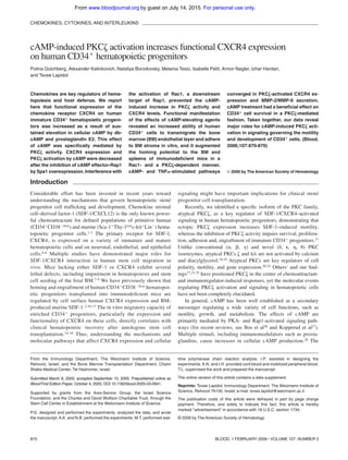
  • 7. Discussion The present study addressed molecular aspects of the regulation of CXCR4 in human hematopoietic progenitors, namely primary normal CD34ϩ cells enriched by MPBL and CB and pre-B ALL G2 cells. Our results showed that CXCR4 expression was increased after elevation of the cellular cAMP level and implicated PKC␨ activity in the cAMP-mediated effects. We showed that in progeni- tor cells, PKC␨ was directly stimulated by cAMP-activated signal- ing. We further found that cAMP-induced and, to a lesser extent, basal CXCR4 expression are PKC␨ dependent because, in the PKC␨-deficient cell line B1, the CXCR4 level was not affected by cAMP stimulation or PKC␨ inhibition, whereas the blocking of PKC␨ activity in CD34ϩ cells completely abrogated membranal CXCR4 expression. Importantly, we found that the inhibition of several other PKC isoenzymes had no effect on CXCR4 expression in hematopoietic progenitors (present work) or on their adhesion, polarization, and chemotaxis.17 Our attempts to elucidate the molecular mechanisms underlying the processes of cAMP-induced CXCR4 elevation led us to Rap1, a member of the Ras family of small GTPases, which is a key regulator of cell polarity, motility, adhesion, and prolifera- tion.26,61,62 Our data indicate that Rap1 activity is required for cAMP-induced CXCR4 up-regulation because the overexpression of Spa1, a principal Rap1GAP in hematopoietic cells, attenuated CXCR4 expression by cAMP. We have also observed reduced membranal PKC␨ translocation in Spa1-overexpressing CD34ϩ progenitors, indicating that interference with Rap1 signaling af- fects PKC␨ activity. During the establishment of cell polarity and intercellular junction formation, atypical PKCs are activated by binding to the Par6/Par3/Cdc42 molecular complex.20,63 Rap1 was positioned upstream of Cdc42 in determining neuronal cell asym- metry mediated by atypical PKC family members.64 It has been shown that Rac1 binds Par6 and potentially activates PKC␨.65 Accordingly, we found that Rac1 inhibition decreased cAMP- induced PKC␨ phosphorylation and had an inhibitory effect on CXCR4 expression identical to that of PKC␨ PS. Therefore, as summarized in the model depicted in Figure 7, our data suggested that Rap1, Rac1, and PKC␨ constitute a signaling pathway of cAMP-induced CXCR4 expression. Our current and previous17 studies also point to a positive feedback loop between PKC␨ activation by cAMP, which results in elevated CXCR4 expression and thus might further enhance PKC␨ activity. We found that the increase in CXCR4 expression in cAMP- stimulated cells resulted from enhanced transcription and, primar- ily, prolonged time of membranal receptor representation. Previous studies29 and our present findings on anti-CXCR4 Ab internaliza- tion suggest that cAMP treatment leads to a decrease in CXCR4 endocytosis. Our preliminary results indicated that PKC␨ inhibition significantly increases membranal CXCR4 turnover; this is under further investigation. Consistent with previous reports, we also observed cAMP-activated CXCR4 mRNA expression attenuated in the presence of PKC␨ inhibitor. We show that CREB phosphoryla- tion was induced by cAMP treatment of CD34ϩ progenitors, whereas PKC␨ inhibition reduced this effect. Given that in T cells cAMP-mediated CXCR4 expression is partially regulated by CREB,31 we suggest that PKC␨ activation increases the CXCR4 Figure 5. cAMP and TNF␣ signaling converge in MMP-2/MMP-9 secretion and induction of CXCR4 expression by CD34؉ progenitors. (A) MMP-2 and MMP-9 levels in the conditioned media of CB and MPBL CD34ϩ cells cultured for 24 hours with the indicated agents, as determined by gelatin zymography. Positive control (contr) is the conditioned medium of HT1080 cells. Representative gel and densitom- etry analysis data are shown as mean Ϯ SD of 3 independent experiments ex- pressed in arbitrary units (AU). Note that the presence of PKC␨ PS (PS␨) abrogates MMP-2/MMP-9 secretion by cAMP and TNF␣ costimulated cells. (B) Membranal CXCR4 expression in MPBL CD34ϩ cells treated as in panel A. Data are shown as mean Ϯ SD of 6 independent experiments. Representative flow cytometry analysis is presented at the bottom. IgG indicates secondary IgG only; CTRL, untreated cells. Figure 6. cAMP increases CD34؉ progenitor cell survival by PKC␨-activated pathway while inhibiting their proliferation. (A) Increased survival of CB CD34ϩ cells cultured for 3 days under cytokine-deprived conditions in the presence of 500 ␮M dbcAMP (cAMP) as analyzed by flow cytometry. Data represented as fold change (mean Ϯ SD of 6 independent experiments each in triplicate) in the number of PI-positive (Dead cells) in cAMP-treated compared with untreated (CTRL) samples. Inset shows representative changes in the proportion of CD34ϩ cells with fragmented DNA (dead cells [%]) cultured for the indicated time periods untreated (CTRL), treated with cAMP, or treated with cAMP and 10 ␮M PKC␨ PS (cAMPϩPS␨). (B) Comparison of the percentage of dividing CD34ϩ cells (mean Ϯ SD of 4 independent experiments) cultured for 3 days in serum-supplemented medium with 500 ␮M cAMP or without (CTRL), fixed, and PI labeled for cell cycle analysis. (C) Ki-67 expression in G2 cells cultured in full growth medium for 3 days either untreated (CTRL) or with 500 ␮M cAMP. Data are shown as mean Ϯ SD of 3 independent experiments. Representative flow cytometry analyses are shown on the right; numbers indicate percentages of Ki-67–positive cells. (D) Immunoblot analysis of ERK1/2 phosphorylation (P-ERK1/2) status compared with the total ERK1/2 level in G2 cells incubated for 24 hours with 500 ␮M cAMP or left untreated (CTRL). Representative of 1 of 3 independent experiments with similar results. 876 GOICHBERG et al BLOOD, 1 FEBRUARY 2006 ⅐ VOLUME 107, NUMBER 3 For personal use only.on July 14, 2015.by guestwww.bloodjournal.orgFrom
  • 8. mRNA level by CREB-mediated transcription. In several cellular systems, CXCR4 expression required NF-␬B activity.66-68 Interest- ingly, however, we found no significant effect of NF-␬B inhibition on CREB phosphorylation or cAMP-induced membranal CXCR4 elevation. Similarly, NF-␬B–independent PKC␨-mediated gene transcription in human epithelial and monocytic cells was demon- strated in response to Toll-like receptor 2 activation.69 Our data also suggest that cAMP-stimulated CXCR4 expression by PKC␨ prob- ably did not require PI3K (Figure S1). Accordingly, it was recently reported that PKC␨ activation by TC10 (a member of the Rho small GTPase family), which mediates insulin-stimulated PKC␨ activa- tion in adipocytes, is PI3K-independent and is, rather, regulated by the guanyl nucleotide exchange factor C3G.70 C3G is also a Rap1-activating protein.71 These findings imply that members of the small GTP-binding protein superfamily might compose a PI3K-independent pathway for PKC␨ activation (Figure 7). Our data suggest that PKC␨ stimulation and CXCR4 expression in cAMP-treated hematopoietic progenitors increased their adhesion and migration in vitro. Indeed, Rap family proteins, effectors of cAMP signaling, are critical regulators of chemokine- induced integrin activation and motility in lymphocytes,61,72 including SDF-1/CXCR4–mediated cell polarization, adhesion, and migration.73,74 It was also shown that along with RhoA, PKC␨ stimulates CCL21-induced adhesion and in vivo homing of lymphocytes.75 Human hematopoietic progenitor cell homing in immunodeficient mice and adhesion to BM endothelial and stromal cells are CXCR4-dependent processes (for a review, see Lapidot and Kollet5). In accordance, we observed facilitated short-term homing of cAMP-treated CD34ϩ cells. However, because of the transient nature of the stimulation by cAMP, reversed within a few hours, we did not detect improved repopulation capacity by these cells. The positive effect of cAMP treatment on homing was abrogated on the inhibition of Rac1 or PKC␨ activity. Our findings imply that functional Rac1 is required for cAMP-induced PKC␨ activation. In murine hematopoietic progenitors, lack of Rac1 results in significant impairment in hematopoietic reconstitution, and the combined Rac1/Rac2 deficiency causes progenitor mobilization.76 Recent reports indicate that a defect in progenitor retention in the stem cell niche at the endosteum occurs in the absence of Rac1 and is more severe in cells lacking both Rac1 and Rac2.77 Reminiscent of our findings on PKC␨ inhibition,17 the injection of the Rac1 inhibitory compound NSC23766 increases the number of circu- lating murine hematopoietic progenitors,77 pointing to the direct role of PKC␨ and RacGTPases in progenitor adhesion and retention. At present, the hematopoietic progenitors of PKC␨ knockout mice have not been characterized. However, PKC␨- deficient mice display profound defects in B-cell development and function,25,78 the processes that are dependent on SDF-1/ CXCR4 signaling. Data from the present study and our previous work17 indicate that a variety of clinical conditions might benefit from approaches targeting PKC␨,79 a key signaling molecule in cAMP- and SDF-1–activated pathways. Numerous studies imply that an increase in cellular cAMP level inhibits immune cell responses to chemotactic stimuli.80,81 In that respect, we found that whereas sustained cAMP elevation activated PKC␨, resulting in increased CXCR4-mediated responses, short (30-minute) priming with dbcAMP antagonized PKC␨ activity and largely inhibited SDF-1–induced PKC␨ phosphorylation, cell polar- ization, and spreading, with no apparent effect on CXCR4 expres- sion (Figure S3). The duality of cell responses to the changes in cellular cAMP might reflect physiologic situations such as inflam- mation, in which chemokines and cytokines act in concert with noncytokine mediators, among them PGE2 and neuromediators, in modulating the immune response.28,82 These ligand-induced changes in cAMP concentration, which vary in duration and amplitude, are integrated and converged on the signaling pathways activated by cytokines, regulating cell trafficking. In accordance, we found that sustained cAMP elevation in CD34ϩ progenitors had additive and synergistic effects on PKC␨ activation by TNF␣, manifested by the increased CXCR4 expression and MMP-2 and MMP-9 production. We further show that cAMP treatment affected progenitor cell survival and proliferation. Cellular cAMP elevation was demon- strated to have an antiapoptotic/survival effect on various experi- mental settings.83-86 Interestingly, enhanced CREB phosphoryla- tion was recently correlated with improved SDF-1–induced survival in the MO7e megakaryocytic cell line.87 In that respect, we observed a significantly better survival of cAMP-stimulated CD34ϩ cells under the cytokine-deprived conditions, and this effect of cAMP was abrogated in the presence of PKC␨ PS or NF-␬B inhibitor (data not shown). These findings, in line with our previous results,17 indicate that cAMP increases hematopoietic progenitor cell survival by activating the PKC␨ pathway. PKC␨ activity is controlled by several adaptor proteins, including the negative regulator Par-4,53 which is shown to antagonize the positive effect of PKC␨ on cell survival.23,88-90 Thus, one may speculate that by decreasing Par-4 expression/function, cAMP stimulates PKC␨ activity and cell survival. In addition, we have found that sustained stimulation with cAMP significantly decreased CD34ϩ progenitor cell proliferation, pointing to a variety of mechanisms affecting the ultimate size of progenitor cell population. It was previously shown that cAMP-elevating agents reduce the proliferation of murine hematopoietic precursors.91 In contrast, deregulated Rap1 activity in Spa-1–deficient mice causes enhanced expansion of the BM hematopoietic progenitors.62 Our data suggest that the effect of cAMP on the cell cycle is mediated by the inhibition of ERK1/2 Figure 7. Schematic representation of a model of the PKC␨ regulation by cAMP and SDF-1/CXCR4. Cellular cAMP accumulation is induced by the ligands (eg, PGE2) that activate G␣s-coupled receptors. Persistent (“prolonged” hours) stimula- tion with the cAMP-elevating agents results in PKC␨ activation in a Rap1/Rac1- dependent manner and a subsequent increase in membranal CXCR4 expression. CXCR4 binding to SDF-1 further activates PKC␨, resulting in a positive feedback loop. On the other hand, SDF-1/CXCR4 interactions might reduce the cellular cAMP level as a result of G␣i activity, establishing a negative feedback loop on the cAMP-induced PKC␨ activation. SDF-1 activates PKC␨ by a distinct from the cAMP, PI3K-dependent pathway.17 PKC␨ activity is required for multiple cellular functions, including MMP-2/MMP-9 secretion, motility, adhesion, and survival. cAMP reduces cell proliferation in a PKC␨-independent manner because of a negative effect on ERK1/2 activity. In contrast to “prolonged” cAMP activation, “short” (minutes) priming with the cAMP-elevating agents antagonizes PKC␨-mediated SDF-1/CXCR4– induced responses. cAMP ACTIVATES PKC␨ IN HEMATOPOIETIC PROGENITORS 877BLOOD, 1 FEBRUARY 2006 ⅐ VOLUME 107, NUMBER 3 For personal use only.on July 14, 2015.by guestwww.bloodjournal.orgFrom
  • 9. activity. Several studies have demonstrated the negative role of Rap1 on cell division and ERK activation,92,93 suggesting that cAMP-induced inhibition of CD34ϩ cell proliferation is Rap1 dependent. Finally, given that we have previously shown that a positive effect of SDF-1 on cell proliferation and ERK activation was mediated by PKC␨,17 we propose that in progenitor cells the cAMP-induced inhibition of cell division and ERK phosphoryla- tion is PKC␨ independent (Figure 7). In conclusion, we present novel evidence of the role and molecular mechanisms of cAMP-induced PKC␨ activation in human immature hematopoietic progenitor cells that result in increased CXCR4 expression and signaling and that affect cell motility and growth. Acknowledgments We thank LoyaAbel andYaronVagima for their excellent technical help and Amiela Globerson for her valuable contribution to manuscript preparation. We also thank Masakazu Hattori and Nagahiro Minato for providing us with the Spa1-GFP–expressing vector.Tsvee Lapidot holds the professional Edith Stein Immunology Chair. References 1. Aiuti A, Webb IJ, Bleul C, Springer T, Gutierrez- Ramos JC. The chemokine SDF-1 is a chemoat- tractant for human CD34ϩ hematopoietic pro- genitor cells and provides a new mechanism to explain the mobilization of CD34ϩ progenitors to peripheral blood. J Exp Med. 1997;185:111-120. 2. Peled A, Petit I, Kollet O, et al. Dependence of human stem cell engraftment and repopulation of NOD/SCID mice on CXCR4. Science. 1999;283: 845-848. 3. Wright DE, Bowman EP, Wagers AJ, Butcher EC, Weissman IL. Hematopoietic stem cells are uniquely selective in their migratory response to chemokines. J Exp Med. 2002;195:1145-1154. 4. Kucia M, Jankowski K, Reca R, et al. CXCR4- SDF-1 signalling, locomotion, chemotaxis and adhesion. J Mol Histol. 2004;35:233-245. 5. Lapidot T, Kollet O. The essential roles of the che- mokine SDF-1 and its receptor CXCR4 in human stem cell homing and repopulation of trans- planted immune-deficient NOD/SCID and NOD/ SCID/B2m(null) mice. Leukemia. 2002;16:1992- 2003. 6. Lataillade JJ, Domenech J, Le Bousse-Kerdiles MC. Stromal cell-derived factor-1 (SDF-1)/ CXCR4 couple plays multiple roles on haemato- poietic progenitors at the border between the old cytokine and new chemokine worlds: survival, cell cycling and trafficking. Eur Cytokine Netw. 2004; 15:177-188. 7. Ara T, Tokoyoda K, Sugiyama T, Egawa T, Kawa- bata K, Nagasawa T. Long-term hematopoietic stem cells require stromal cell-derived factor-1 for colonizing bone marrow during ontogeny. Immu- nity. 2003;19:257-267. 8. Nagasawa T, Hirota S, Tachibana K, et al. Defects of B-cell lymphopoiesis and bone-marrow myelo- poiesis in mice lacking the CXC chemokine PBSF/SDF-1. Nature. 1996;382:635-638. 9. Zou YR, Kottmann AH, Kuroda M, Taniuchi I, Litt- man DR. Function of the chemokine receptor CXCR4 in haematopoiesis and in cerebellar de- velopment. Nature. 1998;393:595-599. 10. Kahn J, Byk T, Jansson-Sjostrand L, et al. Over- expression of CXCR4 on human CD34ϩ progeni- tors increases their proliferation, migration, and NOD/SCID repopulation. Blood. 2004;103:2942- 2949. 11. Kollet O, Petit I, Kahn J, et al. Human CD34(ϩ) CXCR4(Ϫ) sorted cells harbor intracellular CXCR4, which can be functionally expressed and provide NOD/SCID repopulation. Blood. 2002; 100:2778-2786. 12. Kollet O, Spiegel A, Peled A, et al. Rapid and effi- cient homing of human CD34(ϩ)CD38(Ϫ/low) CXCR4(ϩ) stem and progenitor cells to the bone marrow and spleen of NOD/SCID and NOD/ SCID/B2m(null) mice. Blood. 2001;97:3283- 3291. 13. Ponomaryov T, Peled A, Petit I, et al. Induction of the chemokine stromal-derived factor-1 following DNA damage improves human stem cell function. J Clin Invest. 2000;106:1331-1339. 14. Christopherson KW 2nd, Hangoc G, Mantel CR, Broxmeyer HE. Modulation of hematopoietic stem cell homing and engraftment by CD26. Sci- ence. 2004;305:1000-1003. 15. Spencer A, Jackson J, Baulch-Brown C. Enu- meration of bone marrow ’homing’ haemopoietic stem cells from G-CSF-mobilised normal donors and influence on engraftment following allogeneic transplantation. Bone Marrow Transplant. 2001; 28:1019-1022. 16. Voermans C, Kooi ML, Rodenhuis S, van der Le- lie H, van der Schoot CE, Gerritsen WR. In vitro migratory capacity of CD34ϩ cells is related to hematopoietic recovery after autologous stem cell transplantation. Blood. 2001;97:799-804. 17. Petit I, Goichberg P, Spiegel A, et al. Atypical PKC-␨ regulates SDF-1-mediated migration and development of human CD34ϩ progenitor cells. J Clin Invest. 2005;115:168-176. 18. Liu WS, Heckman CA. The sevenfold way of PKC regulation. Cell Signal. 1998;10:529-542. 19. Toker A. Signaling through protein kinase C. Front Biosci. 1998;3:D1134-D1147. 20. Henrique D, Schweisguth F. Cell polarity: the ups and downs of the Par6/aPKC complex. Curr Opin Genet Dev. 2003;13:341-350. 21. Hirai T, Chida K. Protein kinase C␨ (PKC␨): acti- vation mechanisms and cellular functions. J Bio- chem (Tokyo). 2003;133:1-7. 22. Laudanna C, Sorio C, Tecchio C, et al. Motility analysis of pancreatic adenocarcinoma cells re- veals a role for the atypical zeta isoform of protein kinase C in cancer cell movement. Lab Invest. 2003;83:1155-1163. 23. Duran A, Rodriguez A, Martin P, et al. Crosstalk between PKC␨ and the IL4/Stat6 pathway during T-cell-mediated hepatitis. EMBO J. 2004;23: 4595-4605. 24. Laudanna C, Mochly-Rosen D, Liron T, Constan- tin G, Butcher EC. Evidence of zeta protein ki- nase C involvement in polymorphonuclear neu- trophil integrin-dependent adhesion and chemotaxis. J Biol Chem. 1998;273:30306- 30315. 25. Martin P, Duran A, Minguet S, et al. Role of zeta PKC in B-cell signaling and function. EMBO J. 2002;21:4049-4057. 26. Bos JL, de Bruyn K, Enserink J, et al. The role of Rap1 in integrin-mediated cell adhesion. Bio- chem Soc Trans. 2003;31:83-86. 27. Kopperud R, Krakstad C, Selheim F, Doskeland SO. cAMP effector mechanisms: novel twists for an ’old’ signaling system. FEBS Lett. 2003;546: 121-126. 28. Van Miert AS. Present concepts on the inflamma- tory modulators with special reference to cyto- kines. Vet Res Commun. 2002;26:111-126. 29. Cole SW, Jamieson BD, Zack JA. cAMP up-regu- lates cell surface expression of lymphocyte CXCR4: implications for chemotaxis and HIV-1 infection. J Immunol. 1999;162:1392-1400. 30. Cristillo AD, Bierer BE. Regulation of CXCR4 ex- pression in human T lymphocytes by calcium and calcineurin. Mol Immunol. 2003;40:539-553. 31. Cristillo AD, Highbarger HC, Dewar RL, Dimitrov DS, Golding H, Bierer BE. Up-regulation of HIV coreceptor CXCR4 expression in human T lym- phocytes is mediated in part by a cAMP-respon- sive element. FASEB J. 2002;16:354-364. 32. Cristillo AD, Xiao X, Campbell-Malone R, Dimitrov DS, Bierer BE. Differences between CEM and human peripheral blood T lymphocytes in cAMP- dependent HIV viral fusion and CXCR4 expres- sion. Exp Mol Pathol. 2002;73:9-18. 33. Gagliardi MC, De Magistris MT. Maturation of hu- man dendritic cells induced by the adjuvant chol- era toxin: role of cAMP on chemokine receptor expression. Vaccine. 2003;21:856-861. 34. Heystek HC, Thierry AC, Soulard P, Moulon C. Phosphodiesterase 4 inhibitors reduce human dendritic cell inflammatory cytokine production and Th1-polarizing capacity. Int Immunol. 2003; 15:827-835. 35. Salcedo R, Zhang X, Young HA, et al. Angiogenic effects of prostaglandin E2 are mediated by up- regulation of CXCR4 on human microvascular endothelial cells. Blood. 2003;102:1966-1977. 36. Odemis V, Moepps B, Gierschik P, Engele J. In- terleukin-6 and cAMP induce stromal cell-derived factor-1 chemotaxis in astroglia by up-regulating CXCR4 cell surface expression: implications for brain inflammation. J Biol Chem. 2002;277: 39801-39808. 37. Spiegel A, Kollet O, Peled A, et al. Unique SDF-1- induced activation of human precursor-B ALL cells as a result of altered CXCR4 expression and signaling. Blood. 2004;103:2900-2907. 38. Signoret N, Oldridge J, Pelchen-Matthews A, et al. Phorbol esters and SDF-1 induce rapid endo- cytosis and down modulation of the chemokine receptor CXCR4. J Cell Biol. 1997;139:651-664. 39. Harazaki M, Kawai Y, Su L, et al. Specific recruit- ment of SPA-1 to the immunological synapse: involvement of actin-bundling protein actinin. Im- munol Lett. 2004;92:221-226. 40. Nguyen BT, Dessauer CW. Relaxin stimulates protein kinase C Zeta translocation: requirement for cyclic adenosine 3’,5’-monophosphate pro- duction. Mol Endocrinol. 2005;19:1012-1023. 41. Janowska-Wieczorek A, Marquez LA, Nabholtz JM, et al. Growth factors and cytokines upregu- late gelatinase expression in bone marrow CD34(ϩ) cells and their transmigration through reconstituted basement membrane. Blood. 1999; 93:3379-3390. 42. Fossiez F, Banchereau J, Murray R, Van Kooten C, Garrone P, Lebecque S. Interleukin-17. Int Rev Immunol. 1998;16:541-551. 43. Kurachi H, Wada Y, Tsukamoto N, et al. Human SPA-1 gene product selectively expressed in lym- phoid tissues is a specific GTPase-activating pro- tein for Rap1 and Rap2: segregate expression profiles from a rap1GAP gene product. J Biol Chem. 1997;272:28081-28088. 878 GOICHBERG et al BLOOD, 1 FEBRUARY 2006 ⅐ VOLUME 107, NUMBER 3 For personal use only.on July 14, 2015.by guestwww.bloodjournal.orgFrom
  • 10. 44. Foey AD, Brennan FM. Conventional protein ki- nase C and atypical protein kinase C␨ differen- tially regulate macrophage production of tumour necrosis factor-alpha and interleukin-10. Immu- nology. 2004;112:44-53. 45. McConkey M, Gillin H, Webster CR, Anwer MS. Cross-talk between protein kinases C␨ and B in cyclic AMP-mediated sodium taurocholate co- transporting polypeptide translocation in hepato- cytes. J Biol Chem. 2004;279:20882-20888. 46. Maillet M, Robert SJ, Cacquevel M, et al. Crosstalk between Rap1 and Rac regulates se- cretion of sAPPalpha. Nat Cell Biol. 2003;5:633- 639. 47. Arthur WT, Quilliam LA, Cooper JA. Rap1 pro- motes cell spreading by localizing Rac guanine nucleotide exchange factors. J Cell Biol. 2004; 167:111-122. 48. Tunggal JA, Helfrich I, Schmitz A, et al. E-cad- herin is essential for in vivo epidermal barrier function by regulating tight junctions. EMBO J. 2005;24:1146-1156. 49. Gao Y, Dickerson JB, Guo F, Zheng J, Zheng Y. Rational design and characterization of a Rac GTPase-specific small molecule inhibitor. Proc Natl Acad Sci U S A. 2004;101:7618-7623. 50. Gschwendt M, Kittstein W, Johannes FJ. Differ- ential effects of suramin on protein kinase C isoenzymes: a novel tool for discriminating pro- tein kinase C activities. FEBS Lett. 1998;421:165- 168. 51. Johannessen M, Delghandi MP, Moens U. What turns CREB on? Cell Signal. 2004;16:1211-1227. 52. Bleul CC, Fuhlbrigge RC, Casasnovas JM, Aiuti A, Springer TA. A highly efficacious lymphocyte chemoattractant, stromal cell-derived factor 1 (SDF-1). J Exp Med. 1996;184:1101-1109. 53. Moscat J, Diaz-Meco MT. The atypical protein kinase Cs: functional specificity mediated by spe- cific protein adapters. EMBO Rep. 2000;1:399- 403. 54. Esteve PO, Chicoine E, Robledo O, et al. Protein kinase C-␨ regulates transcription of the matrix metalloproteinase-9 gene induced by IL-1 and TNF-alpha in glioma cells via NF-kappa B. J Biol Chem. 2002;277:35150-35155. 55. Byk T, Kahn J, Kollet O, et al. Cycling G1 CD34ϩ/ CD38ϩ cells potentiate the motility and engraft- ment of quiescent G0 CD34ϩ/CD38-/low severe combined immunodeficiency repopulating cells. Stem Cells. 2005;23:561-574. 56. Heissig B, Hattori K, Dias S, et al. Recruitment of stem and progenitor cells from the bone marrow niche requires MMP-9 mediated release of kit- ligand. Cell. 2002;109:625-637. 57. Pruijt JF, Fibbe WE, Laterveer L, et al. Prevention of interleukin-8-induced mobilization of hemato- poietic progenitor cells in rhesus monkeys by in- hibitory antibodies against the metalloproteinase gelatinase B (MMP-9). Proc Natl Acad Sci U S A. 1999;96:10863-10868. 58. Chen W, Yu YL, Lee SF, et al. CREB is one com- ponent of the binding complex of the Ces-2/E2A- HLF binding element and is an integral part of the interleukin-3 survival signal. Mol Cell Biol. 2001; 21:4636-4646. 59. Krakstad C, Christensen AE, Doskeland SO. cAMP protects neutrophils against TNF-alpha- induced apoptosis by activation of cAMP-depen- dent protein kinase, independently of exchange protein directly activated by cAMP (Epac). J Leu- koc Biol. 2004;76:641-647. 60. Stork PJ, Schmitt JM. Crosstalk between cAMP and MAP kinase signaling in the regulation of cell proliferation. Trends Cell Biol. 2002;12:258-266. 61. Kinashi T, Katagiri K. Regulation of lymphocyte adhesion and migration by the small GTPase Rap1 and its effector molecule, RAPL. Immunol Lett. 2004;93:1-5. 62. Kometani K, Ishida D, Hattori M, Minato N. Rap1 and SPA-1 in hematologic malignancy. Trends Mol Med. 2004;10:401-408. 63. Etienne-Manneville S, Hall A. Cell polarity: Par6, aPKC and cytoskeletal crosstalk. Curr Opin Cell Biol. 2003;15:67-72. 64. Schwamborn JC, Puschel AW. The sequential activity of the GTPases Rap1B and Cdc42 deter- mines neuronal polarity. Nat Neurosci. 2004;7: 923-929. 65. Noda Y, Takeya R, Ohno S, Naito S, Ito T, Sumi- moto H. Human homologues of the Caenorhabdi- tis elegans cell polarity protein PAR6 as an adap- tor that links the small GTPases Rac and Cdc42 to atypical protein kinase C. Genes Cells. 2001;6: 107-119. 66. Han KH, Hong KH, Ko J, et al. Lysophosphatidyl- choline up-regulates CXCR4 chemokine receptor expression in human CD4 T cells. J Leukoc Biol. 2004;76:195-202. 67. Helbig G, Christopherson KW 2nd, Bhat-Naksha- tri P, et al. NF-␬B promotes breast cancer cell mi- gration and metastasis by inducing the expres- sion of the chemokine receptor CXCR4. J Biol Chem. 2003;278:21631-21638. 68. Hwang JH, Hwang JH, Chung HK, et al. CXC chemokine receptor 4 expression and function in human anaplastic thyroid cancer cells. J Clin En- docrinol Metab. 2003;88:408-416. 69. Teusch N, Lombardo E, Eddleston J, Knaus UG. The low molecular weight GTPase RhoA and atypical protein kinase C␨ are required for TLR2- mediated gene transcription. J Immunol. 2004; 173:507-514. 70. Kanzaki M, Mora S, Hwang JB, Saltiel AR, Pessin JE. Atypical protein kinase C (PKC␨/␭) is a con- vergent downstream target of the insulin-stimu- lated phosphatidylinositol 3-kinase and TC10 sig- naling pathways. J Cell Biol. 2004;164:279-290. 71. Tsukamoto N, Hattori M, Yang H, Bos JL, Minato N. Rap1 GTPase-activating protein SPA-1 nega- tively regulates cell adhesion. J Biol Chem. 1999; 274:18463-18469. 72. Wittchen ES, van Buul JD, Burridge K, Worthy- lake RA. Trading spaces: Rap, Rac, and Rho as architects of transendothelial migration. Curr Opin Hematol. 2005;12:14-21. 73. McLeod SJ, Li AH, Lee RL, Burgess AE, Gold MR. The Rap GTPases regulate B cell migration toward the chemokine stromal cell-derived fac- tor-1 (CXCL12): potential role for Rap2 in promot- ing B cell migration. J Immunol. 2002;169:1365- 1371. 74. Shimonaka M, Katagiri K, Nakayama T, et al. Rap1 translates chemokine signals to integrin activation, cell polarization, and motility across vascular endothelium under flow. J Cell Biol. 2003;161:417-427. 75. Giagulli C, Scarpini E, Ottoboni L, et al. RhoA and zeta PKC control distinct modalities of LFA-1 acti- vation by chemokines: critical role of LFA-1 affin- ity triggering in lymphocyte in vivo homing. Immu- nity. 2004;20:25-35. 76. Gu Y, Filippi MD, Cancelas JA, et al. Hematopoi- etic cell regulation by Rac1 and Rac2 guanosine triphosphatases. Science. 2003;302:445-449. 77. Cancelas JA, Lee AW, Prabhakar R, Stringer KF, Zheng Y, Williams DA. Rac GTPases differentially integrate signals regulating hematopoietic stem cell localization. Nat Med. 2005;11:886-891. 78. Leitges M, Sanz L, Martin P, et al. Targeted dis- ruption of the ␨PKC gene results in the impair- ment of the NF-␬B pathway. Mol Cell. 2001;8: 771-780. 79. Eaves CJ. SDF-1 tells stem cells to mind their P’s and Z’s. J Clin Invest. 2005;115:27-29. 80. Delgado M, Ganea D. Inhibition of endotoxin-in- duced macrophage chemokine production by va- soactive intestinal peptide and pituitary adenylate cyclase-activating polypeptide in vitro and in vivo. J Immunol. 2001;167:966-975. 81. Laudanna C, Campbell JJ, Butcher EC. Elevation of intracellular cAMP inhibits RhoA activation and integrin-dependent leukocyte adhesion induced by chemoattractants. J Biol Chem. 1997;272: 24141-24144. 82. Rocca B, FitzGerald GA. Cyclooxygenases and prostaglandins: shaping up the immune re- sponse. Int Immunopharmacol. 2002;2:603-630. 83. Chang HS, Jeon KW, Kim YH, Chung IY, Park CS. Role of cAMP-dependent pathway in eosino- phil apoptosis and survival. Cell Immunol. 2000; 203:29-38. 84. Cui Q, So KF. Involvement of cAMP in neuronal survival and axonal regeneration. Anat Sci Int. 2004;79:209-212. 85. Jhala US, Canettieri G, Screaton RA, et al. cAMP promotes pancreatic beta-cell survival via CREB- mediated induction of IRS2. Genes Dev. 2003;17: 1575-1580. 86. Saavedra AP, Tsygankova OM, Prendergast GV, Dworet JH, Cheng G, Meinkoth JL. Role of cAMP, PKA and Rap1A in thyroid follicular cell survival. Oncogene. 2002;21:778-788. 87. Joo EK, Broxmeyer HE, Kwon HJ, et al. En- hancement of cell survival by stromal cell-derived factor-1/CXCL12 involves activation of CREB and induction of Mcl-1 and c-Fos in factor-dependent human cell line MO7e. Stem Cells Dev. 2004;13: 563-570. 88. Berra E, Municio MM, Sanz L, Frutos S, Diaz- Meco MT, Moscat J. Positioning atypical protein kinase C isoforms in the UV-induced apoptotic signaling cascade. Mol Cell Biol. 1997;17:4346- 4354. 89. Diaz-Meco MT, Municio MM, Frutos S, et al. The product of par-4, a gene induced during apopto- sis, interacts selectively with the atypical isoforms of protein kinase C. Cell. 1996;86:777-786. 90. Sanchez P, De Carcer G, Sandoval IV, Moscat J, Diaz-Meco MT. Localization of atypical protein kinase C isoforms into lysosome-targeted endo- somes through interaction with p62. Mol Cell Biol. 1998;18:3069-3080. 91. Rossi GB, Migliaccio AR, Migliaccio G, et al. In vitro interactions of PGE and cAMP with murine and human erythroid precursors. Blood. 1980;56: 74-79. 92. Ishida D, Kometani K, Yang H, et al. Myeloprolif- erative stem cell disorders by deregulated Rap1 activation in SPA-1-deficient mice. Cancer Cell. 2003;4:55-65. 93. Schmitt JM, Stork PJ. PKA phosphorylation of Src mediates cAMP’s inhibition of cell growth via Rap1. Mol Cell. 2002;9:85-94. cAMP ACTIVATES PKC␨ IN HEMATOPOIETIC PROGENITORS 879BLOOD, 1 FEBRUARY 2006 ⅐ VOLUME 107, NUMBER 3 For personal use only.on July 14, 2015.by guestwww.bloodjournal.orgFrom
  • 11. October 4, 2005 originally published onlinedoi:10.1182/blood-2005-03-0941 2006 107: 870-879 Nagler, Izhar Hardan and Tsvee Lapidot Polina Goichberg, Alexander Kalinkovich, Nataliya Borodovsky, Melania Tesio, Isabelle Petit, Arnon hematopoietic progenitors+on human CD34 activation increases functional CXCR4 expressionζcAMP-induced PKC http://www.bloodjournal.org/content/107/3/870.full.html Updated information and services can be found at: (1930 articles)Signal Transduction (3330 articles)Hematopoiesis and Stem Cells (564 articles)Chemokines, Cytokines, and Interleukins Articles on similar topics can be found in the following Blood collections http://www.bloodjournal.org/site/misc/rights.xhtml#repub_requests Information about reproducing this article in parts or in its entirety may be found online at: http://www.bloodjournal.org/site/misc/rights.xhtml#reprints Information about ordering reprints may be found online at: http://www.bloodjournal.org/site/subscriptions/index.xhtml Information about subscriptions and ASH membership may be found online at: Copyright 2011 by The American Society of Hematology; all rights reserved. of Hematology, 2021 L St, NW, Suite 900, Washington DC 20036. Blood (print ISSN 0006-4971, online ISSN 1528-0020), is published weekly by the American Society For personal use only.on July 14, 2015.by guestwww.bloodjournal.orgFrom