Deep Sulcus Sign and Other Key Pulmonary CT Signs
•Descargar como PPTX, PDF•
17 recomendaciones•6,571 vistas

This document discusses several radiographic signs seen on chest imaging: 1. The deep sulcus sign refers to an abnormally deep lateral costophrenic angle on supine chest x-rays and indicates the presence of a pneumothorax. 2. The fallen lung sign describes a collapsed lung that has fallen away from the hilum, either inferiorly and laterally in upright patients or posteriorly in supine patients, and indicates a fractured bronchus. 3. The CT angiogram sign is seen on contrast-enhanced CT scans as enhancing pulmonary vessels within a region of low attenuating lung consolidation and suggests conditions like pneumonia or lung tumors. 4. The flat waist sign is the loss

Recomendados
Recomendados
Más contenido relacionado
La actualidad más candente
La actualidad más candente (20)
Destacado
Destacado (11)
Similar a Deep Sulcus Sign and Other Key Pulmonary CT Signs
Similar a Deep Sulcus Sign and Other Key Pulmonary CT Signs (20)
Más de Minstry of health ,Ibn alnafis hoapital, Damascus
Más de Minstry of health ,Ibn alnafis hoapital, Damascus (9)
Último
Último (20)
Deep Sulcus Sign and Other Key Pulmonary CT Signs
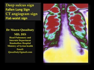
- 1. Deep sulcus sign Fallen Lung Sign CT angiogram sign Flat-waist sign Dr Mazen Qusaibaty MD, DIS Head Pulmonary and Internist Department Ibnalnafisse Hospital Ministry of Syrian health Email: Qusaibaty@gmail.com
- 2. Topic Outline 1. Deep sulcus sign 2. Fallen Lung Sign 3. CT angiogram sign 4. Flat-waist sign 2
- 4. Deep sulcus sign • Refers to a deep lateral costophrenic angle on a supine CXR. 4
- 5. What is your diagnosis?
- 6. Deep sulcus sign • Representing gas collection in a subpulmonary location due to pneumothorax and may be the only sign of it 6
- 7. Deep Sulcus Sign AP position • Bilateral pneumothorax • Lucency of the left lateral costophrenic angle 7
- 8. Supine chest radiograph of a neonate Deep sulcus sign with abnormal deepening and lucency of the left lateral costophrenic angle (∗)8
- 9. ARDS • Deep sulcus sign • Right pneumothorax 9
- 10. Deep Sulcus Sign Left pneumothorax 10
- 11. Conclusion • Deep sulcus sign on a supine radiograph - indicates pneumothorax 11
- 13. Fallen lung sign • This sign refers to the appearance of the collapsed lung occurring with a fractured bronchus
- 14. The bronchial fracture Results in the lung to fall away from the hilum Either inferiorly and laterally in an upright patient 14
- 15. The bronchial fracture Results in the lung to fall away from the hilum Either inferiorly and laterally in an upright patient Or posteriorly, as seen on CT in a supine patient 15
- 16. Fallen lung sign • Either inferiorly and laterally in an upright patient 16
- 17. Fallen lung sign • Or posteriorly, as seen on CT in a supine patient 17
- 18. Conclusion • fallen lung sign - indicates a fractured bronchus
- 20. 20 Terminology • Gray • Low Attenuation • Radiolucent • Skin and Muscles • White • High Attenuation • Radiopaque • Fingres
- 21. The criteria to define the CT angiogram sign are: 1. Pulmonary vessels extending 3 cm or more along a single channel 2. Diffuse homogeneous low attenuation of the consolidated lung parenchyma compared with the attenuation of the chest wall musculature. 21
- 22. The criteria to define the CT angiogram sign • Attenuation of Lung consolidation less than Attenuation of chest wall musculature • 28 HU LC < 74 HU CWM 22
- 23. The CT angiogram sign is seen on contrast material–enhanced scans • Results from the normally enhancing pulmonary vessels
- 24. The CT angiogram sign is seen on contrast material–enhanced scans • Within the low- attenuating consolidated lung parenchyma relative to the chest wall musculature.
- 25. CT Angiogram Sign • The vessels are prominently seen against a background of low- attenuation material.
- 26. CT Angiogram sign The CT angiogram sign was initially described as a specific sign of lobar bronchoalveolar cell carcinoma Specificity can be as high as 92.3% 27
- 27. CT Angiogram sign 1. Pneumonia 2. Pulmonary edema 3. Obstructive pneumonitis due to central lung tumors 4. Lymphoma 5. Metastasis from gastrointestinal carcinomas 28
- 28. Conclusion: CT angiogram sign • Enhancing pulmonary vessels against a background of low attenuation material in the lung 29
- 29. Flat-waist sign المسطح الوسط عالمة
- 30. Flat-waist sign A symmetrical frontal CXR
- 31. Flat-waist sign Refers to the loss of concavity of the left heart border
- 32. Flat-waist sign Due to slight anterior oblique rotation of the heart seen.
- 33. Flat-waist sign Due in some cases of left lower lobe collapse.
- 34. Flat waist Sign
- 35. Flat waist Sign Atelectasis LLL 36
- 36. Conclusion • Flat waist sign- indicates left lower lobe collapse 37
- 37. REFERENCES • 1. Marshall GB, Farnquist BA, MacGregor JH, Burrowes PW. Signs in thoracic imaging. J.Thorac.Imaging 2006;21:76-90 • 2. Webb WR. Thin-section CT of the secondary pulmonary lobule: anatomy and the image—the 2004 Fleischner lecture.Radiology. 2006 May;239(2):322-38 • 3. Austin JH, Muller NL, Friedman PJ, Hansell DM, Naidich DP, Remy-Jardin M, Webb WR, Zerhouni EA. Glossary of terms for CT of the lungs: recommendations of the Nomenclature Committee of the Fleischner Society. Radiology 1996;200(2):327-31 38