Types of atelectasis occupational lung disease-comet tail sign
•Download as PPTX, PDF•
7 likes•1,431 views

Types of atelectasis occupational lung disease comet tail sign

Recommended
Recommended
More Related Content
What's hot
What's hot (20)
Similar to Types of atelectasis occupational lung disease-comet tail sign
Similar to Types of atelectasis occupational lung disease-comet tail sign (20)
Recently uploaded
Recently uploaded (20)
Types of atelectasis occupational lung disease-comet tail sign
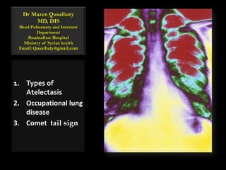
- 1. Dr MazenQusaibatyMD, DISHead Pulmonary and Internist Department Ibnalnafisse HospitalMinistry of Syrian healthEmail: Qusaibaty@gmail.com Types of Atelectasis Occupational lung disease Comet tail sign
- 2. Topic Outline Types of Atelectasis Occupational lung disease Comet tail sign 2
- 3. Types of Atelectasis 3
- 4. 4 Definition of Atelectasis The loss of air in the alveoli Alveoli devoid of air (not replaced) Muller, NL, Fraser, RS, Colman, NC, et al. Radiologic diagnosis of diseases of the chest. Saunders, Philadelphia, 2001.
- 5. 5
- 6. 6
- 7. ObstructiveAtelectasis Atelectasis Right Upper Lobe 7
- 8. 8
- 9. 9
- 10. Relaxation Atelectasis / Mechanical Atelectasis Massive right pneumothorax 10
- 11. 11
- 12. 12
- 13. 13
- 14. 14
- 15. 15 Lead to a diminution in lung volume below the usual resting volume
- 16. Compressive Atelectasis 16 Courtesy of Paul Stark, MD.
- 17. Compressive Atelectasis 17 Courtesy of Paul Stark, MD.
- 18. 18
- 22. Adhesive Atelectasis ARDS is an example of diffuse alveolar atelectasis 22
- 23. 23
- 24. 24 ReplacementAtelectasis Occurswhen the alveoli of an entire lobe are filled by tumor
- 25. Replacement Atelectasis Bronchioloalveolar Cell Carcinoma 25
- 26. 26
- 27. 27 Cicatrization Atelectasis Results from diminution of volume as a sequel of severe parenchymal scarring
- 31. 31
- 32. 32
- 34. Occupational lung disease 34
- 35. Occupational lung disease 35
- 36. Pneumoconiosis Chemical pneumonitis Occupational infection Hypersensitivity pneumonitis Organic dust toxic syndrome 36
- 37. 37
- 38. 38
- 39. 39
- 40. 40
- 41. Chemical pneumonitis The term chemical pneumonitis refers to the aspirationof substances that are toxic to the lower airways, independent of bacterial infection. Mendelson's syndrome. 41 Mendelson, CL. The aspiration of stomach contents into the lungs during obstetric anesthesia. Am J ObstetGynecol 1946; 52:191.
- 42. Occupational infection Viral and Bacterial infections at health workers Parasitic infections : Echinococcus disease 42
- 43. Comet tail sign
- 44. Comets Comets are made up of ice and dust 44
- 45. Comets The center of a comet is called a nucleus and is the only part that is solid. It is made from ice, rock and dust 45
- 46. Comets Usually a comet has 2 tails, a white or yellow dust tail and a blue gas tail. 46
- 54. Rounded Atelectasis It presents as a subpleural mass Segmental or subsegmental atelectasis 54
- 56. 56 Staples CA. Computed tomography in the evaluation of benign asbestos-related disorders. RadiolClin North Am 1992; 30:1191-1207 / Lynch DA, Gamsu G, Ray CS, Aberle DR. Asbestos-related focal lung masses: manifestations on conventional and high-resolution CT scans.Radiology 1988; 169:603-607.
- 57. 57
- 58. 58
- 59. 59
- 60. Rounded Atelectasis All rounded atelectasis is actually round. 60 Lynch DA, Gamsu G, Ray CS, Aberle DR. Asbestos-related focal lung masses: manifestations on conventional and high-resolution CT scans.Radiology 1988; 169:603-607.
- 62. Lentiform
- 63. Or (less often) irregular opacities or attenuation 61 Batra P, Brown K, Hayashi K, Mori M. Rounded atelectasis. J Thorac Imaging 1996; 11:187-197.
- 64. Rounded Atelectasis Volume loss of the affected lobe is uniformly present Often with hyperlucency of the adjacent lung. Serial examination usually shows a stable appearance 62 Batra P, Brown K, Hayashi K, Mori M. Rounded atelectasis. J Thorac Imaging 1996; 11:187-197.
- 65. 63
- 66. 64
- 67. 65
- 68. 66
- 69. 67
- 70. 68 RoundedAtelectasis Low power photomicrograph shows invagination of thickened visceral pleura (arrows). From Colby, TV, Koss, MN, Travis, WD. Tumors of the Lower Respiratory Tract. Armed Forces Institute of Pathology, Washington, DC.
- 71. Rounded atelectasis in the right lung A nodular mass in the right mid-lung (long arrow). 69
- 72. Rounded atelectasis in the right lung A tail-like connection is seen between the mass and the hilum (short arrow). 70
- 73. CT scan of rounded atelectasis in the right lower lobe A subpleural mass in the right lower lobe with tentacle-like extensions towards the hilum (arrow) 71
- 74. Comet tail signBronchovascularbundles (Red arrows) 72
- 76. What is your diagnosis ? Klebsiella pneumonia 74
- 77. Sequential 2-mm, nonenhanced CT scans 75
- 78. Bronchovascular bundles (solid arrow) appear to be pulled into the mass in a curvilinear fashion 76
- 79. Pleural thickening (arrowhead) is present 77
- 80. The major fissure (open arrow) is displaced posteriorly owing to the volume loss in the right lower lobe 78
- 81. CT scans and autopsy findings in an 86-year-old man with silicosis (former metal ore miner). Transverse CT scans obtained with 10-mm collimation. 79 http://radiology.rsna.org/content/236/2/685/F8.expansion.html
- 82. CT scans and autopsy findings in an 86-year-old man with silicosis (former metal ore miner). Lung window scan shows a rounded opacity in the right lower lobe. 80 http://radiology.rsna.org/content/236/2/685/F8.expansion.html
- 83. CT scans and autopsy findings in an 86-year-old man with silicosis (former metal ore miner). Lung volume is reduced 81 http://radiology.rsna.org/content/236/2/685/F8.expansion.html
- 84. CT scans and autopsy findings in an 86-year-old man with silicosis (former metal ore miner) The major fissure is displaced (arrows) 82 http://radiology.rsna.org/content/236/2/685/F8.expansion.html
- 85. Mediastinal window scan at the same level as in a shows a thickened band (arrow) that connects Progressive Massive Fibrosis (PMF) with the thickened pleura. http://radiology.rsna.org/content/236/2/685/F9.expansion.html
- 86. There is a small amount of ipsilateral pleural effusion and multiple calcifications in the PMF http://radiology.rsna.org/content/236/2/685/F9.expansion.html
- 87. RoundedAtelectasis / Silicosis Gross lung specimen from a coronal section shows diffuse pleural thickening in the right lung base and invagination (arrow) into the mass http://radiology.rsna.org/content/236/2/685/F10.expansion.html
- 88. Elastic-Goldner stain, original size / Silicosis Silicotic nodules forming the mass (Blue color) Interstitial fibrosis and atelectasis (Black color) Thickened pleura (arrows) (Blue color) http://radiology.rsna.org/content/236/2/685/F11.expansion.html
- 89. A 50-year-old man with asbestos exposure from working in a brake lining production plant 87
- 91. Curvilinear band opacities in the left lower lung zone88 http://radiographics.rsna.org/content/21/6/1371/F16.expansion.html
- 92. High-resolution CT scan (mediastinal windowing) /Asbestosis pleural plaques on the right side (small white arrows) 89 http://radiographics.rsna.org/content/21/6/1371/F17.expansion.html
- 93. High-resolution CT scan (mediastinal windowing) /Asbestosis Rounded atelectason the left side (large white arrow) 90 http://radiographics.rsna.org/content/21/6/1371/F17.expansion.html
- 94. High-resolution CT scan (mediastinal windowing) /Asbestosis Diffuse pleural thickening (black arrows) on the left side. 91 http://radiographics.rsna.org/content/21/6/1371/F17.expansion.html
- 95. High-resolution CT scan (mediastinal windowing) /Asbestosis Pleural plaques along the diaphragmatic contour (black arrows) 92 http://radiographics.rsna.org/content/21/6/1371/F18.expansion.html
- 96. High-resolution CT scan (mediastinal windowing) /Asbestosis An irregular attenuation pattern, which is typical in rounded atelectasis (white arrows). http://radiographics.rsna.org/content/21/6/1371/F18.expansion.html
- 97. High-resolution CT scan (lung windowing) obtained at the level of the liver dome /Asbestosis A visceral pleural plaque in the right major fissure (arrow) http://radiographics.rsna.org/content/21/6/1371/F19.expansion.html
- 98. High-resolution CT scan (lung windowing) obtained at the level of the liver dome /Asbestosis Curvilinear bands of hyperattenuation in the posterior subpleural area http://radiographics.rsna.org/content/21/6/1371/F19.expansion.html
- 99. High-resolution CT scan (lung windowing) obtained at the level of the liver dome /Asbestosis Rounded atelectasis + posterior displacement of the left major fissure. http://radiographics.rsna.org/content/21/6/1371/F19.expansion.html
- 100. High-resolution CT scan (lung windowing) obtained at the level of the liver dome /Asbestosis The diagnosis of asbestosis was proved at open lung biopsy http://radiographics.rsna.org/content/21/6/1371/F19.expansion.html