
Recomendados
Más contenido relacionado
La actualidad más candente
La actualidad más candente (20)
Similar a Facial Nerve Guide
Similar a Facial Nerve Guide (20)
Último
Último (20)
Facial Nerve Guide
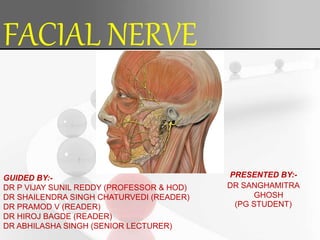
- 1. FACIAL NERVE GUIDED BY:- DR P VIJAY SUNIL REDDY (PROFESSOR & HOD) DR SHAILENDRA SINGH CHATURVEDI (READER) DR PRAMOD V (READER) DR HIROJ BAGDE (READER) DR ABHILASHA SINGH (SENIOR LECTURER) PRESENTED BY:- DR SANGHAMITRA GHOSH (PG STUDENT)
- 2. CONTENTS:- Introduction Key facts Facial Nerve Embryonic Development Surface Marking Facial nerve functional component Origin Nuclei Course Branches and distribution Branches and communication Ganglia associated with the facial nerve Function
- 3. Facial nerve blood supply Applied anatomy Clinical manifestation associated with facial nerve damage Conclusion References
- 4. Seventh cranial nerve Known as queen of face Nerve of the second branchial arch Introduction
- 7. Type – Mixed nerve (motor and sensory) Origin – Pons of brain stem Intracranial branches- Greater petrosal nerve, Communicating branch with otic ganglion, Nerve to stapedius, Chorda tympani KEY FACTS
- 8. Extracranial branches- Post auricular nerve, Branch to posterior diagastric belly Branch to stylomastoid muscles Temporal branch, Zygomatic branch, Buccal branch, Marginal mandibilar branch Cervical branch.
- 9. Field of innervation- Motor : Facial expression muscles, posterior belly of digastric muscle, stylohyoid muscle, stapedius muscle. Special sensory : Taste from anterior 2/3 of the tongue Parasympathetic: Submandibular gland , Sublingual gland, lacrimal glands Clinical relations – Palsy Inferior medial pontine syndrome
- 10. Embryology Pons develops from metencephalon Facial nerve embryonic development
- 11. During first 3 months of prenatal life course, branching pattern and anatomical relationships are established Untill about 4 years of age nerve is not fully developed. At 3rd week of gestation first identifiable nerve tissue is seen (FACIO-ACOUSTIC ) primordium or crest. FACIO-ACOUSTIC primordium gives rise to CN 7th and 8th
- 12. During fourth week:- The facial portion extents to placode The acoustic portion terminates to otocyst. By the end of the 4th week the facial and acoustic portion are more distinct.
- 13. During fifth week:- At early 5th week the geniculate ganglion forms the distal part of primordium. It separates into 2 branches main trunk of facial nerve and chorda tympani.
- 14. During 6th week :- At the end of 5th week the facial motor nucleus is recognizable. The motor nuclei VI and VII cranial nerve initially lie in close proximaty. The internal genu forms as metencephalon, it elongates and CN VI nucleus ascends
- 15. During 7th week:- The geniculate ganglion is well defined and facial nerve root are recognizable Nervus intermedius arises from the geniculate ganglion and passes to brain stem Motor root fibres pass mainly caudal to ganglion Proximal branches form in the 6th week, posterior auricular branch, branch of digastric
- 17. During 8thweek:- Early:- Temporofacial and cervicofacial divisions present . late :- 5 major peripheral Subdivisions present.
- 18. Surface marking Short horizontal line joining :- Middle of the anterior border of the mastoid process. Behind the neck of the mandible
- 20. Functional component Nuclei Distribution Function GVE ( general visceral efferent ) Superior salivatory nucleus (lies in the pons lateral to main motor nucleus of VII) Submandibular and sublingual salivary glands. Preganglionic secretomotor SVE ( special visceral efferent ) Motor nucleus of facial nerve(lies in lower part of pons) Muscles of facial expression,stylohyoid ,posterior belly of digastrics,platysma and stapedius. Facial expression SVA ( special viseral afferent) Nucleus of tractus solitaries (lies in medulla) Taste buds in the ant 2/3rd of the tongue except vallate papillae Taste sensations GSA (general somatic afferent Spinal nucleus of Vth nerve Part of skin of external ear Extroceptive sensation. (pain, temp, touch) Facial nerve functional components :-
- 23. Origin It originates from cerebellopontine angle – lateral part of pontomedullary junction.
- 24. The facial nerve originates from the following nuclei:- 1) Motor nucleus of the facial nerve :- The nerve belongs to the SVE coloum, It lies in the lower part of the pons
- 25. 2) Superior salivatory and lacrimal nuclei (GVE) It lies in the pons lateral to the main motor nucleus of VII and gives rise to secretomotor parasympathetic fibers that pass in greater superficial petrosal nerve and chorda tympani.
- 26. (3) Nucleus of tractus solitarus (SVA):- It lies in the medulla, receives the taste sensation from the anterior 2/3 of the tongue via the central processes of the cells of the geniculate ganglion of the facial nerve
- 27. (4) GSA fibers:- Through these fibers to acoustic meatus & back of auricle through communication from auricular branch of vagus. These fibers terminate in main sensory nucleus & spinal nucleus of 5 th nerve
- 28. Course Intracranial course and branches- Cerebellopontine angle, Internal acoustic meatus (Nervus intermedius joins) Geniculate ganglion (sensory fibres) is deep in internal acoustic meatus Greater petrosal nerve Travels along medial wall of tympanic (middle ear) cavity a) Branch to stapedius (attached to stapes) b) Chorda tympani (This passes anteriorly across tympanic membrane into Infratemporal fossa where it joins lingual nerve) Emerges at stylomastoid foramen.
- 30. Extracranial course and branches:- Outside stylomastoid foramen , 1) Occipital belly of occipitofrontalis, stylohyoid and posterior belly of diagastrics. 2) Cutaneous sensation from skin of external auditory meatus. 3) Nerve enters parotid gland where it forms intricate plexus.
- 34. Branches of distribution • Intracranial :- a. Greater petrosal nerve. b. Nerve to stapedius c. Chorda tympani • Extracranial :- a. Posterior auricular nerve b. Digastrics nerve c. Stylohyoid nerve • Terminal branches:- a. Temporal branch b. Zygomatic branch c. Buccal branch d. Marginal mandibular branch e. Cervical branch
- 37. Geniculate ganglion:- Ganglia associated with the facial nerve Located on the 1st bend of facial nerve, in relation to the medial wall of the middle ear. It is a sensory ganglion. The taste fibers present in the nerve are peripheral processes of pseudounipolar neurons present in the geniculate ganglion.
- 38. Submandibular ganglion:- Parasympathetic ganglion. For relay of secretomotor fibres to the submandibular and sublingual glands.
- 39. Pterygopalatine ganglion:- Parasympathetic ganglion. Secretomotor fibers meant for the lacrimal gland relay in this ganglion.
- 41. Facial nerve blood supply (vasa nervorum) The facial nerve gets its blood from:- 1 Anterior inferior cerebellar artery- At the cerebellopontin angle 2 Labyrinthine artery- Within the internal acoustic meatus. 3 Superficial petrosal-geniculate ganglion and nearby parts. 4.Stylomastoid artery- mastoid segment 5. Posterior auricular artery- Distal to stylomastoid foramen.
- 42. 1 Taste sensation from ant 2/3rd of the tongue and soft palate 2 ( pain ,touch and temperature ) from the auricle ,posterior part of external acoustic meatus and tympanic membrane. 3 Helps in facial expression 4 Controls the secretion of tears ,saliva and mucus in nasal and oral cavities) FUNCTIONS
- 43. Testing of Facial Nerve Branches Testing the Temporal branches of the facial nerve:- To test the function of the temporal branches of the facial nerve, a patient is asked to frown and wrinkle his or her forehead. Testing the Zygomatic branches of the facial nerve:- The patient is asked to close their eyes tightly.
- 44. Testing the buccal branches of the facial nerve :- Puff up cheeks (buccinator) Smile and show teeth (orbicularis oris) Tap with finger over each cheek to detect ease of air expulsion on the affected side
- 45. The marginal mandibular nerve may be injured during surgery in the neck region, especially during excision of the submandibular salivary gland or during neck dissections.
- 46. Applied anatomy Facial nerve paralysis:- Involves paralysis of any structure innervated by the facial nerve. The facial nerve is long and relatively convoluted, so a number of causes may result in facial nerve paralysis . The most common is bell’s palsy
- 47. Motor neuron diseases:- The motor neuron diseases are a hetrogenous group of progressive neurological disorders characterized by degeneration of lower motor neurons, the cells that controls essential voluntary muscle activity such as speaking, walking, breathing and swallowing. The neurons have cell bodies in the cranial nerve nuclei or in the ant horn of the spinal cord and synapse directly on muscle and /or upper motor neurons(which have cell bodies in the brain as synapse on lower motor neuron)
- 48. Upper motor neuron:- Upper motor neuron lesions occur in conditions affecting motor neurons in the brain or the spinal cord such as stroke, multiple sclerosis,traumatic brain injury and cerebral plasy.
- 49. Lower motor neuron:- A lower motor neuron lesion affects nerve fibres travelling from the anterior horn of the spinal cord or the cranial motor nuclei to the relevant muscles.
- 52. Bell’s Palsy:- Most common cause of facial nerve paralysis. There is no known cause of Bell’s Palsy, although it has been associated with herpes simplex infection. Bell’s palsy may develop over several days and may last several months. It is typically diagnosed clinically, in patients with no risk factors for other causes, without vesicles in the ear and with no other neurological signs. Recovery may be delayed in the elderly or in those with a complete paralysis. Bell’s palsy is often treated with corticosteroids.
- 53. Bell's phenomenon:- Is the upward diversion of the eye ball on attempted closure of the lid is seen when eye closure is incomplete.
- 54. Features of Bell’s Palsy:- Unilateral involvement Inability to smile, close eye or raise eyebrow Whistling impossible Drooping of corner of the mouth Inability to close eyelid (Bell’s sign) Inability to wrinkle forehead Loss of blinking reflex Slurred speech Mask like appearance of face Loss/ alteration of taste
- 56. Infection:- 1) Ramsy-Hunt syndrome:- • Reactivation of herpes zoster virus, as well as being associated with bell’s palsy, • Dorsal root ganglion of the facial nerve is associated with vesicles affecting the ear canal and is termed Ramsy Hunt syndrome.
- 58. 2)Otitis media :- • Is an infection in the middle ear which can spread to the facial nerve and inflame it. • Compress the nerve in its canal. • It usually presents in an ear with chronic discharge (otorrhoea) or hearing loss with or without ear pain(otalgia)
- 60. Inflammation The middle ear inflamation can spread to canalis facialis of the temporal bone. The facial nerve travels through this canal together with the statoacousticus nerve. In the case of inflammation, the nerve is exposed to oedema and subsequent high pressure, resulting in a peripheral – type palsy.
- 61. Facial nerve paralysis most commonly during inferior alveolar nerve block:- • It causes due to injection of local anesthesia into the capsule of parotid gland. • It can be prevented by using proper technique and by avoiding insertion of needle into the parotid gland. • Transient treatment ,self correcting within 3 hrs or less.
- 62. Clinical condition /syndrome Symptoms Site of lesion /possible cause(s) Crocodile tears syndrome Also called Bogorad syndrome Gustatolacrimal reflex Paraoxymal lacrimation Spontaneous tearing in parallel with the normal salivation of eating Facial paralysis (due to injury to facial nerve proximal to the geniculate ganglion ), when nerve fibres destined for a salivary gland are damaged and by accident regrow to tear gland. Bilateral facial palsy Flat,expressionless,drooling face Bulbar lesion Marcus gunn phenomenon Nursing infants will have rhythmic upward jerking of their upper eyelid (first described by marcus gunn in 1883) An autosomal dominance condition with incomplete penetrance Congenital ptosis Millard –gubler syndrome Also known as ventral pontine syndrome. Paralysis of the nerve- leads to diplopia, loss of power to rotate the affected eye outwards Disruption of the Facial nerve (VII cranial nerve )leads to the symptoms including flaccid paralysis of the muscles of facial expression and loss of the corneal reflex Damage of the VII nerve in pons a unilateral lesion of the ventrocaudal pons may involve the fibres of VI and VI cranial nerve. Clinical manifestation associated with facial nerve damage:-
- 63. The symptoms according to the level of injury of facial nerve:- At internal auditory meatus; loss of lacrimation, stapedial reflex, taste from most of anterior two-third of tongue, lack of salivation and paralysis of muscles of facial expression. Below geniculate ganglion; loss of stapedial reflex, taste from anterior two- third of tongue, lack of salivation and paralayis of facial expression muscles. Region b/w nerve to stapedius and chorda tympani : loss of taste from anterior two-third of tongue, lack of salivation and paralysis of facial expression muscles. Region below stylomastoid foramen : paralysis of facial expression muscles.
- 65. Facial nerve, the seventh cranial nerve supplies the submandibular, sublingual, lacrimal glands, the mucosal glands of the nose, palate, pharynx and taste fibres, and on being injured it leads to loss of lacrimation, loss of salivation, loss of taste sensation and paralysis of the muscles of facial expression. Hence a complete understanding of its anatomy is essential and care should be taken during surgical procedures. Conclusion
- 66. Refrences 1. Chaurasia’s BD Human Anatomy- Vol 3. 5th edition. CBS Publishers and Distributors Pvt. Ltd. 337-370 2. Monkhouse Stanley cranial nerves 1st edition Cambridge University Press,2006 functional anatomy. 3. DM Kadasne Kadasne’s textbook of anatomy volume 31st edition 1st Jaypee brothers medical publishers (p) ltd. New Delhi. . 996-1001 4. Sembulingam k, Sembulingam p. Essentials of medical physiology, 3rd edition. Jaypee brothers medical publishers (p) ltd. New Delhi. P619-627 5. Singh DR essentials of anatomy for dentistry students.1st edition. Wolters Kluwer India Pvt. Ltd. New Delhi. 291-297. 6. G.J.Romanes Cunningham’s manual of practical anatomy 15th edition volume 3 head, neck and brain oxford medical publications 7. Pal GP Illustrated textbook of Neuroanatomy 8. Sinnatamby chummy s. Last’s anatomy regional and applied 10th edition.