

Recomendados
Recomendados
Más contenido relacionado
La actualidad más candente
La actualidad más candente (20)
Similar a Nasopharyngeal carcinoma
Similar a Nasopharyngeal carcinoma (20)
Más de Springer
Más de Springer (20)
Último
Último (20)
Nasopharyngeal carcinoma
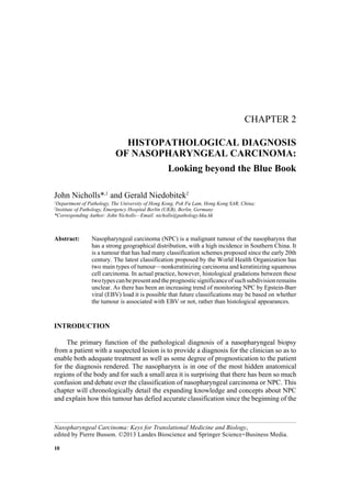
- 1. 10 CHAPTER 2 HISTOPATHOLOGICAL DIAGNOSIS OF NASOPHARYNGEAL CARCINOMA: Looking beyond the Blue Book John Nicholls*,1 and Gerald Niedobitek2 1 Department of Pathology, The University of Hong Kong, Pok Fu Lam, Hong Kong SAR, China; 2 Institute of Pathology, Emergency Hospital Berlin (UKB), Berlin, Germany *Corresponding Author: John Nicholls—Email: nicholls@pathology.hku.hk Abstract: Nasopharyngeal carcinoma (NPC) is a malignant tumour of the nasopharynx that has a strong geographical distribution, with a high incidence in Southern China. It is a tumour that has had many classification schemes proposed since the early 20th century. The latest classification proposed by the World Health Organization has two main types of tumour—nonkeratinizing carcinoma and keratinizing squamous cell carcinoma. In actual practice, however, histological gradations between these twotypescanbepresentandtheprognosticsignificanceofsuchsubdivisionremains unclear. As there has been an increasing trend of monitoring NPC by Epstein-Barr viral (EBV) load it is possible that future classifications may be based on whether the tumour is associated with EBV or not, rather than histological appearances. INTRODUCTION The primary function of the pathological diagnosis of a nasopharyngeal biopsy from a patient with a suspected lesion is to provide a diagnosis for the clinician so as to enable both adequate treatment as well as some degree of prognostication to the patient for the diagnosis rendered. The nasopharynx is in one of the most hidden anatomical regions of the body and for such a small area it is surprising that there has been so much confusion and debate over the classification of nasopharyngeal carcinoma or NPC. This chapter will chronologically detail the expanding knowledge and concepts about NPC and explain how this tumour has defied accurate classification since the beginning of the Nasopharyngeal Carcinoma: Keys for Translational Medicine and Biology, edited by Pierre Busson. ©2013 Landes Bioscience and Springer Science+Business Media.
- 2. 11HISTOPATHOLOGICAL DIAGNOSIS OF NASOPHARYNGEAL CARCINOMA 20th century. It will explain the rationale behind the current World Health Organization classification of NPC and detail the strengths as well as concerns over this classification. The pathologist has a vital role to play in the diagnosis of NPC, both in the primary diagnosis and in the identification of tumour relapses. As NPC occurs in a worldwide distribution, the desirability of a universally satisfactory and acceptable classification schemeisobvioussothatdiagnosiscanbestandardizedandthatcomparisonofdiagnostic, therapeutic and other studies can be comparable from investigators around the world. For most pathologists there have been two series of publications available worldwide thathaveattemptedtoprovidesomedegreeofuniformityontheclassificationoftumours. The Armed Forces Institute of Pathology (AFIP) based in the United States of America has provided a series of fascicles and the International Agency for Research on Cancer (IARC) under the umbrella of World Health Organization (WHO) has also published another series which have colloquially been referred to as the ‘blue books’. In 2005 the latest update to the latter series dealing with tumours of the head and neck was produced.1 This chapter will also critically appraise the latest classification of NPC. EARLY CLASSIFICATION SCHEMES FOR NPC The development of a classification scheme for nasopharyngeal carcinoma has been attempted many times since the early 20th century. Until the early 1900s NPC was consideredtobeararetumourandreferencestothislesionwereusuallyabsentintextbooks of clinical medicine or surgery. Only since the late 1960s has the literature on this unique tumourelucidateditspeculiarclinical,pathological,radiographic,epidemiologic,serologic and therapeutic features. These studies have shown that nasopharyngeal carcinoma is composedofseveralsimilarbutmorphologicallydifferentcarcinomas,thatcertaintypesof this tumour have their highest incidence in populations of Oriental descent, that there are probable environmental factors of significance and that certain types of nasopharyngeal carcinoma are associated with Epstein-Barr virus (EBV). In the early 1900s, the classification of malignant nasopharyngeal neoplasms was simple; a distinction was made only between those arising from the surface epithelium (epitheliomas) and lymphosarcomas arising from the submucous lymphoid tissue. The first report of NPC in the English literature was in 19012 with a detailed clinical series published ten years later3 but a histological classification of the carcinomas was first attempted in 1903 by Citelli and Calamida (reviewed in ref. 4) who initially divided NPC into three groups: a mixed carcinoma, a pure cylindric carcinoma arising from glands and a pure squamous cell carcinoma. This classification was expanded by dividing the cylindric carcinomas into two different groups, carcinoma cylindrocellulare solidum and adenomatosum arising from the surface epithelium and glandular epithelium respectively (reviewed in ref. 4). The carcinomas were further subdivided in 1922 by Duval and Laccasagne into degrees of differentiation but most American and English authorsadoptedtheBrodersclassificationpublishedlater.Thedescriptivetermsproposed by Bang (carcinoma basocellulare, carcinoma planocellulare mucous membrane type, paraketatoticum and cornescens) emphasized the descriptive limits to which the 4 stages of differentiation were being promoted (reviewed in ref. 4). The lymphosarcomas were initally separated from the epitheliomas but later it was realized that the tumours called lymphosarcomas were actually lymph node metastases resulting from a primary tumour located in the nasopharynx. For instance, one of the
- 3. 12 NASOPHARYNGEAL CARCINOMA first studies in Chinese patients in 1923 documented 90 cases of cervical lymphosarcoma but later review considered these to be lymph node metastases of NPC. A new tumour with an intimate relation between epithelial cells and lymphoid tissue was reported in 1921 in two simultaneous works—one in France by Reverchon and Coutard, inspired by Regaud, the other in Germany by Schmincke, the Munich pathologist (reviewed in ref. 4). Both authors stressed the unique histological picture of the tumour and the unique sensitivity to radiotherapy. The tumour was called lymphoepithelioma as there was an intimate mixture of large polygonal cells and lymphoid cells in a syncytial character. The Regaud morphology was defined as consisting of well circumscribed strands of epithelial cells with large, pale staining vesicular nuclei and poorly delineated cytoplasm, embedded in a stroma more or less rich in lymphocytes. The nuclei were round and nucleoli were prominent. No features of keratinization could be identified. Within the epithelial groups were small nuclei considered to be lymphocytes. Sometimes tumour cell nests were separated by a fibrous stroma. The Schmincke type, in contrast, contained epithelial cells in irregular anastomosing trabeculae of ill-defined cells with large vesicular nuclei. In many places the appearance of the epithelial columns was lost and the cells became dissociated from one another giving rise to a reticular mass of round, oval or polygonal cells. All stages of transition could be found between the epithelial cells forming trabeculae and those in the syncytial type masses and it therefore seemed that the two classical types were not separate but actually merged into one another. Only a few years after Regaud and Schmincke’s publication another type of radiosensitive tumour was described by Quick and Cutler5 which was called transitional cell carcinoma. They identified a group of patients with intraoral carcinomas which were both susceptible to radiation treatment and had a peculiar histological appearance— lacking the usual features of squamous cell carcinoma. These cells were small, uniform in size with a relatively large hyperchromatic nucleus and scanty cytoplasm, closely packed with little intercellular ground substance. The cells formed solid sheets, growing in anastomosing columns of opaque granular polyhedral cells with convolutions. Flat, pavementcharacteristics,spines,keratinizationandpearlformation(indicativeofsquamous differentiation) were absent. The authors considered this condition to be markedly different from the routine squamous cell carcinoma of the intraoral region but only 2 of the cited cases were from the nasopharynx. Though the term ‘transitional’ referred to an epithelium not seen in the nasopharynx, the radiosensitivity of this tumour and of the cervical gland metastases6 resulted in this tumour being associated more with the lymphoepithelioma (an entity of which apparently Quick and Cutler were unaware of ) than squamous cell carcinoma. Later Cutler characterized the transitional cell carcinoma as a lymphoepithelioma without the lymphoid elements. In 1929, Ewing addressed the problem of the lymphoepithelioma7 and stated that an epidermoid carcinoma, or transitional cell carcinoma, when associated with lymphoid tissues (as in the case of the nasopharynx) became difficult to separate from lymphoepithelioma and that the separation of the latter tumour from the others remained a somewhat arbitrary decision. Cappell in 19348 and 19389 came to the same conclusion. The transitional cell carcinoma could be distinguished from the other two groups by a more obvious origin from the surface of the epithelium and the pattern of growth. The cells formed broad alveoli in which central necrosis and degeneration were more common. Yet again there was no trace of keratinization in that the cells were devoid of
- 4. 13HISTOPATHOLOGICAL DIAGNOSIS OF NASOPHARYNGEAL CARCINOMA intercellular bridges and squamous pearls. There was an overall absence of the intimate mixture of the lymphocytes with the tumour cells as seen in the ‘lymphoepithelioma’. Even so, he as well as other pathologists of the time found difficulty in separating the transitional cell carcinoma from lymphoepithelioma. Even though Cappell thought that the lymphoepithelioma was more common than the transitional cell carcinoma he considered that there was great interobserver error. The place for transitional cell carcinoma in the 1940s still remained unclear—several workers claimed that it was inseparable from lymphoepithelioma whilst others saw it as a squamous cell carcinoma of low grade differentiation. Clearly there was also much variation between pathologists; the studies of Salinger and Pearlman10 had a number of nasopharyngeal neoplasms judged microscopically by 3 independent pathologists, all of whom came to divergent results concerning the tumours of low grade differentiation. Theseparationofnasopharyngealcarcinomaintothethreeelementslymphoepithelioma, transitionalcellcarcinomaandsquamouscellcarcinomapersistedintothe1950s.Thomson11 reinforced previous observations in his studies on transitional cell carcinoma in that he documented the appearance of central necrosis and an origin from the surface epithelium. He also agreed that though attempts were made to separate transitional cell carcinoma from lymphoepithelioma and lymphosarcoma it was difficult. He considered that intercellular bridges were useful for the diagnosis of epidermoid carcinoma and silver stains for reticulin were helpful for separating lymphoepithelioma from lymphosarcoma. To highlight the difficulty in diagnosis he described one patient who had a diagnosis of lymphosarcoma made which was then changed to epidermoid carcinoma, malignant atypical epithelial cells and finally lymphoepithelioma. Within the 1930s and 1940s the high frequency of NPC in Chinese people became more evident. Originally 90 cases of ‘cervical lymphosarcoma’ were described in 1923 but these were actually considered on further review to be lymph node metastases of nasopharyngeal carcinoma (reviewed in ref. 4). In 1935 Ch’eng12 described 7 clinical cases of lymphoepithelioma and in 1940 Ch’in and Szutu 90 cases.13 Professor Digby from Hong Kong published 240 cases of nasopharyngeal carcinoma occurring in the Chinese of Hong Kongandnotedthattherewasamalepredominance.14 Inhisopinionthegrowthsoriginated fromthecolumnarepitheliumliningtheupperpartofthenasopharynx.Thepresenceofboth lymphoid and epithelial elements was not observed in metastases other than lymph nodes. Teoh in 1957 studied the histopathological features of nasopharyngeal carcinoma in a large autopsy series and noted that within the tumour there was much morphological variation.15 In one case described (Case 1) he stated that in some areas of the tumour there was cornifying squamous cell carcinoma whilst in others there was undifferentiated carcinoma containing both small and large cells in ill defined clumps mixed with lymphocytes and plasma cells. The metastasis from this primary growth showed similar features to the primary growth but without areas of keratinization. In the liver metastases there were nests and trabeculae of closely packed cells without clear cell boundaries. His histological studies of the primary growths gave clear evidence that all the tumours were carcinomas and in 4 of the 31 cases showed frank epidermoid features. In one case glycogen containing clear cells were seen. Tumours similar to the lymphoepithelioma of Schmincke and Regaud were identified and a transition to the transitional cell carcinoma of Quick and Cutler was documented in one case—indicating that pathologists were probably dealing with one type of tumour and not three. In cases where there were metastases to nonlymphoid tissue there were tumour cells only and not lymphocytes,
- 5. 14 NASOPHARYNGEAL CARCINOMA even though lymphocytes were present in the primary growth and in cervical lymph node metastases. Teoh’s observation concluded that lymphocytes were not true components of the tumour and that their association with tumour cells in sites containing lymphoid tissue was incidental. In 1962 Shu Yeh from Taiwan published a large (1,000 cases) series of biopsies from patients with nasopharyngeal carcinoma and divided the tumours into three groups; carcinomas, sarcomas and carcinosarcomas.16 The carcinomas were subdivided into 7 categories depending on histological appearances. Yeh believed that the entity lymphoepithelioma did not really exist and that the 2 criteria which had been used in the past; radiosensitivity and the intimate mixture of lymphocytes were not substantiated. Even though he found that his series of lymphoepitheliomas had a better 5 year survival than the other tumours, because he was very rigid in his inclusion of what should be called a lymphoepithelioma he considered that the low numbers made an accurate assessmentofprognosisdifficult.Heconsideredthelymphoepitheliomatobenothingbut a transitional cell carcinoma originating from deeper crypts of the epithelium, growing down to deeper layers on one hand and infiltrating the superficial lymphoid tissues on the other. As he also found the admixture of lymphocytes and tumour cells to be present in adenocarcinomas, in his opinion the separation of lymphoepithelioma into a separate category was not justified. Liang, Zhong and others from Canton17 classified NPC’s into three groups, undifferentiated, poorly differentiated and well differentiated which they believed also reflected their biological behaviour; the undifferentiated carcinomas showing cranial base invasion and distant metastases, poorly differentiated either spreading to the cranial base or to lymph nodes and squamous cell carcinoma which usually neither invaded the cranial base nor metastasized distally. An electron microscopic study of 3 different types of undifferentiated carcinoma was done by Svoboda18 and this showed cytoplasmic keratin fibrils, features indicative of squamous differentiation. Gazzolo19 looked at normal and abnormal nasopharyngeal epithelium and identified keratin fibrils, tonofilaments and desmosomes in the tumour cells. When the tumour was undifferentiated there were fewer keratin fibrils, but the presence of desmosomes confirmed the epithelial nature of these tumours. Various types of nuclear bodies were also noted but no Herpes type viral particles were identified. Prasad20 examined nasopharyngeal carcinoma specimens using transmission electron microscope and confirmed the presence of desmosomes in all cases, tonofilaments in all but 3 cases, rare secretory granules and cytoplasmic inclusion bodies. His conclusion was that the normal pseudostratified columnar epithelium had undergone metaplastic change to the squamous type before undergoing malignant transformation. Michaels and Hyams21 examined 6 cases and the electron microscopic features correlated well with the histopathological findings. Ultrastructurally, the vesicular nuclei seen histologically revealed small deposits of chromatin beneath the nuclear membrane but little or no chromatin elsewhere. Nucleoli were prominent. Cell processes were not distinct and the cells did not appear to show a definite cell border but appeared to merge into one another. As with previous studies desmosomes were present but tonofilaments uncommon. Both of these investigators, as well as Prasad, attributed the nuclear changes to an increased metabolic activity of the cells. The conclusion was that the undifferentiated carcinoma was a form of squamous cell carcinoma showing minimal differentiation and evidence of high metabolic activity.
- 6. 15HISTOPATHOLOGICAL DIAGNOSIS OF NASOPHARYNGEAL CARCINOMA THE FIRST INTERNATIONAL CLASSIFICATION In 1978 the World Health Organisation proposed a histopathological classification whichdividednasopharyngealcarcinomasintothreecategories;squamouscellcarcinoma, nonkeratinizingcarcinomaandundifferentiatedcarcinoma.22 WHOcorrectlyemphasized, by definition, that NPC was a malignant neoplasm that has its origin from the epithelial layer of the nasopharynx, an epithelial layer that might be stratified squamous type, ciliated respiratory type, or some gradation between these two extremes. Squamous Cell Carcinoma This was a recognized morphologic category of nasopharyngeal carcinoma and has also been labelled WHO Category I. When this entity was proposed in 1978, in geographic areas of low incidence for nasopharyngeal carcinoma, SCC accounted for approximately 25 percent of all nasopharyngeal carcinomas in Caucasian patients but in Orientals living in USA the frequency was 10% or lower. Patients with squamous cell carcinoma of the nasopharynx, in these low incidence regions tended to have a serologicalantibody“profile”toEpstein-Barrvirusthatwassimilartonormal“controls”, though this was not absolute. Nonkeratinizing Carcinoma Nonkeratinizing carcinoma of the nasopharynx (WHO Category II), was microscopically identified as a tumour which is neither anaplastic or undifferentiated nor keratinizing. By description, this tumour produced neoplastic epithelium that was “transitional” in type. It often resembled transitional cell carcinoma of the urinary bladder. Cytologically, the cells were of moderate size, variable between polygonal and spindled and variable in differentiation. Nonkeratinizing carcinomas that were poorly differentiated often had the appearance of the tumour described by Regaud. Many pathologists and authors have suggested that nonkeratinizing carcinoma is but a variant of undifferentiated carcinoma, recognizing that some nonkeratinizing carcinomas are cytologicallybland(andnotundifferentiated).SerologicstudiesofEBVantibodiesshow a similar elevated profile of this tumour with undifferentiated carcinoma suggesting that nonkeratinizing carcinoma should be considered separate from the squamous cell carcinoma. In low incidence North American geographic regions, nonkeratinizing carcinoma accounts for 12 percent of all nasopharyngeal carcinomas. Undifferentiated Carcinoma Undifferentiatednasopharyngealcarcinoma(WHOCategoryIII)isthemostcommon type of nasopharyngeal carcinoma, accounting for approximately 63 percent of all nasopharyngealcarcinomasinlowincidenceareasandaccountingforupto98%percentin highincidenceareas.Itisrecognizedasacarcinomathatfeaturescellswithdistinctcytological characteristics;specificallycellswithsingleprominentnucleoli,indistinctcytoplasmanda lack of discernable cytoplasmic outline. The result is a tumour which appears to grow as a syncytiumincontrasttosquamouscellcarcinoma.Manyundifferentiatedcarcinomasincite a reaction of T-lymphocytes. As described above, this lymphocytic response has led to the term lymphoepithelioma, a term which is descriptively inaccurate as the lymphocytes are
- 7. 16 NASOPHARYNGEAL CARCINOMA not neoplastic. If “lymphoepithelioma” is used in a diagnostic manner, it should be used parenthetically, since these are but variants of undifferentiated nasopharyngeal carcinoma. The term is deeply engrained in pathological literature, however and it is unlikely to be abandonedinthenearfuture.Ofinterest,undifferentiatednasopharyngealcarcinomaswith alymphoepitheliomapatternaresomewhatuniquetocertainanatomicregions,particularly the region of Waldeyers’s ring and the thymus. However, other organ systems (lung, skin, stomach, uterine cervix, breast and bladder) have produced undifferentiated carcinomas with a lymphoid stroma that are microscopically similar to lymphoepithelioma of the nasopharynx. Polymerase chain reaction and in situ hybridization of undifferentiated carcinomas from some of these some of these body sites have been shown to harbour the EB viral genome within the malignant epithelial cells. At the same time as the 1978 WHO classification was being formulated a group of European pathologists met to discuss the classification of NPC. They agreed that the conceptoflymphoepitheliomashouldbepreservedasitdescribedanentitydifferentfrom squamous cell carcinoma, a reliable and reproducible definition of lymphoepithelioma could be given but that the term lymphoepithelioma was misleading and should be replaced by the term undifferentiated carcinoma of nasopharyngeal type (UCNT). This term would have both a solid cell type (Regaud), isolated cell type (Schmincke) and a spindle cell type. The interobserver agreement on this classification was between 80% and 90%. The other category of squamous cell carcinoma was divided into the traditional well differentiated, moderately differentiated and poorly differentiated carcinoma.23 The simplified classification of NPC into squamous cell carcinoma and UCNT showed significant correlation with the EBV serology. The patients with squamous cell carcinoma had low levels of EBV serology while patients with UCNT had elevated titres.24 Though this finding may have stood ground in Caucasian patients the same cannot be said for Asian patients in whom the EBV genome or its products have been detected in squamous cell carcinoma, though in lower numbers. Thus it appears that in Caucasian patients UCNT and SCC may be two distinct disease processes but in Asian patients, the 2 morphological patterns can be seen within the same tumour specimen. The French classification believed that the WHO Type II (nonkeratinizing carcinoma) was not a genuine entity according to EBV serology and a two tier classification of NPC into differentiated and undifferentiated carcinoma appeared to be the most favourable if a classification based on clinical relevance and serology was to be used.25 THE 1991 WHO MODIFICATION The 1991 modification of the WHO Classification26 attempted to include some of the findings by European pathologists.24 The main change was that undifferentiated carcinoma was placed as a subset of nonkeratinizing carcinoma and that the former nonkeratinizing carcinoma was called nonkeratinizing differentiated carcinoma. It is of interest that one of the criteria used for classification in this group was similar to that proposed in the early 1900s—the sensitivity to radiotherapy and that what was called epithelioma in 1903 was now called squamous cell carcinoma (keratinizing squamous cell carcinoma). Another feature of the revised classification was the opinion that the nonkeratinizing tumours have a stronger relationship with the Epstein-Barr virus, a feature which has geographical variation. The 1991 modified scheme still placed what may be considered an undue emphasis on the term lymphoepithelioma. Pathologists
- 8. 17HISTOPATHOLOGICAL DIAGNOSIS OF NASOPHARYNGEAL CARCINOMA should be aware that this histological subtype is only seen in less than 10% of cases in an endemic area and that the presence of a small number of lymphocytes does not imply that the tumour is a lymphoepithelioma. THE 2005 WHO CLASSIFICATION This update to the 1991 WHO classification still maintains the separation between nonkeratinizing carcinoma and keratinizing squamous cell carcinoma but includes as a separate entity a rare tumour called basaloid squamous cell carcinoma. The alternative labelling of the tumours into Types I, II and III has not been emphasized. Within this simplified classification there are a number of statements that bear further investigation, of which the most important relates to the significance of subclassification, as the 2005 classificationstatesthatwithinthenonkeratinizingcarcinoma“…subclassificationintothe undifferentiated and differentiated types is optional, since their distinction is of no clinical orprognosticsignificanceanddifferentareasofthesametumourordifferentbiopsiestaken at different time intervals from the same patient may exhibit features of one or the other subtype …”.1 As the main reason for classification is to determine treatment options and prognosis,thisstatementimpliesthatprognosisofnonkeratinizingNPCsisnotdetermined by whether the tumour has a lymphoid stroma or is transitional in appearance (Fig. 1). Figure 1. The many faces of nonkeratinizing carcinoma. The tumour may be composed of small isolated tumour cells set among a lymphoid stroma (A). In this case immunohistochemistry for the epithelial marker AE1/AE3 delineates the tumour cells (B). The tumour cells may appear slightly more cohesive (C) and then form nests (D). A B C D
- 9. 18 NASOPHARYNGEAL CARCINOMA Giventhattherehasbeenconsiderableconfusionsincethe1900sovertheclassification of NPC it is not surprising that the data and results of response to treatment have led to uncertainty regarding whether there is a relationship between the histological subtype of NPC and prognosis. As there have been many classification schemes it is difficult to compare studies as the criteria for sub-classifying a tumour vary from study to study. The2005classificationcorrectlystatesthat“…thedensityoflymphocytesandplasma cells is highly variable. At one extreme, there are no or few lymphocytes within the tumour islands … and … at the other extreme abundant lymphocytes and plasma cells infiltrate the tumour islands, breaking them up into tiny clusters or single cells and obscuring the epithelial nature of the tumour …”.27 Thus from a purely morphological point of view the justification of Regaud versus Schminke does not appear warranted, but is there a relation between lymphoid infiltrate and prognosis? In 1979 Shanmugaratnam et al stated that the 3 year survival rate for tumours with a marked lymphocytic infiltrate (45.8%) was significantlybetterthanthosewithamoderatelymphocyticinfiltrate(32%)andthosewithno lymphocytic infiltrate (20.7%).28 However, as this study was done before CT scanning was routinely performed for accurate staging the real significance of these findings is not clear. In 1986 a report from Japan by Nomori et al focused not on the lymphocytes but the histiocytic infiltrate in NPC. Using the antibodies S100 and lysozyme, the survival of patients with NPC was related to the density of T-zone histiocytes (Langerhan’s cells and their precursors); the more S100 positive cells there were within the tumour, the better the prognosis. The intensity of lysozyme positive cells however was not considered to be related to prognosis.29 This finding, however, was quickly refuted by a group of European pathologists30 who found no statistical correlation between the number of S100 positive cells and patient survival but upheld by another European group led by Giannini.31 These authors first looked at lymphocytic infiltration; finding it to be of no prognostic significance but cases in which a moderate to marked density of dendritic and monocyte/macrophage cells were present showed a prolonged survival. Studies from the Guangzhou region in Southern China27 have looked in detail at the stromal response or reaction to the tumour and the findings are in closer agreement with Nomori et al and Giannini et al. This study has shown prognostic significance of the undifferentiated carcinoma when the infiltrate amongst the tumour cells is considered. When the stroma contained abundant lymphocytes, the 5 year survival rate was 59.5%, which was higher than that of the moderate lymphocytic type (51.5%, or when scanty lymphocytes were present (40.1%). This study in effect states that the Schmincke pattern withanabundantlymphocyticinfiltratewithinthetumourshoulddobetterthantheRegaud typeandnotonlythelymphocyticinfiltrationbutalsotheinfiltrationofmacrophages(using lysozyme and S100 protein immunohistochemistry) appears to confer a better prognosis to the patient. The plasma cell population is also not insignificant as the VCA-IgA titre rises with the intensity of the plasma cell infiltrate. Tumours with abundant plasma cells have a geometric mean titre (GMT) of 1:66, those with a moderate plasma cell infiltrate a GMT of 1:30 and a scanty plasma cell infiltrate a GMT of 1:13. When the plasma cells are mature the titre tends to be higher than when the plasma cells are immature.32 In conclusion, with regard to the nonkeratinizing tumours there appears to be an impasseoversubclassification.Ononehand,the“lumpers”willmergethenonkeratinizing differentiated and nonkeratinizing undifferentiated into one entity, arguing that both patterns can be seen within the tumour and the subclassification has no relation to prognosis (Fig. 2). The “splitters” will argue that the lymphoid stroma does appear to be related to prognosis. If the 2005 classification is adhered to then this distinction will be
- 10. 19HISTOPATHOLOGICAL DIAGNOSIS OF NASOPHARYNGEAL CARCINOMA lost and future prospective studies will have difficulty in determining prognosis related to histology. In this respect, a large multicentre study from Hong Kong found that T staging was more predictive of tumour relapse and histopathology was not reported to be associated with this relapse, thus supporting the concept of the “lumpers” over the “splitters”.33 Selek and others also found that differentiating between nonkeratinizing undifferentiated and differentiated carcinomas (referred to as WHO II and III) did not appear to have prognostic significance.34 KERATINIZING SQUAMOUS CELL CARCINOMA The 2005 WHO update still separates keratinizing SCC from undifferentiated carcinoma and states that “… the degree of differentiation can be further graded as: well differentiated (most common), moderately differentiated and poorly differentiated…The surfaceepitheliumisfrequentlyinvolved,apparentlyrepresentingcarcinomain-situ…”.1 TheseparationofSCCfromundifferentiatedcarcinomahasbeenonprognosis,butaswith undifferentiated carcinoma this is still controversial (Fig. 3). The work of Liang17 showed that SCC (WHO I, 1978) was mainly locally invasive, without lymph node metastases but appeared to have a poorer prognosis that the other types of NPC. Similar findings werereportedbyMeyerandWang,35 showingthatnonkeratinizingcarcinomahasthebest prognosis and SCC the least favourable. These results are a reflection of the sensitivity of the carcinoma to radiation therapy which remains the primary form of treatment. From a practical diagnostic point of view when the tumour has well formed squamous pearls and a surface in-situ dysplastic component the diagnosis will not be contentious, but what happens when the tumour is undifferentiated with small foci of apparent squamous differentiation? (Fig. 4) Should it be called a poorly differentiated squamous cell carcinoma or an undifferentiated carcinoma with focal squamous differentiation? The 2005 classification states that within nonkeratinizing carcinomas there “… can be small foci of primitive squamous differentiation, where small groups of tumour cells exhibit greater amount of lightly eosinophilic cytoplasm and slightly Figure 2. Nonkeratinizing differentiated carcinoma. This tumour characteristically has well delineated nests of tumour cells, often with surface spread. The tumour cells at the periphery of the nests appear palisaded (A). In-situ hybridization for EBER demonstrates that all the tumour cells contain positive signal in the nucleus (B). A B
- 11. 20 NASOPHARYNGEAL CARCINOMA more distinct cell borders …”.1 To the authors, this is an unresolved grey zone and we tend to compromise by stating that this is an “undifferentiated to poorly differentiated squamous cell carcinoma” realizing that this is perhaps not an ideal situation but Figure 3. Keratinizing squamous cell carcinoma. The tumour cells show increased eosinophilic cytoplasm with a glassy appearance in keeping with keratin production. Often the stroma is more fibrotic and less lymphoid (A). Orangeophilic keratin is seen in this focus (B). Figure 4. The contentious tumour. In this biopsy most of the tumour is nonkeratinizing but small foci of cytoplasmic eosinophilia and pearl like arrangement are seen. Whether this tumour should be called poorly differentiated squamous cell carcinoma or undifferentiated carcinoma with focal squamous differentiation is subjective. A B
- 12. 21HISTOPATHOLOGICAL DIAGNOSIS OF NASOPHARYNGEAL CARCINOMA with the understanding that this is not going to affect patient management as there is no difference in treatment for the squamous cell carcinomas compared with the undifferentiated carcinomas. In this respect in situ hybridization studies for EBER will not be rewarding as the squamous cell carcinomas in Hong Kong and other regions with a high incidence of NPC do contain EBV and there is positive staining for EBER in these contentious tumours.36 We envisage that it is possible that future studies may concentrate on whether the presence or absence of EBV in these tumours may be more of a prognostic guide rather than pure histopathology. ANCILLARY TECHNIQUES One of the benefits of the 2005 WHO classification is the inclusion of ancillary immunohistochemical and in-situ hybridization (ISH) techniques for the diagnosis of NPC and the separation from non-epithelial tumours—primarily lymphomas. The cytokeratins are beneficial for separating NPC from lymphoid lesions and commercially available EBER ISH probes can distinguish reactive epithelial lesions from neoplastic ones. The WHO 2005 monograph states that p63, a basal cell marker is useful37 but the authors have also found that Bcl-2 is another marker that is useful for separating reactive epithelial lesions from neoplastic ones. CONCLUSION The 2005 WHO Head and Neck monograph present a crucial decision point for the classification of NPC. Just under 100 years have passed since the first classification of NPC was attempted. Since that time, several classification schemes have been proposed, all failing to find acceptance and utilization by all pathologists around the world. If the current recommended classification by WHO is accepted then the previous attempts at sub-classification will be relegated to historical curiosity as the contention is that they have no diagnostic importance. It is probably not possible to develop a classification scheme, based solely on light microscopy, which totally eliminates subjectivity and inter-observer discrepancies. For the individual patient it is the authors’ opinion that the clinician is mainly concernedwithseparationofNPCfromlymphomaandsubclassificationdoesnotaffectinitial patientmanagement.Whetherthedifferentsubtypes(squamousversusundifferentiated)do haveanyprognosticimportancewillmostlikelyonlybeansweredbylargescale,multi-centre meta-analyses where retrospective analysis of biopsies can be performed. REFERENCES 1. Chan JKC, Bray F, McCarron P et al. Nasopharyngeal carcinoma. In: Barnes L, Eveson JW, Reichart P, Sidransky D, eds. Pathology and Genetics. Head and Neck Tumours. Lyon: IARC Press, 2005:85-97. 2. Jackson C. Primary carcinoma of the nasopharynx: A table of cases. JAMA 1901; 37:371-377. 3. Trotter W. On certain clinically obscure malignant tumours of the nasopharyngeal wall. Brit Med J 1911; 2:1057-1058. 4. Godtfredsen E. On the histopathology of malignant nasopharyngeal tumours. Acta Path et Microbiol Scand Supp 1944; 55:38-319. 5. Quick D, Cutler M. Surg Gynec Obst 1927; 45:320-331.
- 13. 22 NASOPHARYNGEAL CARCINOMA 6. Quick D, Cutler M. Radiation reaction of metastatic squamous cell carcinoma in cervical lymph nodes. Am J Roentgenol 1925; 14:529-540. 7. Ewing J. Lymphoepithelioma. Am J Path 1929; 5:99-107. 8. Cappell D. On lymphoepitheliomas of the nasopharynx and tonsils. J Pathol and Bact 1934; 39:49-64. 9. Cappell D. The pathology of nasopharyngeal tumours. J Laryngol Otol 1938; 53:558-580. 10. Salinger S, Pearlman S. Malignant tumours of the epipharynx. Tr Am Acad Ophth 1935; 41:281-316. 11. Thompson E. Lymphoepithelioma of the nasopharynx. AMA Arch Otolaryngol 1951; 54:390-408. 12. Ch‘eng Y. Lymphoepithelioma of the nasopharynx with involvement of the nervous system. Chin Med J 1935; 49:1075-1091. 13. Ch‘in K, Szutu C. Lymphoepithelioma; pathologic study of 97 cases. Chin Med J Supp 1940; 3:94-119. 14. Digby K, Fook W, Che Y. Nasopharyngeal carcinoma. Br J Surg 1941; 28:517-537. 15. Teoh T. Epidermoid carcinoma of the nasopharynx among Chinese: a study of 31 necropsies. J Pathol Bacteriol 1957; 73:451-465. 16. Yeh S. A histological classification of carcinomas of the nasopharynx with a critical review as to the existence of lymphoepitheliomas. Cancer 1962; 15:895-920. 17. Liang PC, Ch’En CC, Chu CCb et al. The histopathologic classification, biologic characteristics and histogenesis of nasopharyngeal carcinomas. Chin Med J 1962; 81:629-658. 18. Svoboda D, Kirchner F, Shanmugaratnam K. Ultrastructure of nasopharyngeal carcinomas in american and chinese patients; an application of electron microscopy to geographic pathology. Exp Mol Pathol 1965; 28:189-204. 19. Gazzolo L, De-The G, Vuillaume M et al. Nasopharyngeal carcinoma. II. Ultrastructure of normal mucosa, tumor biopsies and subsequent epithelial growth in vitro. J Natl Cancer Inst 1972; 48(1):73-86. 20. Prasad U. Cells of origin of nasopharyngeal carcinoma: an electron microscopic study. J Laryngol Otol 1974; 88(11):1087-1094. 21. Michaels L, Hyams VJ. Undifferentiated carcinoma of the nasopharynx: a light and electron microscopical study. Clin Otolaryngol Allied Sci 1977; 2(2):105-114. 22. ShanmugaratnamK,SobinL.Histologicaltypingofupperrespiratorytracttumours.Internationalhistological classification of tumours. Geneva: World Health Organisation 1978; No 19:32-33. 23. Micheau C, Rilke F, Pilotti S. Proposal for a new histopathological classification of the carcinomas of the nasopharynx. Tumori 1978; 64(5):513-518. 24. Micheau C. What’s new in histological classification and recognition of naso-pharyngeal carcinoma (NPC). Pathol Res Pract 1986; 181(2):249-253. 25. Krueger GR, Kottaridis SD, Wolf H et al. Histological types of nasopharyngeal carcinoma as compared to EBV serology. Anticancer Res 1981; 1(4):187-194. 26. ShanmugaratnamK.Histologicaltypingoftumoursoftheupperrespiratorytractandear.sShanmugaratnam K, ed. International histological classification of tumours. 2 ed. Geneva: World Health Organization, 1991:32-33. 27. Zong YS, Zhang CQ, Zhang F et al. Infiltrating lymphocytes and accessory cells in nasopharyngeal carcinoma. Jpn J Cancer Res 1993; 84(8):900-905. 28. Shanmugaratnam K, Chan SH, de-The G et al. Histopathology of nasopharyngeal carcinoma: correlations with epidemiology, survival rates and other biological characteristics. Cancer 1979; 44(3):1029-1044. 29. Nomori H, Watanabe S, Nakajima T et al. Histiocytes in nasopharyngeal carcinoma in relation to prognosis. Cancer 1986; 57(1):100-105. 30. Vera-SempereFJ,MicheauC,Llombart-BoschA.S-100proteinpositivecellsinnasopharyngealcarcinoma (NPC): absence of prognostic significance. A clinicopathological and immunohistochemical study of 40 cases. Virchows Arch A Pathol Anat Histopathol 1987; 411(3):233-237. 31. Giannini A, Bianchi S, Messerini L et al. Prognostic significance of accessory cells and lymphocytes in nasopharyngeal carcinoma. Pathol Res Pract 1991; 187(4):496-502. 32. Li J, Y Z. A study on the relationship between the serum IgA antibody titers to EB VCA and the pathological changes of the untreated NPC patients. Acta Acad Med Zhongshan 1983; 4:17-26. 33. Yu KH, Leung SF, Tung SY et al. Survival outcome of patients with nasopharyngeal carcinoma with first local failure: a study by the hong kong nasopharyngeal carcinoma study group. Head Neck 2005; 27(5):397-405. 34. Selek U, Ozyar E, Ozyigit G et al. Treatment results of 59 young patients with nasopharyngeal carcinoma. Int J Pediatr Otorhinolaryngol 2005; 69(2):201-207. 35. Meyer JE, Wang CC. Carcinoma of the nasopharynx. Factors influencing results of therapy. Radiology 1971; 100(2):385-388. 36. Nicholls JM, Agathanggelou A, Fung K et al. The association of squamous cell carcinomas of the nasopharynx with epstein-barr virus shows geographical variation reminiscent of burkitt’s lymphoma. J Pathol 1997; 183(2):164-168. 37. Crook T, Nicholls JM, Brooks L et al. High level expression of deltaN-p63: a mechanism for the inactivation of p53 in undifferentiated nasopharyngeal carcinoma (NPC)? Oncogene 2000; 19(30):3439-3444.