HSG
•Download as PPTX, PDF•
10 likes•2,348 views

HYSTEROSALPINGOGRAPHY - It is the radiological procedure in which the contrast is injected into the uterus to study the uterine tube and fallopian tube

Recommended
More Related Content
What's hot
What's hot (20)
Similar to HSG
Similar to HSG (20)
Recently uploaded
Recently uploaded (20)
HSG
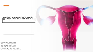
- 1. HYSTEROSALPINGOGRAPH Y SWAPNIL SHETTY 1st YEAR MSc.MIT MCHP. MAHE. MANIPAL
- 2. Contents 2 Female Reproductive System Indications & Contraindication, Equipments, Procedure, Technique, Aftercare & Complications Basic anatomy and its blood supply Falloposcopy & Sono Salpingography (Sion Test) Other Procedures 1 2 3
- 3. 3 Anatomy The female reproductive organs can be subdivided into the internal and external genitalia.
- 4. 4 External Organ The external genitalia lie outside the true pelvis. These include the perineum, mons pubis, clitoris, urethral (urinary) meatus, labia majora and minora, vestibule, greater vestibular (Bartholin) glands, Skene glands, and periurethral area.
- 5. The internal genitalia are those organs that are within the true pelvis. These include the vagina, uterus, cervix, uterine tubes (oviducts or fallopian tubes), and ovaries. 5 Internal Organ
- 6. Vagina: ● The vagina extends from the vulva externally to the uterine cervix internally. It is located within the pelvis, anterior to the rectum and posterior to the urinary bladder. The vagina lies at a 90º angle in relation to the uterus. The vagina is held in place by endopelvic fascia and ligaments. ● The vascular supply to the vagina is primarily from the vaginal artery, a branch of the anterior division of the internal iliac artery. Uterus ● The uterus is the inverted pear-shaped female reproductive organ that lies in the midline of the body, within the pelvis between the bladder and the rectum. ● The uterus can be divided into 2 parts: the most inferior aspect is the cervix, and the bulk of the organ is called the body of the uterus. ● The vasculature of the uterus is derived from the uterine arteries and veins. The uterine vessels arise from the anterior division of the internal iliac, and branches of the uterine artery anastomose with the ovarian artery along the uterine tube. 6
- 7. Cervix: ● The cervix is the inferior portion of the uterus, separating the body of the uterus from the vagina. ● The cervix is cylindrical in shape, with an endocervical canal located in the midline, allowing passage of semen into the uterus. ● The external opening into the vagina is termed the external os, and the internal opening into the endometrial cavity is termed the internal os. ● The internal os is the portion of a female cervix that dilates to allow delivery of the fetus during labor. ● The average length of the cervix is 3-5 cm. ● The vasculature is supplied by descending branches of the uterine artery. 7
- 8. Uterine tubes: ● The uterine tubes (also referred to as oviducts or fallopian tubes) are uterine appendages located bilaterally at the superior portion of the cavity. ● Their primary function is to transport sperm toward the egg, which is released by the ovary, and then to allow passage of the fertilized egg back to the uterus for implantation. ● Each tube is approximately 10 cm in length and 1 cm in diameter and is situated within a portion of the broad ligament called the mesosalpinx. ● The uterine tube has 3 parts. The first segment, closest to the uterus, is called the isthmus. The second segment is the ampulla. The final segment, furthest from the uterus, is the infundibulum. ● The infundibulum gives rise to the fimbriae, fingerlike projections that are responsible for catching the egg that is released by the ovary. ● The arterial supply to the uterine tubes is from branches of the uterine and ovarian arteries. 8
- 9. Ovaries: ● The ovaries are paired organs located on either side of the uterus. ● The ovaries are responsible for housing and releasing the ova, or eggs, necessary for reproduction. ● The ovaries are small and oval-shaped, exhibit a grayish color, and have an uneven surface. ● the ovaries are approximately 3-5 cm in length during childbearing years and become much smaller and atrophic once menopause occurs. ● Blood supply to the ovary is via the ovarian artery; both right and left ovarian arteries originate directly from the descending aorta at the level of the L2 vertebra. 9
- 10. It is the radiological procedure in which the contrast is injected into the uterus to study the uterine tube and fallopian tube 10 What is Hysterosalpingography?
- 11. Menstruation The mean duration of the MC • Mean 28days(only in 15% of the females) ranging from 21 to 35 yrs Average duration : 3-8days The normal estimated blood loss • approximately 30ml . The ovulation occurs • Usually day 14. The mean age • Menarche-12.7 • Menopause-51.4
- 12. When can this procedure be performed? 12 28 days rule Menstrual cycle varies, generally 28 days. If patient is to be exposed to ionizing radiation for diagnostic purposes and the patient is of child-bearing age, postpone exposure for 28 days from first day of menstrual cycle to next to rule out pregnancy. 10 days rule If patient is to be exposed to ionizing radiation for diagnostic purposes If patient is to be exposed to ionizing radiation for diagnostic purposes and the patient is of child-bearing age, she should be booked in the first 10 days of the menstrual cycle, when conception is unlikely to have occurred.
- 13. Indications 13 Infertility Infertility means not being able to become pregnant after a year of trying. Recurrent abortion Recurrent pregnancy loss, defined as 3 consecutive pregnancy losses prior to 20 weeks from the last menstrual period. Following tubal surgery During tubal ligation, the fallopian tubes are cut, tied or blocked to permanently prevent pregnancy. Uterine and tubal lesions tuberculosis, sub mucous fibroids, polyps etc. Migrated IUCD Migration of the IUD to the pelvic and abdominal cavity or adjacent organs may be seen following perforation of the uterus. Congenital or acquired uterine anomalies including septa and adhesions or synechiae, as in Asherman’s syndrome
- 14. Contraindications Active pelvic sepsis Sensitive to contrast media Severe renal or cardiac disease The week prior to and the week following onset of menstruation Pregnancy 14 Cervicitis/purule nt vaginal discharge
- 15. Equipments 15 1. Fluoroscopy 2. Sims speculum 3. Uterine sound and dilator 4. 20cc syringe 5. Foley’s catheter
- 16. Contrast Media Water soluble Urograffin 60%, Conray 280, trivideo 280, angiografin. Volume: • Average volume 5-6 ml. • In nulliparous women 3-4 ml, if there is hydrosalphyx >10 ml 16
- 17. Preparation & premedications 17 Ideal time of procedure: between 8th and 10th day of menstrual cycle. • patient should be advised to abstain from intercourse between booking the appointment and the time of examination • 4 hours of fasting prior to the procedure. • Ask the patient to void the urine before procedure • If patient is anxious – 5 to 10 mg of I.V diazepam 30 min before the procedure • 0.6mg atropine sulphate in 1ml ampoule can given I.V 10 - 15 min before starting the procedure
- 18. Techniques 18 1. Using Cannula 2. Using Foleys catheter 1. Leech Wilkinson canula 2. Jercho type 3. Spackman
- 19. 19 Cannula method • The patient is placed in lithotomy position at the edge of the x-ray table. • A speculum is introduced into the vagina and the anterior lip of the cervix is held with tenaculum and gentle traction is applied.
- 20. 20 Cannula method • The canula is inserted into the cervical canal under direct vision. • The speculum is then removed, and patient is carefully moved to supine position • Under fluoroscopic control, 2ml of the contrast media is injected to outline uterine cavity. • To prevent the leak from cervix, a downward traction should be kept on the tenaculum while keeping an upward pressure to the canula. cannula
- 21. 21 Cannula method Disadvantage: Causes cervical trauma and bleeding
- 22. 22
- 23. 23 Foley’s catheter method • 8F Foley’s catheter is used. • The cervix is exposed with a vaginal speculum and swabbed with an antiseptic solution. • The lumen of the catheter is filled with contrast to prevent air bubbles.
- 24. 24 Foley’s catheter method The catheter is inserted through the cervical os using a cervical forceps to guide it when the ballon lies within uterine cavity ,it is gently inflated with water (2-3ml) Before injection of contrast, the ballon is pulled downwards against the internal os. The speculum is withdrawn, and catheter is attached to syringe and contrast is injected
- 25. 25
- 26. Filming 26 As the tube begin to fill When peritoneal spill has occurred Maximum x-ray screening time must not exceed 30 sec. Only 3-4 spot exposures are permitted in order to minimize radiation to gonads
- 27. b.Tubal Filling Phase a.Early Filling Phase c.Uterus Fully Distended d.Peritoneal Spillage
- 28. Aftercare 28 It must be ensured that patient is in no serious discomfort before she leaves. She must be cautioned that there may be mild bleeding for 1-2 days
- 29. 1.Pain may occur • Using the vulsellum forceps. • During insertion of canula. • With tubal distension and distension of uterus. • Generalized lower abdominal pain due to peritoneal irritation by the contrast media 2.Trauma to the uterus due to canula causing perforation. 3.Exacerbation of pelvic infection (0.25-3% infection rate after procedure) 29 Complication
- 31. 31 Falloposcopy • Falloposcopy is a recent development, pioneered by Dr. Kerin of USA. • In this method, a very fine flexible fiberoptic tube is guided through the cervix and uterus into each fallopian tube, thus allowing the visualization of the inner lining of the entire length of the fallopian tube. • This can provide useful information about the extent of tubal damage, and the possibility for successful repair.
- 32. 32 Sonosalpingography (Sion test) • Foley's catheter (SF) is introduced into uterine cavity with the patient in supine position. The bulb of the catheter is inflated with 2 ml of normal saline. • Transvaginal sonography of uterus with catheter insitu is performed in sagittal and coronal planes. • After scanning the uterus and ovaries, the area between the cornua of uterus and the ovary on one side is focused upon. • A mixture of normal saline and air is pushed with moderate force into uterine cavity using a 20 cc syringe fixed to the metallic adaptor. • be seen distending in case of tubal block.
- 33. 33 Sonosalpingography (Sion test) • A slight traction is given to the catheter while injecting to occlude internal os with the bulb. If the fallopian tube is patent the flow can be seen as a gush of fluid cascading past the 'surprised' ovary and this phenomenon is called the 'Waterfall Sign’. • Then the same procedure is repeated with the other side focused. • When the tubes are blocked, the patient complains of acute pain in the suprapubic region and , the reflux of fluid and air is seen in the stem of the catheter. • Also uterine cavity can be seen distending in case of tubal block.
- 34. 34 Sonosalpingography (Sion test) Advantages: • Can demonstrate the tubal block, its site and extent with higher accuracy and reliability. • No radiation exposure. Disadvantages: • Individual tube evaluation sometimes become difficult.
- 35. Abnormal findings in HSG UTERINE 1. Uterine anomalies : Unicornuate, Didelphys, Bicornuate, Septate, Arcuate 2. Fibroid ( submucosal) 3. Adenomyosis 4. Endometrial polyp 5. Intrauterine adhesions/synechiae 6. Endometrial TB 7. Cervical incompetence 8. Uterine myoma TUBAL 1. Tubal block 2. Tubal spasm 3. Tubal polyp 4. Hydrosalpinx 5. Salpingitis isthmic nodosum (SIN) 6. Peritubular adhesions 7. TB salpingitis
- 36. 36 Unicornuate Uterus Unicornuate uterus is a rare genetic condition in which only one half of a girl's uterus forms.
- 37. 37 Uterus didelphys Uterus didelphys represents a uterine malformation where the uterus is present as a paired organ when the embryogenetic fusion of the Müllerian ducts fails to occur.
- 38. 38 Bicornuate uterus A bicornuate uterus is a type of congenital uterine malformation or müllerian duct anomaly in which the uterus appears to be heart- shaped.
- 39. 39 Septate Uterus A septate uterus is a deformity of the uterus, which happens during fetal development before birth. A membrane called the septum divides the inner portion of uterus, at its middle.
- 40. 40 Arcuate Uterus An arcuate uterus is a mildly variant shape of the uterus. It is technically one of the Müllerian duct anomalies but is often classified as a normal variant.
- 41. 41 Uterine Fibroid Uterine fibroids are noncancerous growths of the uterus that often appear during childbearing years.
- 42. 42 Adenomyosis Adenomyosis is a condition in which the inner lining of the uterus (the endometrium) breaks through the muscle wall of the uterus (the myometrium). Adenomyosis can cause menstrual cramps, lower abdominal pressure, and bloating before menstrual periods and can result in heavy periods.
- 43. 43 Endometrial Polyps Overgrowth of cells in the lining of the uterus (endometrium) leads to the formation of uterine polyps, also known as endometrial polyps.
- 44. 44 Intrauterine synechiae Intrauterine synechiae, also known as Asherman syndrome, is a condition characterized by the formation of intrauterine adhesions, which are usually sequela from injury to the endometrium, and is often associated with infertility.
- 45. 45 Endometrial TB Endometrial tuberculosis is a rare diagnosis in the postmenopausal period, and it can mimic a carcinoma.
- 46. 46 Cervical insufficiency Cervical insufficiency is the inability of the cervix to retain fetus, in the absence of uterine contractions or labor (painless cervical dilatation), owing to a functional or structural defect.
- 47. 47 Uterine myoma Myomas are tumors of the smooth muscle found in the wall of the uterus. Myomas range in size and might not grow or grow slowly.
- 48. 48 Tubal Block Fallopian tube obstruction is a major cause of female infertility. Blocked fallopian tubes are unable to let the ovum and the sperm converge, thus making fertilization impossible.
- 49. 49 Tubal Spasm Fallopian tube spasm is a transient functional anomaly that can mimic a true mechanical tubal occlusion
- 50. 50 Tubal Polyp A Fallopian tube polyp refers to a small focal lesion of ectopic endometrial tissue located at the intramural portion of the fallopian tube.
- 51. 51 Hydrosalpinx Hydrosalpinx refers to a fallopian tube that's blocked with a watery fluid. This condition is typically caused by a previous pelvic or sexually transmitted infection, a condition like endometriosis, or previous surgery.
- 52. 52 Salpingitis isthmic nodosum (SIN) Salpingitis isthmica nodosa (SIN), sometimes also referred to as diverticulosis of the fallopian tube, refers to nodular scarring of the fallopian tubes.
- 53. 53 Peritubular Adhesion Adhesions, also called scar tissue, can block or distort the fallopian tubes. Anything that leads to an inflammatory response can start adhesion formation.
- 54. 54 TB salpingitis Tuberculous salpingitis is an infection in human's body, which develops hematogenously from the primary lesions (lung and intestine) to the reproductive system.
- 55. Does anyone have a any questions? 55 Thanks!
Editor's Notes
- • The uterine cavity is shown during HSG as a triangular contrast-filled structure, with its base on top and the apex caudally (inverted triangle) and the uterine fundus on top, which can be flattened, concave or slightly convex . -free spillage of the contrast to the peritoneum noted.
- 2 uterus 2 cervix 2 vagina
- 2 uterus 2 cervix 1 vagina
- Irregular outline, multiple diverticulum (arrows)
- Pelvic tuberculosis in patient with chronic genital TB. Uterine cavity is small, deformed with irregular contour. Both tubes are occluded. Several calcified lymph nodes in the pelvis and intravasation of contrast into the veins are visualized.
- Right fallopian tube does not opacify beyond the cornual portion
- small outpouchings or diverticula from the isthmic portion of the fallopian tubes. SIN can be either unilateral or (as in this case) bilateral.
- sterosaipingogram In patient with bilateral peritubal adhesins shows peritubal halo effects (curved arrows) with IOcuIatIOn of spillage of contrast medium (straight arrows).