Department of Botany B.Sc (Bot) First Year First Sem Notes (UNIT-III) Phycology
•
1 like•482 views

1. The document provides information on the general characteristics, structure, reproduction, and life cycle of the green algae Volvox. 2. Volvox forms spherical or oval colonies composed of hundreds to tens of thousands of cells arranged in a single layer. Each cell contains flagella, chloroplasts and other organelles. 3. Volvox reproduces asexually through the formation of gonidia - reproductive cells that divide to form daughter colonies inside the parent colony. The daughter colonies eventually invert and are released into the water.

Recommended
More Related Content
What's hot
What's hot (20)
Similar to Department of Botany B.Sc (Bot) First Year First Sem Notes (UNIT-III) Phycology
Similar to Department of Botany B.Sc (Bot) First Year First Sem Notes (UNIT-III) Phycology (20)
More from Head Department of Botany Govt Degree College Mahabubaba
More from Head Department of Botany Govt Degree College Mahabubaba (20)
Recently uploaded
Recently uploaded (20)
Department of Botany B.Sc (Bot) First Year First Sem Notes (UNIT-III) Phycology
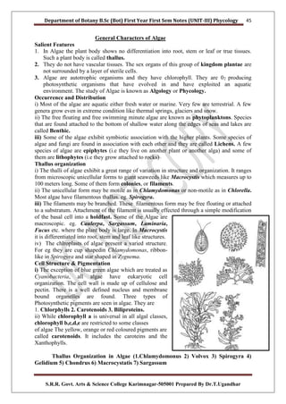
- 1. Department of Botany B.Sc (Bot) First Year First Sem Notes (UNIT-III) Phycology S.R.R. Govt. Arts & Science College Karimnagar-505001 Prepared By Dr.T.Ugandhar 45 General Characters of Algae Salient Features 1. In Algae the plant body shows no differentiation into root, stem or leaf or true tissues. Such a plant body is called thallus. 2. They do not have vascular tissues. The sex organs of this group of kingdom plantae are not surrounded by a layer of sterile cells. 3. Algae are autotrophic organisms and they have chlorophyll. They are 02 producing photosynthetic organisms that have evolved in and have exploited an aquatic environment. The study of Algae is known as Algology or Phycology. Occurrence and Distribution i) Most of the algae are aquatic either fresh water or marine. Very few are terrestrial. A few genera grow even in extreme condition like thermal springs, glaciers and snow. ii) The free floating and free swimming minute algae are known as phytoplanktons. Species that are found attached to the bottom of shallow water along the edges of seas and lakes are called Benthic. iii) Some of the algae exhibit symbiotic association with the higher plants. Some species of algae and fungi are found in association with each other and they are called Lichens. A few species of algae are epiphytes (i.e they live on another plant or another alga) and some of them are lithophytes (i.e they grow attached to rocks) Thallus organization i) The thalli of algae exhibit a great range of variation in structure and organization. It ranges from microscopic unicellular forms to giant seaweeds like Macrocystis which measures up to 100 meters long. Some of them form colonies, or filaments. ii) The unicellular form may be motile as in Chlamydomonas or non-motile as in Chlorella. Most algae have filamentous thallus. eg. Spirogyra. iii) The filaments may be branched. These filamentous form may be free floating or attached to a substratum. Attachment of the filament is usually effected through a simple modification of the basal cell into a holdfast. Some of the Algae are macroscopic. eg. Caulerpa, Sargassum, Laminaria, Fucus etc. where the plant body is large. In Macrocystis it is differentiated into root, stem and leaf like structures. iv) The chlroplasts of algae present a varied structure. For eg they are cup shapedin Chlamydomonas, ribbon- like in Spirogyra and star shaped in Zygnema. Cell Structure & Pigmentation i) The exception of blue green algae which are treated as Cyanobacteria, all algae have eukaryotic cell organization. The cell wall is made up of cellulose and pectin. There is a well defined nucleus and membrane bound organelles are found. Three types of Photosynthetic pigments are seen in algae. They are 1. Chlorphylls 2. Carotenoids 3. Biliproteins. ii) While chlorophyll a is universal in all algal classes, chlorophyll b,c,d,e are restricted to some classes of algae The yellow, orange or red coloured pigments are called carotenoids. It includes the caroteins and the Xanthophylls. Thallus Organization in Algae (1.Chlamydomonus 2) Volvox 3) Spirogyra 4) Gelidium 5) Chondrus 6) Macrocystatis 7) Sargassum
- 2. Department of Botany B.Sc (Bot) First Year First Sem Notes (UNIT-III) Phycology S.R.R. Govt. Arts & Science College Karimnagar-505001 Prepared By Dr.T.Ugandhar 46 iii) The water soluble biliproteins called phycoerythrin (red) and phycocyanin (blue) occur generally in the Rhodophyceae and Cyanophyceae and the latter is now called cyanobacteria. iv) These pigments absorb sunlight at different wavelengths mainly in blue and red range and help in photosynthesis. v) Pigmentation in algae is an important criterion for classification. The colour of the algae is mainly due to the dominance of some of the pigments. For example in red algae(class Rhodophyceae) the red pigment phycoerythrin is dominant over the others. vi) The pigments are located in the membranes of chloroplasts. In each chloroplast one or few spherical bodies called pyrenoids are present. They are the centres of starch formation. Nutrition and reserve food materials in Algae Algae are autotrophic in their mode of nutrition. The carbohydrate reserves of algae are various forms of starch in different classes of Algae. For example, in Chlorophyceae, the reserve food is starch and in Rhodophyceae it is Floridean starch, in Phaeophyceae it is laminarian starch while in Euglenophyceae it is paramylon. Members of Phaeophyceae store mannitol in addition to carbohydrate. Members of Xanthophyceae and Bacillariophyceae store fats, oils and lipids. The nature of reserve food material is also another important criterion used in classification. Arranagement of Flagella i) Flagella or cilia( sing.flagellum / cilium) are organs of locomotion that occur in a majority of algal classes. ii) There are two types of flagella namely whiplash (Acronematic) and tinsel (pantonematic). iii) The whiplash flagellum has a smooth surface while the tinsel flagellum has fine minute hairs along the axis. iv) The number, insertion, pattern and kind of flagella appear to be consistent in each class of algae and it is an important criterion for classification of algae. Motile cells of the Algae are typically biflagellate. When both flagella are of equal length and appearance, they are described as isokont. Heterokont forms have dissimilar flagella with reference to their length and types. Types of Whiplash Flagella v) Red algae(Rhodophyta) and Blue green algae(Cyanophyta) lack flagella. Each flagellum consists of two central microtubles surrounded by a peripheral layer of nine doublet microtubles. This is called 9+2 pattern of arrangement which is a characteristic feature of eukaryotic flagellum. The entire group of microtubles is surrounded by a membrane. Reproduction Three common methods of reproduction found in Algae are 1. Vegetative 2. Asexual and 3. Sexual Vegetative reproduction It lakes place by fragmentation or by the formation of adventitious branches. Asexual reproduction: i) It takes place by means of different kinds of spores like Zoospores, Aplanospores and Akinetes. ii) Zoospores are naked, flagellated and motile. eg.(Chlamydomonas) Aplanospores are thin walled and non motile (eg Chlorella) Akineties are thick walled and non motile spores (eg Pithophora) Sexual Reproduction: - Sexual reproduction involves fusion of two gametes. If fusing gametes belong to the same thallus it is called homothallic and if they belong to different thalli it is heterothallic. Fusing gametes may be isogametes or heterogametes.
- 3. Department of Botany B.Sc (Bot) First Year First Sem Notes (UNIT-III) Phycology S.R.R. Govt. Arts & Science College Karimnagar-505001 Prepared By Dr.T.Ugandhar 47 Isogamy: - It is the fusion of two morphologically and physiologically similar gametes.eg. Spirogyra and some species of Chlamydomonas . Heterogamy:- This refers to the fusion of dissimilar gametes. It is of two types 1. Anisogamy 2. Oogamy 1. Anisogamy is the fusion of two gametes which are morphologically dissimilar but physiologically similar (both motile or both non-motile) 2. Oogamy refers to the fusion of gametes which are both morphologically and physiologically dissimilar. In this type of fusion the male gamete is usually referred to as antherozoid which is usually motile and smaller in size and the female gamete which is usually non- motile and bigger in size is referred to as egg. The sex organ which produces the antherozoids is called antheridium and the egg is produced in oogonium. The fusion product of antherozoid and egg is called Zygote. The zygote may germinate directly after meiosis or may produce meiospores which in turn will germinate. Classification F.E. Fritsch (1944-45) classified algae into 11 classes in his book “Structure and Reproduction of Algae” based on the following characteristics. 1). Pigmentation 2). Reserve food 3). Flagellar arrangement 4). Thallus organization 5). Reproduction. The 11 classes of algae are: 1. Chlorophyceae 2. Xanthophyceae 3. Chrysophyceae 4. Bacillariophyceae 5. Cryptophyceae 6. Dinophyceae 7. Chlromonodineae 8.Euglenophyceae 9. Phaeophyceae 10. Rhodophyceae and 11. Myxophyceae Table : Characteristics of Major Groups of Algae Class Pigments Flagella Reserve Food Material Chlorophyceae (green algae) Chlorophyll-a,b Carotene XanthophyII Two identical flagella per cell Starch Xanthophyceae Chlorophyll-a, b Carotene XanthophyII Heterokont type, one whiplash type and other tinsel Fats and Leucosin Chrysophyceae (diatoms, golden algae) ChlorophyII-a, b Carotenoids One,two or more unequal flagella Oils and Leucosin Bacillariophyceae Chlorophyll-a, c Carotenes Very rare Leucosin and fats Phaeophyceae (brown algae ChlorophyII-a Xanthophyll Two dissimilar lateral flagella Laminarin, fats Rhodophyceae (Red algae) Chlorophyll-a Phycocyanin Phycoerythrin Non-motile Starch Myxophyceae Chlorophyll-a, carotene, phycocyanin, phycoerythrin Non-motile Cyanophyce an starch
- 4. Department of Botany B.Sc (Bot) First Year First Sem Notes (UNIT-III) Phycology S.R.R. Govt. Arts & Science College Karimnagar-505001 Prepared By Dr.T.Ugandhar 48 Volvox: Occurrence, Structure and Reproduction (With Diagrams) Volvox is represented by about 20 species:Some common Indian species are—Volvox globator, V aureus, V. prolificus, V. africanus and V. rousseletii Occurrence of Volvox: 1. Volvox is free floating fresh water green algae. 2. Volvox grows as planktons on surface of water bodies like temporary and permanent ponds, lakes and water tanks. 3. During rainy season due to its fast growth the surface of water bodies become green. 4. The Volvox colonies appear as green rolling balls on surface of water. Structure of Volvox: i) Volvox thallus is a motile colony with definite shape and number of cells. This habit of thallus is called coenobium. ii) The colony is hollow, spherical or oval in shape and the size of colony is about the size of a pin head. iii) The number of cells in a colony is fixed. Depending upon the species of Volvox the cells can be 500-50,000. iv) The central part of colony is mucilaginous and the cells are arranged in a single layer on periphery of the colony. v) The cells of anterior end possess bigger eye spots than those of posterior end cells. vi) The cells of posterior side become reproductive on maturity. Thus, spherical or round colony of Volvox shows clear polarity. vii) The cells of Volvox colony are Chlamydomonas type. Every cell has its own mucilage sheath. viii) The mucilage envelope of colony appears angular due to compression between cells.
- 5. Department of Botany B.Sc (Bot) First Year First Sem Notes (UNIT-III) Phycology S.R.R. Govt. Arts & Science College Karimnagar-505001 Prepared By Dr.T.Ugandhar 49 ix) The cells are connected to each other through cytoplasmic strands. In some species of Volvox the cytoplasmic connections or strands are not present. x) The cells of colony are usually pyriform with narrow anterior end and broad posterior end. xi) The cells are biflagellate; the two flagella are equal, whiplash type and project outwards. xii) The protoplasm of cell is enclosed within psma membrane. xiii) Each cell contains one nucleus, a cup shaped chloroplast with one or more pyrenoids, an eye spot and 2-6 contractile vacuoles. In some species of Volvox e.g., in V. globator and V. rousseletii the cells are of Sphaerella type. xiv) The cells of colony are independent for functions like photosynthesis, respiration and excretion. xv) The movement of colony takes place by co-ordinated flagellar movement.The reproduction is common to the coenobium. Reproduction in Volvox: i) Volvox reproduces both asexually and sexually. ii) The asexual reproduction takes place under favourable conditions during spring and early summer. iii) In Volvox mostly the cells of posterior part of colony take part in reproduction. iv) These reproductive cells can be recognized by their larger size, prominent nuclei, dense granular cytoplasm, more pyrenoids and absence of flagella (Gonidia). Asexual Reproduction: i) During asexual reproduction some cells of the posterior part of colony become reproductive. ii) These cells enlarge up to ten times, become rounded and lose flagella. iii) These cells are called gonidia (Sing, gonidium). The gonidia lose eye spot. Pyrenoids increase in number. iv) The gonidia are pushed towards interior of the colony. v) The first division of gonidium is longitudinal to the plane of coenobium and this forms 2 cells (Fig. 2 A). . .
- 6. Department of Botany B.Sc (Bot) First Year First Sem Notes (UNIT-III) Phycology S.R.R. Govt. Arts & Science College Karimnagar-505001 Prepared By Dr.T.Ugandhar 50 vi) The second division is also longitudinal and at right angle to the first, forming 4 cells (Fig. 2 B). By third longitudinal division all the four cells divide to make 8 cells of which 4 cells are central and 4 are peripheral vii) These 8 cells are arranged in curved plate-like structure and are called plakea stage (Fig. 2 C, D). Each of these 8 cells divides by longitudinal division forming 16 cells arranged in the form of a hollow-sphere (Fig. 2 E). viii) The sphere is open on exterior side as a small aperture called phialopore (Fig. 2 F). ix) The cells at this stage continue to divide till the number of cells reaches the characteristic of that species. The cells at this stage are naked and in close contact with each other. The pointed anterior end of cells is directed towards inside. x) The next step is called inversion of colony (Fig. 2 G-H). As cells become opposite in direction, their anterior pointed end has to face the periphery of colony xi) The inversion of colony starts with formation of a constriction opposite to phialopore. xii) The cells of posterior end along with constriction are pushed inside the sphere, till the whole structure comes out of the phialopore. xiii) After inversion, the anterior pointed end of the cell faces periphery. xiv) The phialopore gets closed, and makes the anterior part of the colony. After inversion the cells develop cell wall, flagella and eye spot. xv) The cells become separated due to development of gelatinous sheath around each cell. This newly developed colony is called daughter colony (Fig. 2 I). xvi) The daughter colonies initially remain attached to gelatinized wall of parent colony and later become free in gelatinous matrix of parent colony. xvii) The daughter colonies are released in water after the disintegration of parent colony or through the pores. Sexual Reproduction: 1. The sexual reproduction in Volvox is oogamous type. 2. Some species of Volvox e.g., V. globator are monoecious or homothallic (Fig. 3) i.e., the antheridia and oogonia develop on same colony. Other Volvox species e.g., V. rousseletii are dioecious or heterothallic i.e., antheridia and oogonia develop on different colonies. 3. Monoecious species are usually protandrous i.e., antheridia mature before oogonia but some species are protogynous i.e., oogonia develop before antheridia. V aureus is mostly dioecious but sometimes can be monoecious. 4. Reproductive cells mostly differentiate in the posterior part of colony. These cells enlarge, lose flagella and are called gametangia. The male reproductive cells are called antheridia or androgonidia and female reproductive cells are called oogonia or gynogonidia. Development of Antheridium: i) The development of antheridium starts with formation of antheridial initial or androgonidial cell mostly in posterior side of the colony. ii) The initial cells enlarge, lose flagella, protoplasm becomes dense and nucleus becomes larger. iii) The antheridial initial shifts inside towards cavity and remains connected to other vegetative cells through cytoplasmic strands. iv) The protoplast of antheridial initial divides, longitudinally to form 16-512 elongated cells. The cells remain in plate like structure or arrange in a hollow sphere. v) The inversion of cells also takes place as in asexual reproduction. Each cell differentiates in antherozoid or spermatozoid (Fig. 3, 4). vi) The antherozoid is spindle shaped, elongated, bi-flagellated structure containing two contractile vacuoles, nucleus, cup shape chloroplast, pyrenoid and eye spot. It is pale yellow or green in colour.
- 7. Department of Botany B.Sc (Bot) First Year First Sem Notes (UNIT-III) Phycology S.R.R. Govt. Arts & Science College Karimnagar-505001 Prepared By Dr.T.Ugandhar 51 vii) The antherozoids are released individually or sometimes in groups. Development of Oogonium: i) The oogonia also differentiate mostly in posterior side of the colony. ii) The oogonial initials enlarge, nucleus becomes larger, protoplast becomes dense, flagella are lost, eye spot disappears and many pyrenoids appear. iii) The mature oosphere or ovum is round or flask shaped structure. The egg is uninucleate structure, the beak of flask shape oogonium functions as receptive spot (Fig. 5 A, B). Fertilization of Volvox: i) After liberation from antheridium, the anther ozoids swim freely on surface of water. ii) Due to chemotactic response the anthero zoids reach the oogonia. iii) Some antherozoids enter each oogonium. Only one antherozoid enters inside the oogonium through receptive spot. iv) After this plasmogainy i.e., fusion of male and female cytoplasm and karyogamy i.e., fusion of male and female nuclei take place. v) This results in formation of diploid zygote (Fig. 5 D). The diploid zygote secretes a three layered thick wall. vi) The layers of the wall are exospore, mesospore and endospore (Fig. 6 A, B). vii) The outer exospore is thick. It may be smooth e.g., V. aureus (Fig. 6 A) or spiny e.g., V. globator (Fig. 6 B). viii) The mesospores and endospores are thin and smooth. The walls contain nucleus pigment haematochrome which imparts red colour to the zygote. x) The zygotes are released by the disintegration of parent colony. Then zygotes undergo a period of dormancy. Germination of Zygote: i) The dormant zygote germinates on approach of favourable climatic conditions. ii) The diploid nucleus of zygote undergoes meiotic division forming four haploid cells. The outer two layers of zygote burst and the inner layer comes out as vesicle. iii) The four haploid cells migrate with the vesicle (Fig. 6 C, D). The development of new colony from zygote differs in different species of Volvox. In V. aureus and V. minor the
- 8. Department of Botany B.Sc (Bot) First Year First Sem Notes (UNIT-III) Phycology S.R.R. Govt. Arts & Science College Karimnagar-505001 Prepared By Dr.T.Ugandhar 52 protoplasm of zygote divides repeatedly until the cell number of colony is reached and new colony is formed as in asexual reproduction process. iv) In V. campensis the protoplast of zygote divides to make many biflagellate zoospores. Only one zoospore survives and all other disintegrate. v) This zoospore comes out of the vesicle it divides to make many cells which arrange to form a colony. In V. rousseletii the zygote forms a single biflagellate zoospore; the protoplast of zoospore divides and forms a colony. vi) In all the methods the cells divide and undergo inversion to make a mature colony (Fig. 6 E-H). Life Cycle of Volvox: Volvox is haploid (n) algae, the haploid gametes fertilize to make diploid zygote (2n) which divides by meiosis to make haploid cells (n) which mature into haploid Volvox colony (Fig. 7, 8). Oedogonium Systematic Position: Occurrence of Oedogoniales: i) The genus Oedogonium (Oedos-swollen, gonas - reproductive) structure is the only genus of the family with un-branched filaments. ii) It is represented by about 400 species. The common Indian species are: i) O. cardiacum. O. elegans, O. obolongellum and O. tenuis. ii) Oedogonium occurs as the fresh water filamentous alga found in ditches, ponds, pools and lakes. iii) It occurs as epiphytic alga found attached on leaves and twigs of other plants. iv) It is also found attached on other algae such as Cladophora.
- 9. Department of Botany B.Sc (Bot) First Year First Sem Notes (UNIT-III) Phycology S.R.R. Govt. Arts & Science College Karimnagar-505001 Prepared By Dr.T.Ugandhar 53 v) Some species of Oedogonium are terrestrial, found growing on moist soils. It is common in stagnant water and less common in running water. Thallus: The thallus is made of green, multicellular un-branched filaments. The filament is made of three types of cells (Fig. 1): (i) The lower-most basal cell or holdfast. (ii) The intercalary cells. (iii) The upper-most apical cell. Hold Fast: i) The filament is attached by means of specially differentiated basal cell. ii) The holdfast is found in aquatic species and it rarely occurs in terrestrial forms. In terrestrial forms it may give out rhizoid like outgrowths. iii) The hold fast or basal cell is club shaped, broad, round in upper part and narrow in lower part. iv) The lower terminal part of basal cells is multi-lobed, disc like or finger shaped which attaches the filament to substratum. Chloroplasts are absent or poorly developed in basal cell hence it does not take part in photosynthesis. Intercalary Cells: i) All cells of the filament in between apical cell and the basal cell are intercalary cells. ii) The intercalary cells of filaments have base-apex polarity and this is maintained even when filaments break and become free floating. iii) All intercalary cells are alike, only some cells after division develop cap in upper part. Such cells are called cap cells. Apical Cell: i) The terminal cell of the filament called apical cell. It is round or dome shaped. In some species e.g., O. ciliata, the apical cell is tapering and gives rise to narrow hair like structure. The apical cell is green due to chlorophyll and takes part in photosynthesis. Cell Structure of Oedogoniales: i) The cells are elongated and cylindrical. The cell wall is generally thick, rough and rigid. It is made up of three concentric layers, the inner cellulose, middle pectose and the outer layer is chitinous in nature. ii) The protoplasm consists of thin plasma membrane, cytoplasm, central vacuole, reticulate chloroplast and the nucleus. iii) The centre of the cell is occupied by a large central vacuole which contains the cell sap. The cell sap contains excretions, secretions and inorganic compounds. The protoplast occurs as thin layer between the central vacuole and the inner cell wall. iv) The chloroplast is characteristically reticulate, extending or covering the whole cell and encircling the protoplast (Fig. 2 A, B). v) The strands of reticulum may be broad or narrow depending upon the species. In most of the cases the strands are parallel to the long axis of the cell. Many pyrenoids are present at the intersections of the reticulum, Pyrenoids are covered with starch plates.
- 10. Department of Botany B.Sc (Bot) First Year First Sem Notes (UNIT-III) Phycology S.R.R. Govt. Arts & Science College Karimnagar-505001 Prepared By Dr.T.Ugandhar 54 vi) There is single large nucleus, it is biscuit shaped or biconvex and lies in the centre of cell, internal to the chloroplast. vii) The nucleus possesses 1-2 nucleoli, and thread like or elongated chromosomes. The cell also contains mitochondria, Golgi bodies, endoplasmic reticulum and the other cell organelles. Growth: The growth of Oedogonium filaments takes place by cell division in intercalary cells but sometimes the apical cell also divides and takes part in the elongation of filaments. Cell Division: The process of cell division in Oedogonium takes place in following stages: (i) The nucleus from periphery moves towards the centre and slightly towards the upper part of the cell (Fig. 3 A). (ii) Since the wall does not elongate in the usual manner, the cytoplasmic wall material gathers in the form of a“ring” round the inner wall at the upper end of the cell (Fig. 3 A). (iii) The nucleus divides mitotically and there is formation of a groove in the ring (Fig. 3B, C). (iv) The next stage is the elongation or stretching of the daughter cells by breaking up of the wall layers round the groove of the ring. The lower daughter cell elongates to the former level of the ring. The upper one also elongates to the same extent. The process of elongation is completed within 15 minutes (Fig. 3 D). (v) Along with the completion of elongation, a transverse wall formation between the two is also completed. The distal end of the upper cell contains a small portion of the old parent wall which appears as the apical cap. Reproduction in Oedogonum The reproduction in Oedogonium takes place by vegetative, asexual and sexual methods. (i) Vegetative Reproduction: Vegetative reproduction takes place by fragmentation and akinete formation. (A) Fragmentation: Oedogonium filament breaks into many small fragments which have capability to grow into complete filaments under favourable conditions. Fragmentation takes place due to any of the following reasons: (a) Accidental breaking of the filaments. (b) Dying or dehydration of intercalary cells. (c) Disintegration of intercalary cells due to conversion in sporangia. (d) Mechanical injury to the filament. (e) Change in the environmental conditions. (B) Akinete formation: i) The akinetes are formed under unfavorable conditions. ii) Akinetes are modified vegetative cells which become swollen, round or oval, reddish brown and thick walled. iii) These are rich in reserve starch and orange-red coloured oil. Akinetes are formed in chains of 10 to 40 (Fig. 4). Akinetes germinate directly under favourable conditions.
- 11. Department of Botany B.Sc (Bot) First Year First Sem Notes (UNIT-III) Phycology S.R.R. Govt. Arts & Science College Karimnagar-505001 Prepared By Dr.T.Ugandhar 55 (ii) Asexual Reproduction: i) Asexual reproduction takes place by means of multi-flagellate zoospores produced singly in intercalary cap cell. Mostly the newly formed cap cell functions as the zoosporangium. ii) Several factors control zoospore formation of which high pH and CO2 concentration of medium and a diurnal rhythm of light and darkness are significant. iii) The zoospores are not formed in chains and one sterile cell is always present between two zoosporangia. iv) The cell which functions as zoosporangium gets filled with abundant reserve food and a slight contraction of the protoplast from the cell wall takes place (Fig. 5 A, B). v) The central vacuole disappears the chloroplast frees itself from one end of the cell and becomes conical. v) The nucleus comes to lie near this chloroplast. A small lens shaped hyaline region is formed between the wall and the nucleus. This hyaline bald spot later forms the anterior end of the zoospore. vi) At the base of this hyaline area a ring of basal granules appears and from each basal granule or blepharoplast a flagellum arises. vii) The basal granules are connected to each other by fibrous strand. A crown of about 30 flagella is formed around the hyaline spot (Fig. 5 C). viii) The mature zoospore is oval, spherical or pear shaped structure. The zoospore is uninucleate and contains a ring shaped chloroplast. The zoospore is dark green in colour except at the hyaline pointed apical end. A sub apical ring of flagella is present and such flagellation is called stephanokontic type (Fig. 5 F).When the zoospore is mature, the wall of the zoosporangium splits near the apical region and the adjacent cell moves apart to make a gap for the liberation of zoospore (Fig. 5 D).
- 12. Department of Botany B.Sc (Bot) First Year First Sem Notes (UNIT-III) Phycology S.R.R. Govt. Arts & Science College Karimnagar-505001 Prepared By Dr.T.Ugandhar 56 The mucilage substance is secreted at the base of the zoospore which helps in the liberation of zoospore. The zoospore comes out of the zoosporangium in a delicate mucilaginous vesicle which soon gets dissolved and the zoospores are liberated in water (Fig. 5 D, E). Germination of Zoospore: After liberation, the zoospore swims for about an hour. Then it settles and attaches itself to a solid substratum with its anterior end downwards. After attachment flagella are withdrawn and it starts elongation. The lower hyaline part elongates to make holdfast and the upper part divides repeatedly to make new filament (Fig. 5 G-I). Sexual Reproduction: i) The sexual reproduction in Oedogonium is of advanced oogamous type. ii) Sexual reproduction is more frequent in still waters than in running water. iii) The factors influencing sexual reproduction are alkaline medium, deficiency of nutrition, light and dark periods and increased temperature. iv) The genus Oedogonium exhibits sexual dimorphism because the male and the female gametes differ morphologically as well as physiologically. The male gametes are produced in antheridia and the female gametes are produced in oogonia. Depending upon the nature of antheridia producing plants, Oedogonium species are of two types: (i) Macrandrous: i) If antheridia are produced on normal size plant, Oedogonium forms are called macrandrous. Macrandrous species may be monoecious or dioecious. ii) In monoecious macrandrous species antheridia and oogonia are produced on the same plant e.g., O. fragile, O. hirnii, O. kurzii and O. nodulosum. iii) In dioecious macrandrous species antheridia and oogonia are produced on separate male and female plants of normal size. (ii) Nannandrous: i) The female or oogonia bearing plants are normal. The antheridia are produced on special type of small or dwarf plants, known as Dwarf males or Nannandria. ii) The dwarf males are formed by androspores which are produced in androsporangia. If androsporangia and oogonia are formed on same plant, the Oedogonium forms are called gynandrosporous e.g., O. concatinatum. iii) If androsporangia and oogonia are formed on different plants, Oedogonium forms are called idioandrosporous e.g., O. confertum, O. iyengarii and O. setigerum. iv) According to some algologists, nannondrous species are more primitive. Antheridia: (i) In Macrandrous forms: a) The antheridia develop on normal filaments, terminal or intercalary in position. b) The initial cell which gives rise to antheridia is called antheridial mother cell. c) It is normally a cap cell. The antheridial mother cell divides by transverse division to form an upper smaller cell called antheridium and a lower larger cell called sister cell. The sister cell divides repeatedly to form a row of 2-40 antheridia (Fig. 6 A). d) Rarely the antheridia are produced singly. The antheridia are broad, flat, short cylindrical, uninuleate cells. The contents of an antheridial cells divide either longitudinally or transversely into two.
- 13. Department of Botany B.Sc (Bot) First Year First Sem Notes (UNIT-III) Phycology S.R.R. Govt. Arts & Science College Karimnagar-505001 Prepared By Dr.T.Ugandhar 57 The two antherozoids are positioned side-by-side or one above the other if divisions are longitudinal and transverse respectively. The antherozoids are liberated in the same fashion as zoospores (Fig. 6 B).The liberated antherozoids or sperm atozoids or sperms are pale green or yellow green, oval or pear shaped. The antherozoids are motile about 30 sub-apical flagella present at the base of beak or hyaline spot (Fig. 6 C). The flagella are sometimes longer than the body of spermatozoid e.g., in O. crassum and O. kurzii. The antherozoids swim freely in water before they reach oogonia and take part in fertilization. The antherozoids are similar to zoospores in structure but these are smaller than zoospores. (ii) In Nannandrous forms: i) The antheridia are formed on short or dwarf male plants called dwarf males or nannandria (Fig. 7 G). The dwarf male filament is produced by the germination of a special type of spore known as androspore. ii) The androspore is produced singly within an androsporangium. Androporangia are more or less similar looking to the antheridia of macrandrous forms and are produced in a similar manner from a mother cell (Fig. 7 A, B). iii) The androsporangia are flat, discoid cells slightly larger than antheridia. Each androsporangium produces a single androspore just as in the case of zoospore. Liberation of androspore is similar to that of a zoospore. iv) The androspores look similar to zoospore except for the smaller size. The androspores are motile and have a subpolar ring of flagella. v) After swimming about for some time, the androspore settles on oogonial wall e.g., O. ciliatum or on the supporting cell e.g., O. concatenatum. vi) The androspore germinates into a dwarf male or nannandrium. Germlings at one celled stage may divide and produce two antherozoids e.g., O. deplandrum, O. perspicuum (Fig. 7 C-G). vii) The nannandrium or dwarf male can be a few cells long. It has a basal attaching cell the stipe and all others cells are antheridial cells. viii) In many cases cap is present at the top of the apical antheridium. The protoplasm of each antheridial cell divides to form two sperms or antherozoids which are similar to antherozoids of macrandrous species. Oogonia: i) In Oedogonium the female sex organ oogonia are highly differentiated female gametangia. These are mostly intercalary but sometimes can be terminal e.g., O. palaiense. ii) The structure and development of oogonium is identical in macrandrous and nannandrous species. Like antheridia any freely divided or actively growing cap cell functions as the oogonial mother cell.
- 14. Department of Botany B.Sc (Bot) First Year First Sem Notes (UNIT-III) Phycology S.R.R. Govt. Arts & Science College Karimnagar-505001 Prepared By Dr.T.Ugandhar 58 iii) The oogonial mother cell divides by transverse division into two unequal cells, the upper cell and the lower cell. iv) The upper larger cell forms oogonium and the lower smaller cell function as supporting cell or suffultory cell. In some species the oogonial mother cells directly forms the oogonium. Supporting cell is absent is O. americanum. v) If any of the two divided cells again functions as oogonial mother cell many oogonia are formed in chain. vi) In monoecious species the suffultory cell may divide to form antheridia. The upper cell contains more cytoplasm, food and enlarges into spherical or flask shaped oogonium. vi) The oogonium also secretes growth hormones which induce suffultory cell to increase in size (Fig. 8 A-C). vii) The protoplast in oogonium metamorphosis’s into a single egg or oosphere.
- 15. Department of Botany B.Sc (Bot) First Year First Sem Notes (UNIT-III) Phycology S.R.R. Govt. Arts & Science College Karimnagar-505001 Prepared By Dr.T.Ugandhar 59 The oosphere is non-motile, green due to chlorophyll and has a central nucleus. viii) As the ovum matures, the nucleus moves to periphery, the oosphere retracts slightly from the oogonial wall and develops a hyaline or receptive spot just outside the nucleus. The receptive spot receives antherozoids for fertilization. ix) At receptive spot a pore is formed by gelatinization of wall in proliferous species and a transverse slit is formed in operculate species. x) In both species a thin membrane is deposited on the inner node of the exit which functions as a channel leading down to ovum. In some species a mucilage drop is extruded through opening to attract antherozoids. xi) In macrandrous monoecious species, where antheridia and oogonia develop on the same plant, the Oedogonium species are protogynous i.e., the development of oogonia takes place before development of antheridia to ensure cross-fertilization. Fertilization: i) The mature egg secretes chemical substance or mucilage to attract antherozoids or the antherozoids may enter oogonium through the slit. ii) The antherozoids swim through the opening of oogonial wall and enter the egg through hyaline receptive spot (Fig. 8 D-F). iii) Only one male antherozoid is able to fuse with ovum. After plasmogamy and karyogamy the male nucleus and female nucleus fuse to form a diploid zygote nucleus. iv) The zygote secretes a thick wall around itself and forms oospore. The colour of the oospore changes from green to reddish brown. v) The oospore is liberated by the disintegration of oogonial wall. Structure of oospore: i) The oospore is globular reddish brown structure. The oogonial wall is made of three and sometimes two layers. ii) The outermost layer may be smooth in some cases but in most cases it is ornamented with pits, reticulations, spines, ribs or flanges. iii) The ornamentation of oospore is of taxonomic importance. The oospore is red in colour due to accumulation of red oil. Oospore contains a diploid nucleus and cytoplasm rich in proteins. Germination of oospore: i) Oospore is a resting spore but sometimes it can germinate directly. The period of rest for oospore may be a year or more. ii) According to Mainx (1931) the zygote may require chilling before germination. The diploid oospore nucleus undergoes zygotic meiosis to form four haploid nuclei before germination. iii) The diploid oospore divides to form four haploid daughter protoplasts. Each daughter protoplast metamorphosis into a zoospore also called as zoomeiospore. iv) The zoomeiospores are liberated in a vesicle (Fig. 9 A). Soon the vesicle disappears and as in asexual reproduction the zoospores develop to make Oedogonium plants. In some cases out of four nuclei a few may degenerate forming less than four zoomeiospores. In heterothallic forms e.g., O. plagiostomum, two swarmer’s give rise to male and the two swarmer’s give rise to female plants. Under certain conditions meioaplanospores are formed instead of zoomeiospores (Fig. 9 B, C).
- 16. Department of Botany B.Sc (Bot) First Year First Sem Notes (UNIT-III) Phycology S.R.R. Govt. Arts & Science College Karimnagar-505001 Prepared By Dr.T.Ugandhar 60 Life Cycle in Oedogonium: In Oedogonium the thallus is haploid and the life cycle is haplontic type. The diploid stage in life cycle is only zygote. It occurs for a short period. The zygote or oospore undergoes meiosis to make four meiozoospores which again form haploid Oedogonium thalli. The variations in life cycles of Oedogonium are due to macrandrous and nannandrous nature of Oedogonium species. Macrandrous Forms: i) Oedogonium macrandrous species can be monoecious or homothallic, if antheridia and oogonia are produced on same filament (Fig. 10, 11). Oedogonium macrandrous species can be dioecious or heterothallic if antheridia are produced on male plants and oogonia are produced on separate female plants. (Fig. 12). Nannandrous Forms The nannandrium or dwarf male plants are produced by germination of androspores which are produced in androsporangia. In gynandrosporous nannandrium forms the androsporangia and oogonia are formed on same filaments (Fig. 13, 14). In idioandrosporous nannandrium forms, the androsporangia and oogonia are formed on different plants
- 17. Department of Botany B.Sc (Bot) First Year First Sem Notes (UNIT-III) Phycology S.R.R. Govt. Arts & Science College Karimnagar-505001 Prepared By Dr.T.Ugandhar 61 Chara Life Cycle Systemic Position: - Kingdom: Plantea,Division: Thallophyta/Charophyta,Subdivision: Algae, Class: Chlorophyceae/Charophyceae, Order: Charales: Family: Characeae, Genus: Chara Occurrence: i) Chara is fresh water; green alga found submerged in shallow water ponds, tanks, lakes and slow running water. C. baltica is found growing is brackish water and C. fragilis is found in hot springs. ii) Chara is found mostly in hard fresh water, rich in organic matter, calcium and deficient in oxygen. iii) Chara plants are often encrusted with calcium carbonate and hence are commonly called stone wort. Chara often emits disagreeable onion like odour due to presence of sulphur compounds. C. hatei grows trailing on the soil C. nuda and C. grovesii are found on mountains, C. wallichii and C. liydropitys are found in plains. In India Chara is represented by about 30 species of which common Indian species are: C. zeylanica, C. braunii, C. gracilis, C. hatei and C sgymnoptiy etc. Thallus Structure of Chara i) The thallus of Chara is branched, multicellular and macroscopic. The thallus is normally 20-30 cm. in height but often may be up to 90 cm to l m. Some species like C. hatei are small and may be 2-3 cm. long. ii) The plants in appearance resemble Equisetum hence Chara is commonly called as aquatic horsetail. The thallus is mainly differentiated into rhizoids and main axis Rhizoids: i) The rhizoids are white, thread like, multicellular, uniseriate and branched structures. ii) The rhizoids arise from rhizoidal plates which are formed at the base of main axis or from peripheral cells of lower nodes. iii) The rhizoids are characterized by presence of oblique septa. iv)The tips of rhizoids possess minute solid particles which function as statoliths. v) The rhizoids show apical growth. Rhizoids help in attachment of plant to substratum i.e., mud or sand, in absorption of minerals and in vegetative multiplication of plants by forming bulbils and secondary protonema. Main Axis: i) The main axis is erect, long, branched and differentiated into nodes and internodes. ii) The internode consists of single, much elongated or oblong cell. The inter-nodal cells in some species may be surrounded by one celled thick layer called cortex and such species are called as corticate species. iii) The species in which cortical layer is absent are called ecorticate species. iv) The node consists of a pair of central small cells surrounded by 6-20 peripheral cells. The central cells and peripheral cells arise from a single nodal initial cell. Vegetative structure: i) The plant is always found attached to the substratum by a well developed rhizoidal system. The rhizoids are uniseriately branched and obliquely septate. The rhizoids may or may not be differentiated into nodes and internodes. ii) The axis has nodes and internodes. Several branches of limited growth also known as leaves grow in whorls from the nodes of the axis. iii) These leaves do not grow further after attaining a definite length. The leaves may or may not be differentiated into nodes and internodes. iv) They may be branched or simple. This ineternodal cell is sufficiently long and ensheathed by a cortex of vertically elongated cells.
- 18. Department of Botany B.Sc (Bot) First Year First Sem Notes (UNIT-III) Phycology S.R.R. Govt. Arts & Science College Karimnagar-505001 Prepared By Dr.T.Ugandhar 62 v) The cortex is one celled in thickness and consists of the cells of lesser diameter. The cell wall: The cell wall consists of homogeneous cellulose. It is not multilayered. Outer to the cellulose wall there is a gelatinous layer, and this is the sheath for the deposition of calcium. The main axis of Chara consists of mainly two types of cells: (i) Nodal cells (ii) Inter-nodal cells. i) The nodal cells are smaller in size and isodiametric. The cells are dense cytoplasmic, uninucleate with few small ellipsoidal chloroplasts. ii) The central vacuole is not developed instead many small vacuoles may be present. iii) The cytoplasm can be differentiated in outer exoplasm and inner endoplasm. The inter- nodal cells are much elongated. iv) The cytoplasm is present around a large central vacuole. The cells are multinucleate and contain many discoid chloroplasts. v) The cytoplasm is also differentiated into outer exoplasm and inner endoplasm. The endoplasm shows streaming movements. vi) The cell walls between the nodal cell and inter-nodal cells are porous to help in cytoplasmic continuity between cells. Reproduction: The reproduction takes place by vegetative and sexual methods. Asexual reproduction is not found. 1. Vegetative reproduction: The vegetative reproduction takes place by (a) tubers; (b) amylum stars and (c) secondary protomema. (a) By tubers and bulbils: i) The tubers are commonly formed on rhizoids or sometimes even on buried nodes. The whole structure is full of starch. ii) Sometimes the globule divides and becomes multicellular and known as ‘simple tuber’. When the tuber appears on the node, some of the peripheral cells go on dividing and massive structure is developed. Each starch filled tuber and bulbil may develop into a separate plant. (b) By amylum or starch stars: The cells of some subterranean nodes become star-shaped and very much laid in by starch are called amylum stars. Each such structure develops into new plants. (c) By secondary protonema: The protonemata like outgrowths come out from a node. Each such outgrowth is capable to develop into a new plant.
- 19. Department of Botany B.Sc (Bot) First Year First Sem Notes (UNIT-III) Phycology S.R.R. Govt. Arts & Science College Karimnagar-505001 Prepared By Dr.T.Ugandhar 63 2. Sexual reproduction: i) The sexual reproudction is oogamous. A very advanced and specialized type of oogamy is found. There is a special terminology for the sex organs. ii) The male fruiting body is called globule and the female nucule. Most of the species are homothallic (monoecious) and few are heterothallic (dioecious). iii) The globule is borne on secondary lateral of limited growth. iv) One globule and one nucule bome on one node of the leaf. v) In homothallic species both the fructifications borne on the same node. In Chara the nucule is borne above the globule. Development of globule: i) A single superficial nodal cell of the adaxial side of the leaf acts as the initial of both the fructifications, i.e., nucule and globule. ii) This superficial cell divides into two derivatives by a transverse wall. One cell derivative of the superficial cell is the initial cell of the globule and the other is the initial cell of the nucule. iii) The globule initial cell divides transversely and two daughter cells are formed. The lower daughter cell does not divide further and converts into the pedicel cell. iv) The upper daughter cell divides twice successively and four cells are formed arranged in quadrants. v) Each of these quadrants divides transversely and eight cells are produced thus attaining octant stage. Each of these eight cells divides periclinally and thus produced eight outer cells which divide further periclinaily. vi) The outermost eight cells are called shield cells. The middle cells are known as manubrial cells and the innermost eight cells are primary capitulum cells. The shield cells become very much enlarged and expanded. vii) The manubrial cells become very much radially elongated, but the primary capitulum cells are arranged compactly to each other in the centre of the globule. viii) The outer walls of the shield cells fold inward and the shield cells appear multicellular structures. ix) The infoldings are incomplete. The shield cells develop red pigments in them and so the globules appear orange red in colour from each primary capitulum cell six secondary capitulum cells are cut off inside the globule. x) These secondary capitulum cells rarely develop tertiary cells. On the secondary capitulum cells the initials of antheridial filaments are produced. These initials may also be produced upon primary or even tertiary capitulum cells. xi) Each antheridial initial develops into a branched or unbranched antheridial filament. Each antheridial filament has many compartments or cells in it. Each cell is supposed to be an antheridium. x) The protoplast of each antheridium metamorphoses into a single antherozoid. The antherozoid is elongated, coiled and biflagellate. The flagella are sub-terminal in origin.
- 20. Department of Botany B.Sc (Bot) First Year First Sem Notes (UNIT-III) Phycology S.R.R. Govt. Arts & Science College Karimnagar-505001 Prepared By Dr.T.Ugandhar 64 The nucleus is elongated and coiled. Some unused cytoplasm is found in the tail of the antherozoid. xi) On the maturity of the antherozoids the shield cells of the globule somewhat separate from each other, the antheridial filaments protrude out through these openings and the antherozoids liberate in the water. The liberation of antherozoids usually takes place in the morning. Development of nucule: i) The nucule develops from the adaxial cell of basal node of the globule. ii) The globule is homologous with the branch of limited growth and the nucule with the branch of unlimited growth. iii) The nucule initial divides twice and a row of three cells is formed. The terminal cell acts as oogonial mother cell which elongates sufficiently in
- 21. Department of Botany B.Sc (Bot) First Year First Sem Notes (UNIT-III) Phycology S.R.R. Govt. Arts & Science College Karimnagar-505001 Prepared By Dr.T.Ugandhar 65 vertical direction and transverse wall develops in the lower region of it dividing it into two cells. The lower small cell and the upper one is oogonium which contains an egg. iv) The lowermost cell of the row of three cells does not divide and acts as a pedicel. v) The middle cell divides vertically in such a way so that a single central cell and five sheath initials are produced. vi) The sheath initials surround the central cell. The sheath initials elongate vertically sometimes even before the vertical elongation of the oogonial mother cell and encircle it. vii) Each of the sheath initials divides transversely forming the upper tier of coronary cells and lower tier of tube cells. viii) The tube cells elongate several times to their original length and become spirally coiled around the oogonium. The coronary cells do not elongate much and act collectively as the corona of the nucule. Fertilization: i) Prior to fertilization the elongated and twisted tube cells become separated from each other and five small slits are developed just below the corona. ii) The swimming antherozoids around the nucule try to enter through these openings. iii) The flagella are withdrawn and one of the antherozoids penetrates the egg. The male nucleus travels downwards and fuses with the egg nucleus developing a diploid (2n) nucleus. This diploid nucleus situates in the bottom of the zygote. The zygote settles down in the mud, secretes a thick wall and germinates on the approach of favourable conditions. The zygote and its germination: i) In favourable conditions the zygote germinates. The diploid (2n) nucleus moves to the top of the zygote and divides meiotically producing four halpoid nuclei. ii) Simultaneously a septum divides the zygote into two unequal cells. The small distal cell is lenticular cell and contains one functional nucleus in it. iii) The remaining big cell is called storage cell; possessing three nuclei in it disintegrate very soon. The outer wall of the ornamented zygote cracks and the lenticular cell exposes. iv) The lenticular cell divides by a vertical wall giving rise to a protonematal initial and a rhizoidal initial. v) The protonematal initial develops into a primary protonema which later on differentiates into nodes and internodes. The rhizoidal initial gives rise to a colourless rhizoid having nodes and internodes.From the lowermost node of the protonema the appendages are given out which develop into secondary protonema or rhizoids. From the second node of the protonema a whorl of appendages is
- 22. Department of Botany B.Sc (Bot) First Year First Sem Notes (UNIT-III) Phycology S.R.R. Govt. Arts & Science College Karimnagar-505001 Prepared By Dr.T.Ugandhar 66 given out. All the appendages except one develop into green filaments. The life cycle is of Haplontic type. All phases but zygote are haploid. Advanced features of the Charales: i) The position of the Charales is controversial on account of the multicellular female organ, the complex antheridium, strong apical growth by vegetative shoots and a degree of specialization which is generally not found in other green algae. ii) The female sex organ possesses a sterile jacket of cells, which is not found in the typical oogonium of other green algae. In 1875, Sachs referred the oogonium of Chara as a nucule. iii) The presence of sterile jacket of cells is a new thing for the algae whereas on the other hand it is characteristic of the archegonium in the Embryophyta. But the jackets of these two groups are not supposed to be homologous. iv) A parallel or analogous situation is found in the Rhodophyceae, where the female organ is a fairly complex structure. v) In the same way the antheridium of the Charales is a much more complex structure than the typical antheridia and does not resemble the antheridia of either Bryophyta or Tracheophyta. Sachs (1875) called the antheridium as a globule. ___________________________________________________________________________ The main characteristics of phaeophyceae are: 1. The algae of this family are commonly known asbrown algae. 2. The members of phaeophyceae are mostlymarine. 3. Most of them are large sized and multicellular;simple forms are absent. 4. In addition to the golden brown carotene pigmentit also possesses chlorophyll a, chlorophyll c. 5. The reserve food material is present as Laminarin and Mannitol. 6. It possesses double layered cell wall; the inner layer of cellulose and outer layer of phycocolloids and fucoxanthin. 7. Many of the cells possess a characteristic fucosan vesicle. 8. The plant body is attached to the substratum by ahold fast, has a stalk, a stipe and leaf like photosynthetic part. 9. Reproduction occurs both by asexual and sexual methods. 10. Asexual reproduction occurs by fragmentation, zoospores and aplanospores. 11. Sexual reproduction takes place by isogamy, anisogamy or oogamy. 12. The large brown algae are called trees of seas or Kelps. Life Cycle of Ectocarpus Systematic Position: Class: Phaeophyceae Order: Ectocarpales Family: Ectocarpaceae Genus: Ectocarpus Occurrence i) Ectocarpus is a brown alga. It is abundantly found throughout the world in cold waters. A few species occur in fresh waters. ii) The plant grows attached to rocks and stones along coasts. Some species are epiphytes on other algae like members of Fucales and Laminaria. Ectocarpus fasciculatus grows on the fins of certain fish in Sweden. iii) Ectecarpus dermonemcnis is endophytic. Ectocarpus carver and Ectocarpus spongiosus are free- floating. Vegetative Structure Structure of thallus: i) Genetically the thalli may be haploid or diploid. But both the types are morphologically alike. The thallus consists of profusely branched uniseriate filaments. ii) It shows heterotrichous habit. There are two systems of filaments. These are prostrate and projecting system. The filaments of the projecting system arise from the filaments of prostrate system
- 23. Department of Botany B.Sc (Bot) First Year First Sem Notes (UNIT-III) Phycology S.R.R. Govt. Arts & Science College Karimnagar-505001 Prepared By Dr.T.Ugandhar 67 a) Prostate system: i) The prostrate system consist of creeping, leptate, irregularly branched filaments. ii) These filaments are attached to the substratum with the help of rhizoids. This system penetrates the host tissues in epiphytic conditions. iii) Prostrate system is poorly developed in free floating species. b) Projecting system: i) The projecting system arises from the prostrate system. It consists of well branched filaments. ii) Each branch arises beneath the septa. The main axis and the branches of the projecting system are uniseriate. iii) In this case, rens are joined end to end in a single series. iv) The branches terminate into an acute point to form a hair. v) In some species the older portions of main axis are ensheathed (corticated). This sheath is formed of a layer of descending rhizoidal branches. Cell Structure The cells are small. They are cylindrical or rectangular and uninucleate. i) The cell wall is thick It is composed of three layers composed of pectic-cellulose. Algin and fucoidan are also present in the cell wall.
- 24. Department of Botany B.Sc (Bot) First Year First Sem Notes (UNIT-III) Phycology S.R.R. Govt. Arts & Science College Karimnagar-505001 Prepared By Dr.T.Ugandhar 68 ii) These are characteristic gelatinous substances of tne walls of brown algae. iii) The chromatophores may ribbon-like with irregular outline or disc- shaped. iv) The dominant of Ectocarpus is fucoxanthin. v) It gives this algae golden brown colour. vi) The other photosynthetic Pigments are chlorophyll-a,-c, beta.carotene and other xanthophylls. vii) Pyrenoid-like bodies-are associated with the chromatophores viii) All other eukaryotic organelle are present ix) Intercalary: In some species, an intercalary meristem ir present it the base of the hair. It is called trichothallic meristem x) it increases the length of the terminal hair and vegetative cell of the branch. This growth is called trichothallic growth. The growth in the prostrate system is apical Reproduction Ectocarpus reproduces by both asexual and sexual methods. Asexual reproduction i) The asexual reproduction takes place by the formation of biflagellate zoospores. ii) These zoospores may be haploid produced in one-celled Unilocular Sporangia. Or they may be diploid formed in many celled Plurilocular Sporangia. iii) Both kinds of sporangia are present on the same diploid sporophyte plant. iv) The sporangia are borne terminally and singly on lateral branches. (a) Unilocular Sporangia i) A unilocular sporangium develops from a terminal cell of a short lateral branch. ii) The sporangial initial enlarges in size. It becomes globose or ellipsoidal. iii) The number of chromatophores also increases in it. The nucleus of the sporangium divides meiotically to produce four haploid nuclei. iv) These nuclei undergo repeated mitotic divisions to produce 32-64 daughter nuclei. v) A small amount of cytoplasm surrounds a nucleus and a chromatophore to produce daughter protoplasts. vi) Each daughter protoplast metamorphoses into a meiozoospore (produced by meiosis). Meiozoospore is pyriforrn and biflagellate. vii) The flagella are laterally inserted and are of unequal size. The larger one directed forward and the smaller one is directed backward. An apical pore is formed in the gelatinous mass of sporangia. The meiozoospores come out of this pore. viii) These are separated from each other after few moments. They swim freely in all directions. A new sporangium may be nroduced within the old sporangial wall after the liberation of zoospores. (b) Plurilocular Sporangia i) The plurilocular sporangia are stalked or sessile. These are elongated, cone-like multicellular structures. ii) These also develop from a terminal cell of a short lateral branch. The sporangial initial enlarges in size. iii) It undergoes repeated transverse mitotic divisions. It produces a
- 25. Department of Botany B.Sc (Bot) First Year First Sem Notes (UNIT-III) Phycology S.R.R. Govt. Arts & Science College Karimnagar-505001 Prepared By Dr.T.Ugandhar 69 vertical row of 6-12: cells. iv) These cells then divide by vertical and transverse divisions repeatedly. They form a cone-like structure. v) This cone consists of hundreds of small cubical cells. These cells are arranged in 20-40 transverse tiers. Each cell represents a sporangium. vi) The protoplast of each cell metamorphoses into single mitozoospore (produced by mitosis). vii) The mitozoospore is pear-shaped, diploid and biflagellate. The flagella are of unequal size they are laterally inserted. viii) The mitozoospores are liberated through a terminal or a lateral pore. This pore is formed in the wall of the sporangium. Germination of Zoospores a) Germination of meioszoospores: i) The zoospores formed in unilocular sporangia (meioszoospores) swarm for same time. ii) They then come to rest on some solid object. They withdraw their flagella and secrete a membrane around then. iii) They germinate and form a small germ tube. This tube is separated prom the meiozoospore cell through a septum. This germ tube divides and redivides. iv) It forms the prostrate system of plant. The projecting system arises from the filaments of the prostrate system. v) The new plant form is haploid. Therefore, it is gametophyte. vi) The meiozoospores develop into a gameiophytic olant. Therefore, these spores are also called as gonozoospores. b) Germination of mitozoospores: The zoospores proauced in olurilocular sporangia are mitozoospores. They develop in the same manner as the meiozoospores. But they are diploid. Therefore, they develop into a diploid sporophyte. Therefore, the mitozoospores are also called as neutral spores. Sexual Reproduction i) Sexual reproduction takes place by isogamy or anisogamy. Majority of the species are isogamous and homothallic. ii) Some species are anisogamous. Ectocarpus secundus is heterothallic and anisogamous. iii) The gametes are produced in Plurilocular gametangia. These gametangia are many-celled, elongated, and sessile or shortly stalked structures. iv) These gametangia are produced on the haploid plants developing from the meiozoospores. The development of gametangia is similar to that of plurilocular sporangia. v) These develop from terminal cell of a lateral branch. The gametangial initial gets inflated. It divides mitotically by repeated transverse divisions. It produces a vertical row of flat cells. vi) These cells undergo repeated vertical and transverse divisions. They form many hundred small cubical cells. vii) These cells are arranged in 24-40 transverse rows. viii) The protoplast of each cell metatnorphoses into a single, pyriform, biflagellate, haploid zoogamete.
- 26. Department of Botany B.Sc (Bot) First Year First Sem Notes (UNIT-III) Phycology S.R.R. Govt. Arts & Science College Karimnagar-505001 Prepared By Dr.T.Ugandhar 70 ix) The flagella are laterally attached. The zoospores and the gametes are similar in structure. But the gametes are relatively smaller in size. x) The gametes are liberated from the gametangium an apical pore formed in the cell of the sporangium. Forms of sexual reproductions 1. Isogamy: Isogamous species are E. pusilus and E. globifer etc. In these species, the fusion takes place between alike gametes. These gametes belong to the same plant or even to toe same gametangium. 2. Physiological anisogamy or Clump Formation: i) It occurs in species like E. siliculosis. The fusing gametes are identical morphologically. But they show different sexual behaviour. ii) One is less active and is called female gamete. iii) The other is more active and is called male gamete. The female gamete soon comes to rest. It settles on a substratum. It becomes surrounded by active male gametes. iv) The male gametes attach themselves to the female gamete through their anterior flagella. The anchoring flagellum contracts. v) Therefore, the body of the male gamete comes in contact with that of the female gamete and the fusion takes place. This phenomenon is called clump formation. 3. Morphological Anisogamy: It occurs in species like E. secundus. In this case, the two fusing gametes are dissimilar in size. They are produced in different gametangia: The smaller ones are produced in microgametangia. The larger ones are produced in megagametangia Fertilization Ferelization occurs and diploid zygote is formed. There is no zygotic meiosis. The zygote germinates into a diploid sporophyte. Alternation of Generations Ectocarpus shows isomorphic alternation of generations. a) Sporophyte: i) The sporophyte is diploid. It develops two types of sporangia. Zoospores are produced in these sporangia. ii) Zoospores are produced by mitosis (mitozoospores) in plurilocular sporangia. iii) The zoospores in unilocular sporangia are produced meiotically (meiozoospores). The mitozoospores germinate into a diploid sporophyte. iv) These spores cause reduplication of sporophyte generation. The meiozoospores germinate to give rise a haploid gametophyte plant. Gametophyte: i) It develops plurilocular gametangia. These gametophytes are similar to the sporophyte in morphology.
- 27. Department of Botany B.Sc (Bot) First Year First Sem Notes (UNIT-III) Phycology S.R.R. Govt. Arts & Science College Karimnagar-505001 Prepared By Dr.T.Ugandhar 71 ii) Haploid gametes are produced in the gametangia. These gametes fuse to form a diploid zygote. iii) Zygote germinates into a diploid sporophyte plant. In some species the gametophyte generation is also reduplicated by the parthenogenesis. In this case, the gametes from plurilocular sporangia form new gametophyte generation. Occurrence of Polysiphonia: i) Polysiphonia is a large genus with about 200 species. ii) The genus is represented in India by about 16 species found is southern and western coasts of India. Some common Indian species are P. ferulacea, P. urceolata and P. variegata. iii) Most of the species are lithophytes
- 28. Department of Botany B.Sc (Bot) First Year First Sem Notes (UNIT-III) Phycology S.R.R. Govt. Arts & Science College Karimnagar-505001 Prepared By Dr.T.Ugandhar 72 i.e., found growing on rocks. Some species are epiphytic, found growing on other plants and algae e.g., P. ferulacea grows on Gelidium pusillum. P. variegata grows on the roots of mangroves. iv) Some species are semi parasitic e.g., P. fastigiata is semiparasiite on Ascophyllum nodosum and Fucus. Thallus Structure of Polysiphonia: i) The thallus is filamentous, red or purple red in colour. The thallus is multi-axial and all cells are connected by pit connections hence, the name given is Polysiphonia. Due to continuous branching and re- branching the thallus has feathery appearance. ii) The thalli may reach the length of about 30 cm. The thallus is heterotrichous and is differentiated into a basal prostrate system and erect aerial system. iii) The prostrate system creeps over the substratum. Its functions are attachment of the thallus to the substratum and perennation. In many species of Polysiphonia e.g., in P. nigrescens, the prostrate system is well developed and multi-axial in structure. iv) In some species e.g., in P. elongata and P. violacea the multi-axial prostrate system is absent. The plants remain attached to the substratum by: (a) Unicellular richly branched hizoids arising from multi-axial prostrate system. (b) Rhizoids arising from the erect system, forming, an attachment disc or hapteron. (c) By the unicellular rhizoids arising in groups from the prostrate system e.g., P. fastgata. i) The erect aerial system arises from the prostrate system. It is made of multi-axial branched filaments. ii) The main axis and long branches have similar structure. iii) These are made of a central large filament or central siphon of cylindrical cells. iv) The central siphon is surrounded by a number of pericentral cells or pericentral siphons. The number of pericentral siphons varies from species to species. v) The length of central and pericentral siphons is equal hence, the filaments appear to be divided in nodes and internodes like. vi) Each pericentral siphon remains connected with central siphons through pit connections. v) The successive central siphon cells and all peripheral cells are also connected to each other through pit connections. Hence the complete thallus makes a polysiphonaceous structure (Fig. 2 C). Branching: The thallus of Polysiphonia bears two types of branches (a) Short branches (b) Long branches. The branches are lateral and monopodial. The branching starts from the cell lying 2-5 cells below the apical cell.
- 29. Department of Botany B.Sc (Bot) First Year First Sem Notes (UNIT-III) Phycology S.R.R. Govt. Arts & Science College Karimnagar-505001 Prepared By Dr.T.Ugandhar 73 (A) Short Branches or Trichoblasts: The short branches or trichoblasts are branches of limited growth. These are uniaxial in structure and lack pericentral siphons. The cells are connected to each other by pit connections. These branches arise on main axis and on long branches in spiral manner. Their cells contain very few chromatophores. These branches are deciduous, perennial species shed these branches before winter and develop again in spring season. The basal cell of the last trichoblast is retained as scar cell by the pericentral siphon. Development of Trichoblast: The trichoblast initial is differentiated from a cell 2-5 cells below the apical cell (Fig. 3 A, B). It starts as a small cell and divides repeatedly to form dichotomously branched, uniseriate multicellular hair like trichoblast (Fig. 4 C, D). The trichoblast may bear male and female reproductive structures or remain sterile. (B) Long Lateral Branches: The long lateral branches are branches of unlimited growth are polysiphonous at the base and monosiphonous in terminal parts. These branches develop from the basal cells of short branches. In species like P. violacea they develop as outgrowth from trichoblast initial. They develop along with trichoblast and after few divisions the trichoblasts are pushed aside so they appear to arise from trichoblast dichotomously. The outgrowth functions as the apical cell of the Long Branch which after repeated division forms the central siphon. The central siphon later on develops pericentral siphons. In species like P. elongata the long branches arise directly from the main axis. The outgrowth develops from a cell 2-5 cells below the apical cell. The outgrowth forms central siphon and later pericentral siphon in normal way. Cell Structure of Polysiphonia: i) The cells of central and pericentral siphons are cylindrical and elongated. The cell wall is differentiated into outer pectic and inner cellulosic layer. ii) The cell contains a large central vacuole which is delimited by a membrane tonoplast. The cytoplasm is present between the cell wall and the central vacuole. iii) The cell contains a number of red discoid chromatophores which lack pyrenoids. The chromatophores contain pigments chlorophyll a, chlorophyll d, a carotene, (3 carotene, r- phycoerythrin and r-phycocyanin. iv) The chromatophores are parietal in position (Fig. 2A). The central siphon cells and pericentral siphon cells posses’ single peripheral nucleus. The cytoplasm contains granules of floridean starch as food reserve. Growth of Polysiphonia: The growth takes place by the dome shaped apical cell located on the tip of central siphon. The apical cell cuts many cells on lower side by transverse divisions which form the central siphon. Some of the lower cells divide vertically to form pericentral cells. Reproduction in Polysiphonia: Polysiphonia is mainly heterothallic. In the life cycle of Polysiphonia three kinds of thalli are found. These are:
- 30. Department of Botany B.Sc (Bot) First Year First Sem Notes (UNIT-III) Phycology S.R.R. Govt. Arts & Science College Karimnagar-505001 Prepared By Dr.T.Ugandhar 74 (a) The gametophytic thalli which are haploid free living and dioecious. The male sex organs spermatangia are formed on male gametophytic plant and the female sex organs carpogonia are formed on female gametophytic plant. (b) The carposporophytes are diploid, depend upon the female gametophyte. They develop after fertilization from zygote and later bear carposporangia. The carposporangia form diploid carpospores. (c) The tetrasporophytic plant which is formed by germination of diploid carpospores is diploid and independent. Then plant bears tetrasporangia which form four haploid tetraspores which again give rise to male and female gametophytic plants. In life cycle of Polysiphonia both asexual and sexual reproduction takes place. The life cycle is example of triphasic alternation of generation. Sexual Reproduction: Sexual reproduction is oogamous type and plants are dioecious i.e., male and female sex organs are produced on different male and female gametophytic plants. Male Gametophyte: i) The male sex organs, spermatangia or antheridia develop on fertile trichoblasts present on tips of male gametophytic plant. ii) The male trichoblast when only 2-3 celled divides dichotomously. In most of the species one branch remains sterile and the other bears spermatangia, in some specie both branches become fertile. iii) The sterile branch may divide again to form fertile trichoblasts. The cells of fertile uniaxial trichoblast except the 2-3 divide periclinally to form pericentral cells. iv) The pericentral cells form spermatangial mother cells on outer- side (Fig. 4B). Each spermatangial mother cell cuts off 2-4 sporangia on outer side. The complete structure makes cone shaped cluster of spermatangia (Fig. 4 A). v)The mature spermatangium is a globular or oblong, unicellular structure.
- 31. Department of Botany B.Sc (Bot) First Year First Sem Notes (UNIT-III) Phycology S.R.R. Govt. Arts & Science College Karimnagar-505001 Prepared By Dr.T.Ugandhar 75 vi) Its cell wall is differentiated into three layers, inner refractive middle, gelatinous and outer thick layer. vii)The uninucleate protoplast of spermatagnium forms a male gamete or spermatium. The spermatium is non-motile and is released through an apical pore in the spermatangium (Fig. 4 C). Female Gametophyte: i) The female sex organ of Polysiphonia is called as carpogonium. (Fig. 5 F). ii) The carpogonium develops on trichoblast on female gametophytic plant. iii) The trichoblast initial arises from a cell, 2-4 cells behind the apical cell. iv) It develops into 5-7 celled female trichoblast. The three lower cells form 5 pericentral cells of which there is one adaxial, two lateral and two abaxial cells (Fig. 5 C-E). v) These cells surround the central cell. The adaxial cell called supporting cell, forms a basal sterile filament initial, a lateral sterile filament initial and a curved four celled carpogonial branch. vi) The basal swollen flask shaped cell of the carpogonial branch functions as carpogonium or egg cell and the upper tubular elongated part is called trichogyne (Fig. 5 C). vii) The lateral sterile filament initial divides to form two celled lateral sterile filament. The pericentral cells surrounding carpogonium form outgrowths to cover the carpogonium. The sterile sheath around carpogonium is called pericarp (Fig. 5 F). Fertilization: i) The spermatia are carried to the trichogyne of carpogonium through water currents. The spermatium adheres to the trichogyne by the mucilage around it. ii) The walls between spermatium and the trichogyne dissolve. The male protoplasm enters carpogonium through trichogyne. After fertilization of male and female nuclei, a diploid zygote cell is formed. Post fertilization changes: i) After fertilization many changes take place within and around the female reproductive structure. ii) The basal sterile initial divides to form basal sterile filaments which are 2-4 celled. The lateral sterile initials divide to make lateral sterile filaments which are 4- 10 celled. The sterile filaments are of nutritive nature. iii) The supporting cell divides transversely to form an auxiliary cell between itself and the carpogonium. A tubular protoplasmic connection is established between auxiliary cell and carpogonium (Fig. 6A, B). iv) The diploid zygote nucleus divides mitotically and forms two diploid
- 32. Department of Botany B.Sc (Bot) First Year First Sem Notes (UNIT-III) Phycology S.R.R. Govt. Arts & Science College Karimnagar-505001 Prepared By Dr.T.Ugandhar 76 nuclei of which one nucleus remains in the carpogonium and the other nucleus migrates into the auxiliary cell. v) The auxiliary eel which contains one haploid nucleus receives this diploid nucleus. vi) The haploid nucleus of the auxiliary cell degenerates and it then contains diploid nucleus only. vii) The trichogyne at this time degenerates, the carpogonium, auxiliary cell and supporting cell fuse and form irregular shaped placental cell. viii) The diploid nucleus of the auxiliary cells divides mitotically forming many diploid nuclei in the placental cell. ix) A number of gonimoblast initials arise from the placental cell and each initial receives a diploid nucleus from placental cell. Each gonimobalst initial forms a two celled gonimoblast filament or gonimalobe. x) The lower cell of gonimoblast filament can also give rise to new gonimoblast filaments. All the gonimoblast filaments make a compact mass and this structure arising from diploid zygote cell is V called the carposporophyte (Fig. 6 B-D). Carposporophyte: i) This is diploid sporophytic phase in life cycle of Polysiphonia and it is dependent upon the gametophytic haploid phase. ii) The carposporophyte or cystocarp or gonimocarp is made of many gonimoblast filaments attached on the placental cell which remain covered by Sterile pericarp. (Fig. 6 B-D). iii) It is urn shaped structure. The terminal cell of the gonimoblast filament Carpogonium develops into a carposporangium which forms a single diploid carpospore. iv) The diploid carpospores are liberated through the ostiole of carposporophyte (Fig. 6E-F). The catpospores are carried away by water and germinate on suitable substratum. The carpospore develops a wall around itself and then divides by mitotic division to make a small lower cell and the larger apical cell. The two celled filament divides to make four celled filament. v) The lowermost cell of the filament differentiates into rhizoidal cell and the uppermost cell makes the apical cell. The apical cell divides transversely to make central siphon cell which divide periclinally to make pericentral cells. The germination of diploid carpospore results in the formation of diploid tetrasporophytic plant (Fig. 6G-I). Tetra sporophyte: i) The tetra sporophytes are free living diploid plants in the life cycle of Polysiphonia. ii) Morphologically these plants are similar to haploid gametophytic plants but they do not bear male or female sex organs like gametophytic plants.
- 33. Department of Botany B.Sc (Bot) First Year First Sem Notes (UNIT-III) Phycology S.R.R. Govt. Arts & Science College Karimnagar-505001 Prepared By Dr.T.Ugandhar 77 iii) Some pericentral cells of tetrasporophytic plant function as tetra sporangial initials. These are smaller than other pericentral cells and only one in each tier. iv) The tetra sporangial initial divides by vertical division to make an outer cover cell (Fig. 7 A-C) and the inner sporangial mother cell. The cover cell divides further to make two or more cover cells. v) The sporangial mother cell divides by transverse division to make a lower stalk cell and the upper sporangial cell. vi) The sporangial cell enlarges and makes tetra sporangium. The branches bearing tetra sporangia become twisted and swollen and are called stichidia. vii) The diploid nucleus of tetra sporangium divides meiotically forming four haploid nuclei followed by the division of protoplast. The four uninucleate segments develop into four haploid tetra spores or meiospores which are arranged tetrahedrally. viii) The tetra spores on maturity are liberated by splitting of sporangial wall accompanied by lifting of the cover cell. Two of the four tetra spores germinate to make haploid male gametophytic plant and the two make haploid female gametophytic plants (Fig. 7 D-I). Hence the asexual reproduction in Polysiphonia take place by means of haploid tetra spores which are formed on tetrasporophytic plant. Alternation of Generation: The life cycle of Polysiphonia exhibits triphasic alternation of generation. In the life cycle three distinct phases occur. These are: 1. Gametophytic phase. 2. Carposporophyte phase. 3. Tetra sporophyte phase. i) Polysiphonia is dioecious plant. The male gametophytic plants and the female gametophytic plants are distinct. ii) The haploid male gametophytic plant bears sex organs spermatangia which produce haploid spermatia. iii)The haploid female gametophytic plant bears sex organs carpogonium. iv) The fertilization takes place in situ and diploid zygote nucleus is formed. v) The zygote develops in second phase of life cycle, the carposporophyte is dependent upon female gametophytic plant. vi) The carporophyte is um shaped structure and forms diploid carpospores in carposporangia. vii) The carpospores germinate to make diploid tetrasporophytic plants. The tetrasporophytic plant bear tetra sporangia. viii) The diploid tetra sporangial nucleus divides meiotically to form four haploid tetra spores which again make gametophytic male and female plants.
- 34. Department of Botany B.Sc (Bot) First Year First Sem Notes (UNIT-III) Phycology S.R.R. Govt. Arts & Science College Karimnagar-505001 Prepared By Dr.T.Ugandhar 78 ix) In life cycle of Polysiphonia two diploid phases carposprophyte and tetra sporophyte alternate with one haploid gametophytic phase. The life cycle of Polysiphonia can be called as triphasic diplobiontic with isomorphic alternation of generation (Figs. 8, 9). 9. Economic importance of algae in Agriculture and Industry. Economic Importance of Algae Recent estimates show that nearly half the world’s productivity that is carbon fixation, comes from the oceans. This is contributed by the algae, the only vegetation in the sea. Algae are vital as primary producers being at the start of most of aquatic food chains. Algae as Food: Algae are important as a source of food for human beings, domestic animals and fishes. Species of Porphyra are eaten in Japan, England and USA. Ulva, Laminaria, Sargassum and Chlorella are also used as food in several countries. Sea weeds (Laminaria, Fucus, Ascophyllum) are used as fodder for domestic animals. Algae in Agriculture: i) Various blue green algae such as Oscillatoria, Anabaena, Nostoc, Aulosira increase the soil fertility by fixing the atmospheric nitrogen. ii) In view of the increasing energy demands and rising costs of chemically making nitrogenous fertilizers, much attention is now being given to nitrogen fixing bacteria and blue green algae.
- 35. Department of Botany B.Sc (Bot) First Year First Sem Notes (UNIT-III) Phycology S.R.R. Govt. Arts & Science College Karimnagar-505001 Prepared By Dr.T.Ugandhar 79 iii) Many species of sea weeds are used as fertilizers in China and Japan. Algae in Industry a. Agar – agar: This substance is used as a culture medium while growing bacteria and fungi in the laboratory. It is also used in the preparations of some medicines and cosmetics. It is obtained from the red algae Gelidium and Gracillaria. b. A phycocolloid Alginic acid is obtained from brown algae. Algin is used as emulisifier in ice creams, tooth pastes and cosmetics. c. Idodine: It is obtained from kelps (brown algae) especially from speicies of Laminaria. d. Diatomite: It is a rock-like deposit formed on the siliceous walls of diatoms (algae of Chrysophyceae). When they die they sediment, so that on the seabed and lake bottom extensive deposits can be built up over long periods of time. The resulting ‘diatomaceous earth’ has a high proportion of silica. Diatomite is used as a fire proof material and also as an absorbent. It is used in sound and fire proof rooms. It is also used in packing of corrosive materials and also in the manufacture of dynamite. Algae in space travel: Chlorella pyrenoidosa is used in space travel to get rid of Co2 and other body wastes. The algae multiply rapidly and utilize the Co2 and liberate 02 during photosynthesis. It decomposes human urine and faeces to get N2 for protein synthesis. Single cell protein (SCP): Chlorella and Spirulina which are unicellular algae are rich in protein and they are used as protein source. Besides, Chlorella is a source of vitamin also. The rich protein and aminoacid content of chlorella and Spirullina make them ideal for single cell protein production. An antibiotic Chlorellin is extracted from Chlorella. Sewage Disposal:Algae like Chlorella are grown in large shallow tanks, containing sewage. These algae produce abundant oxygen by rapid photosynthesis. Microorganisms like aerobic bacteria use these oxygen and decompose the organic matter and thus the sewage gets purified. Harmful effects of Algae i) Under certain conditions algae produce ‘blooms’, that is dense masses of material. ii) This is especially true in relatively warm conditions when there is high nutrient availability, which sometimes is induced by man as and when sewage is added to water or inorganic fertilizers run off from agricultural land into rivers and lakes. iii) As a result of this a sudden and explosive growth of these primary producers (algae) occurs. They are produced in such a huge quantity that they die before being eaten. iv) The process of decomposition is carried out by aerobic bacteria which in turn multiply rapidly and deplete the water of oxygen. v) The lack of oxygen leads to the death of fish and other animals and plants in the lakes. vi) The increase of nutrients which starts off the entire process is called eutrophication and if rapid it constitutes a major problem of pollution. vii) The toxins produced by algal bloom can also lead to mortality. viii) This can be a serious problem in lakes and oceans. Sometimes the toxins may be stored by shellfish feeding on the algae and be passed on to man causing the disease called paralytic shellfish poisoning. ix) Algae also cause problems in water storage reservoirs where they may taint the water and block the beds of sand used as filters.