Complete Denture insertion
•
220 recomendaciones•66,632 vistas

Clinical Removable Prosthodontics Forth Year

Recomendados
Recomendados
Más contenido relacionado
La actualidad más candente
La actualidad más candente (20)
Destacado
Destacado (20)
Similar a Complete Denture insertion
Similar a Complete Denture insertion (20)
Más de IAU Dent
Más de IAU Dent (20)
Último
Último (20)
Complete Denture insertion
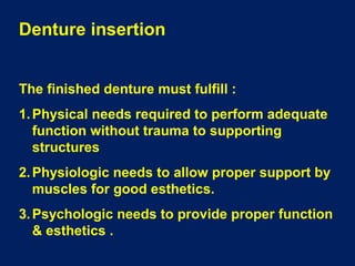
- 1. Denture insertion The finished denture must fulfill : 1. Physical needs required to perform adequate function without trauma to supporting structures 2. Physiologic needs to allow proper support by muscles for good esthetics. 3. Psychologic needs to provide proper function & esthetics .
- 2. Objectives : 1. To identify and correct : -- Any area of denture base causing pain or discomfort. -- Any area of denture interfering with retention & stability of dentures. -- Any part of denture that is esthetically unpleasing 2. To modify occlusal surfaces to harmonize occlusion (refine occlusion ) 3. To instruct the pt. how to use & care his denture . 4. To instruct the pt. in proper care of denture supporting tissues . 5. To advise the pt. on limitations of dentures to be expected
- 3. Spatula try-in The well adapted rims surfaces are paralels, lay onto each other, and bases are attached to the maxilla and mandibule
- 5. Transversal Christensen phenomenon Means that when the patient bites with well-adapted occlusal rims laterally, rims are in occlusion only on the workingside. On the balancing side an open, wedge-shaped gap occurs at the molars, between the upper and lower rims.
- 6. Sagittal Christensen phenomenon Means that when the patient bites with well-adapted occlusal rims, an open, wedge-shaped gap occurs at the molars.
- 7. Insertion procedures : 1. Extraoral examination of the finished denture: A. Examination of impression surface B. Evaluation of denture borders. C. evaluation of polished surface. 2. Intraoral examination of the finished denture : A. Location & relief of pressure areas in denture base B. Identification & reduction of overextended borders C. Evaluation of retention & stability D. Evaluation of esthetics & facial contours E. Refinement of occlusion F. Patient’s instructions
- 9. ARTCULATOR PIP Rubber bowl , mouth wash Hand mirror Completed dentures & study casts Straight handpiece & burs Occlusal indicating wax Articulating paper Mouth mirror & napkin
- 10. DENTURE ASSESSMENT Testing maxillary denture retention Apply a tipping force to the incisors in an attempt to break seal.
- 11. Testing maxillary denture stability Apply unilateral force to posterior occlusal surface of denture.
- 12. Testing stability and retention of mandibular denture Alternately apply unilateral force to posterior occlusal surfaces.
- 13. Removal of sharp edges & roughness as they will traumatize mucosa because dentures in function are continuously shifting
- 14. Prior to insertion , denture fitting surfaces must be inspected by palpation & any sharp projections must be ground
- 15. Checking labial notch As the denture is seated ,labial frenum is too narrow & shallow
- 16. Avoid sharp edges when trimming labial frenum notch . Frenum must be able to “ roll over” the denture
- 17. If stretched cheek is released , buccal frenum will lie tightly against functional border of the denture. As mouth is opened , buccal frena are stretched back & down. So, frena contribute to a well adapted border
- 18. To check retention of upper denture , pull down with 2 fingers . As denture moves down, then holds well, air is trapped under denture base upon insertion. To check retention of lower denture , push gently against lower anteriors with closed pliers as tongue filling floor of the mouth
- 19. Retentive denture is removed by breaking border seal with index fingers or by pulling out the cheeks Demonstrating masticatory stability when a closing force is exerted in the area of posterior teeth
- 20. Cleaning the dentures Denture brushes are advised ( specially important for elderly & disabled pt. )
- 21. Pt. must not hold lower denture using a squeezing action during cleaning Recommended method to hold a lower denture during cleaning
- 22. Pt. must not use hot water ( above 70 C ) to clean dentures. Elevated temperature crazes the denture surface resulting in a bleached appearance.
- 23. Upper denture with a clear palate. It enables areas of high pressure under the denture to be seen & preferred by some pts. due to its lighter appearance
- 24. PIP is used to identify areas on fitting surfaces of dentures , which exert heavier pressures on the tissues
- 25. Thin layer of PIP is painted on denture fitting surface Denture is seated in mouth & removed . Areas of high pressure are identified
- 26. Dynamic relationships of teeth as the jaw is moved to right & left and when protruded . This is checked with articulating paper of a different color
- 27. Pattern of occlusal contacts produced by sliding the mandible to the right ( working side ) & the left as the balancing side . Even occlusal contacts , such as those , are produced by grinding cusps .
- 28. Occlusal indicator wax may be used instead of articulating paper to indicate the location & extent of occlusal contacts , or near contacts .
- 29. Wax has a shiny , mildly adhesive coating on one side . Pencil is used to mark the teeth where the opposing arch is penetrating the wax
- 30. Areas of heavy tooth contact will cause penetration of wax , that allows identification of near contacts . Teeth must be marked where wax is penetrating Areas of heavy occlusal contacts
- 31. Dentures with well-balanced form function sufficiently well
- 32. At first , insert upper denture & observe the length of denture border for proper extension . Frenum may be displaced while making impression , so the frenal area must be checked
- 33. Labial frenum is displaced by notched border of the denture . So, notch must be slightly deepened & widened vertically with a large fissure bur. Also, bevel the inner margin of the notch
- 34. Border of lower denture is checked & adjusted . In mentalis muscle & retromolar pad areas impression tends to be overextended , so examination must be carefully performed
- 35. PIP is used as a thin layer painted on denture fitting surface so that brush marks are visible
- 36. Denture is inserted & heavy pressure is applied with fingers. Location of pressure spots in denture base that displace soft tissue can be determined & eliminated .
- 37. An area where the paste is very thin or completely displaced indicates pressure spots
- 38. Lower denture displays a pressure point on mylohyoid ridge. This pressure spot is removed with a bur
- 39. This recording & trimming is repeated until denture base surface do not show through the paste. The paste layer is even
- 40. PIP layer may be displaced due to brushing against the residual ridge on insertion & removal of denture. It must be determined as to whether areas with displaced paste are pressure areas or due to accidental contact during insertion & removal
- 41. PIP must be wiped off with cotton using firm , uni-directional stokes , not in a back & forth motion
- 42. Even paste record. No more adjustments are needed Areas roughened during adjusting the basal surface must be smoothed with a sandpaper cone & polished with silicone point
- 43. After adjusting the basal surface , occlusion refinement is done at chair-side . Prior to occlusal adjustment , cotton rolls are placed on both sides between upper & lower posterior teeth
- 44. Occlusal adjustment is always needed as inserting new dentures. Occlusal contacts in CO must be checked using thin articulating paper Heavy contacts in CO must be corrected by grinding the fossae & cusp inclines
- 45. These procedures must be repeated until posterior teeth have even occlusal contacts in CO
- 46. If interferences are found as the jaw is moved to right & left ,or protruded , they must be eliminated ( like selective grinding on articulator )
- 47. Occlusal registration paste A separating medium is placed on upper teeth & relationship of dentures recorded using occlusal registration paste .
- 48. An even layer of carborundum paste is placed on occlusal surface of lower posterior teeth . Pt. is instructed to slowly move the jaw to right & left and anteroposteriorly.
- 49. * Adjustment of the occlusion is necessary 1. to account for inherent errors caused by processing changes 2. to eliminate errors apparent at the try-in stage.
- 50. Causes of Occlusal Disharmony 1- Undetected errors in registering jaw relations. 2- Errors in mounting the master casts on the articulator. 3- Processing errors. 4. Dimensional changes of acrylic denture base material
- 51. 5- Differences in tissue adaptation between the processed denture bases & the record bases that were used in recording maxillo- mandibular relations. 6- Changes in the supporting structures since the impression is made ( as pt .using ill-fitting denture )
- 52. Correction of Occlusal Disharmony * Selective Grinding . To provide balanced contacts between the teeth in the retruded jaw relationship, . and in lateral & protrusive contact relations, . and free sliding contact movements to eccentric positions without cuspal interferences. . The occlusal vertical dimension must be maintained.
- 55. Occlusal Discrepancies may be corrected by either:1- Intra-oral Adjustment Techniques a- Articulating paper b- Occlusal indicator waxes c- Central bearing devices d- Abrasive paste.
- 56. 2- Extra-oral Adjustment Techniques a- Laboratory Remounting b- Clinical Remounting
- 57. Intra-oral Adjustment Techniques a- Articulating paper * It will not give an accurate indication of premature contacts because the resiliency of the supporting tissues allows the denture to shift producing markings which are frequently false.
- 58. Articulator paper detects premature occlusal contacts either on articulator or in the pt’s mouth
- 59. Articulating paper of a different color must be used to distinguish contacts marked in eccentric positions from those marked in centric position
- 60. When selective grinding in lateral occlusions is completed , incisal pin usually stays in contact with incisal table during lateral excursions
- 61. Marking & grinding procedure is repeated for both lateral movements until markings indicate uniform contacts on working & balancing sides
- 62. After completing selective grinding , marks made by movements in all directions must show uniform contacts. Red marks show contacts made in centric position & blue marks show contacts made during lateral and protrusive movements
- 63. b- Occlusal Indicator Wax * Two strips of adhesive green occlusal indicator wax 6 mm. wide are placed on the occlusal surfaces of the mandibular denture. The dentures are placed in the patient’ s mouth & the patient’s is guided into retruded contact position.
- 64. Wax must be carefully adapted to occlusal surfaces of teeth Mandible is gently guided so that teeth make contact with the lower jaw maximally retruded
- 65. c- Central- bearing Devices * When a centralbearing device assembled, bearing is the pin is adjusted to permit an evaluation of the occlusion
- 66. D- Abrasive Paste * Should not be used to eliminate errors in occlusion of cusp teeth. * The shifting of the denture bases as a result of premature contact may result in altering the occlusion so that centric occlusion does not correspond to centric relation.
- 67. Extra-oral Adjustment Techniques * Extra-oral adjustment of occlusion is carried out by a procedure known as remounting & selective grinding. It includes:1- Laboratory Remounting 2- Clinical Remounting
- 68. Laboratory Remounting * Objectives: 1- Restore or re-establish the vertical dimension of occlusion. 2- Perfect working and balancing occlusion 3- Establish protrusive balanced occlusion.
- 70. Supporting or Centric Holding Cusps * The vertical dimension of occlusion is maintained by occlusion of the palatal cusps of the maxillary teeth & the buccal cusps of the mandibular teeth.
- 71. Rules of adjustment a- If the cusp is high in centric & eccentric positions. Reduce the cusp. b- If the cusp is high in centric & not in eccentric positions. Deepen the opposing fossa or marginal ridge.
- 72. * After all interceptive contacts have been eliminated in centric & eccentric positions: a- Don’t reduce upper palatal cusps or lower buccal cusps b- Don’t deepen the fossa or marginal ridge of any tooth
- 73. II- Occlusal Balance in Lateral Excursions * Rules of Adjustment A- On the Working Side Adjust the buccal cusps of the upper teeth & the lingual cusps of the lower teeth ( B.U.L.L. rule ) to eliminate deflective contacts.
- 75. B- On the Balancing side Reduce inner inclines of lower buccal cusps , don’t reduce the cusp tip as it is a centric holding cusp
- 76. III- Selective Grinding for Protrusive Balance * In protrusive balance, the anterior teeth should make incisal edge contact at the same time that the tips of the buccal & lingual cusps of the posterior teeth contact.
- 77. Rules of Adjustment a- If anterior teeth have heavy contact with no posterior contact: * Reduce the labio-incisal surfaces of the lower teeth & the palatal surfaces of the upper teeth.
- 78. b. If posterior teeth have heavy contact with no anterior teeth contact. Reduce distal inclines of upper cusps & mesial inclines of lower cusps
- 79. Clinical Remounting * It consists of remounting the finished denture on an articulator by using interocclusal records in the patient’s mouth * The occlusion is then adjusted on the articulator to remove discrepancies & interferences.
- 80. Step by Step Procedure I- Preserve the orientation of the Maxillary cast to the Articulator:* A plaster remount index is an occlusal registration of the maxillary denture which is recorded on a remount jig attached to the lower member of the articulator.
- 82. II- Preparation of the Remount Casts Casts should be constructed to facilitate the positioning of the complete denture on articulator & the process of occlusal correction.
- 84. III- Centric Interocclusal Record * The centric interocclusal record is used to mount the mandibular denture on the articulator as a part of the clinical remount & selective grinding procedure.
- 85. Advantages of Clinical Remounting 1- It reduces patient participation. 2- It permits the dentist to see better what he is doing. 3- It provides a stable working foundation; denture bases are not shifting .
- 86. 4- The absence of saliva makes possible more accurate markings with the articulating paper. 5- Corrections can be made away from the patient .