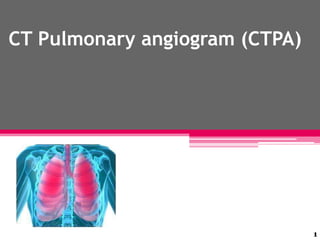
2. At a glance …
Medical diagnostic test that employs computed
tomography to obtain an image of the pulmonary
arteries.
CT offers a method of direct visualization of the
pulmonary vessels and lung parenchyma (structure in
the lungs that permits gas exchange) using diagnostic
x-ray images.
Main use is to diagnose pulmonary embolism(PE).
Uses a special dye (contrast material) and x-rays to see
how blood flows through the lungs.
2
3. Why the Test is Performed?
The test is used to detect blood clots (pulmonary
embolism)
May also be used to help your doctor diagnose:
• AV malformations of the lung
• Congenital (present from birth) narrowing of the
pulmonary vessels
• Pulmonary artery aneurysms
• Pulmonary hypertension - high blood pressure in the
arteries of the lungs
3
5. Arteriovenous malformation of the lung
Arteriovenous malformation or AVM is an abnormal
connection between arteries and veins, bypassing
the capillary system
5
7. 7
How to Prepare for the Test?
You should not eat or drink anything for 6
- 8 hours before the test.
Health care provider need to be informed :
• If the patient is pregnant.
• If the patient ever had any allergic reactions to
x-ray contrast material or iodine substances.
• If the patient is allergic to any medications.
• Which medications patient is taking .
• If the patient ever had any bleeding problems.
8. How the Test is Performed ?
Performed in the radiology department scanning room
Patient lying flat (supine) on a CT table
A timed intravenous injection of contrast media allows for
evaluation for pulmonary emboli.
CT scanner gantry (donut) passes over and around the
patient to perform the scan. The gantry travels over the
patient from the neck to the abdomen to create the scan.
8
9. How the Test is Performed ?(cont..)
Timing is essential and the radiographer will use
instructions to indicate when to breathe in, breath out
or hold breath.
Intravenous contrast is used to highlight the pulmonary
vessels, and to determine the presence of a clot in the
lung. If the patient lies very still, and the chest is
stationary (breath hold) this optimizes the quality of the
scan.
9
11. Example of a CTPA
demonstrating a saddle embolus. The white area above the
center is the pulmonary artery, opacified by radio contrast.
Inside it, the grey matter is blood clot.
The black areas on either side are the lungs, with around it the
chest wall.
11
12. Risks
Allergic reaction to the contrast dye.
Damage to the blood vessel as the needle and catheter
are inserted.
Blood clot traveling to the lungs, causing an embolism
Excessive bleeding or a blood clot where the catheter is
inserted, which can reduce blood flow to the leg.
Heart attack or stroke.
Hematoma (a collection of blood at the site of the needle
puncture).
Injury to the nerves at the puncture site
Kidney damage from the dye.
12