SlideShare una empresa de Scribd logo
1 de 64
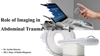
Role of Imaging in
Abdominal Trauma
• Dr. Sachin Sharma
• JR-1, Dept. of Radio-Diagnosis
Introduction
• Blunt trauma is a major cause of abdominal trauma and usually occurs
due to road traffic accidents or falls, with resulting compression and
deceleration injuries often associated with injuries to the head, spine
and limbs.
• In this situation, abdominal injuries can be missed, with up to 70% of
patients having either neurological impairment or a distracting injury.
• Although less common, penetrating trauma is also on the increase,
particularly in urban areas.
• Simultaneous therapeutic and diagnostic measures need to be
instituted.
• Plain abdominal radiographs have no role in assessment of blunt
abdominal trauma but may be useful in penetrating injury to
demonstrate bullets or fragments.
• FAST scanning can be performed rapidly and concurrently with other
procedures to look for free intraperitoneal or intrathoracic free fluid
and can triage a hemodynamically unstable patient to surgery.
• However, it is insufficiently sensitive to exclude solid organ,
mesenteric or retroperitoneal injury.
FAST
(FOCUSED ASSESSMENT WITH SONOGRAPHY IN TRAUMA)
• A focused, goal directed, sonographic examination of the abdomen.
• Goal is to see the presence of hemoperitoneum or hemopericardium.
• An extension of clinical examination.
• Part of the Primary Survey of any patient with signs of shock or
suspicion of abdominal injury.
• FAST examines four areas for free fluid:
oPerihepatic & hepato-renal space.
oPeri-splenic.
oPelvic.
oPericardial.
The Peri-hepatic Space
The Peri-splenic Space
The Pelvis Scan
• The pelvic examination visualizes :
• The cul-de-sac: the Pouch of Douglas in female sand the rectovesical
pouch in the male.
• Bladder
• Uterus and prostrate
A B
The pericardial scan
• The pericardial examination screens for fluid between the fibrous
pericardium and the heart.
• The transducer is placed just to the left of the xiphisternum and
angled upwards under the costal margin.
• FAST can detect between 100-250ml
• 0.5 cm in Morison's Pouch = 500ml
• 1 cm in Morison's Pouch = 1000ml
• CT can detect volumes of free fluid as low as 100ml
Some Important Points about FAST
Extended FAST (eFAST)
• Evaluation of pneumothorax and hemothorax in addition to
intraperitoneal injuries.
• Hemothorax:
Ultrasound is much more sensitive for detecting pleural fluid and
can identify as little as 20mL in the pleural space
• Pneumothorax :
M-MODE USG is used to detect pneumothorax.
eFAST: Anterior Thoracic Views
• Probe is usually placed on the anterior chest in the 3-4th intercostal
space and midclavicular line.
• When "Sliding sign" (seashore sign){NORMAL} is not present, a
pneumothorax is suspected.
• Comparing one side of the chest to the other may be helpful.
MDCT SCAN
• The mainstay of imaging following abdominal trauma is MDCT.
• CT should be done in all hemodynamically stable patients with
evidence of abdominal trauma (including a positive FAST scan).
• MDCT has a high accuracy (over 95%) for significant abdominopelvic
injury and is one of the major factors responsible for the increased
use of non-operative management in trauma patients.
MDCT PROTOCOLS
• Abdominal images are usually obtained around 60–65s after an
intravenous bolus injection of iodinated contrast
• The 60–65 s delay represents the portal venous phase of imaging and
gives an optimal trade-off between vascular opacification and solid
organ enhancement.
• The CT raw data are then reconstructed on
• A soft tissue algorithm.
• Bony algorithm
• A soft tissue algorithm at 1.5, 5-mm sections and multiplanar reformats
(MPRs) are also routinely reconstructed at 5 mm sections in the coronal
plane. MPRs are useful in assessment of disruption in the cephalocaudal
plane, e.g.- diaphragmatic rupture.
• Bony algorithm :- axial images should also be obtained and may be
used to provide coronal and sagittal MPRs of the spine and pelvis.
• Thin-section sagittal or coronal MPRs, MIPs are useful for CT
angiography and 3D reformats such as volume-rendered images, can be
subsequently generated.
• Delayed scans 5–10min after contrast are not performed routinely, can
be useful to differentiate
• Active bleeding from contained vascular injury, and
• To assess the integrity of the urinary tract, which should be opacified with
contrast on delayed images.
CT findings of shock
• Collapse of inferior vena cava
• Small aorta
• Persistent nephrogram without excretion
• Hypodense spleen, without enhancement and normal vascular pedicle
• Increased enhancement of the small bowel wall
• Increased enhancement of the adrenal glands
• Sometimes findings of right cardiac insufficiency with reflux into the
hepatic veins
ABDOMINAL WALL TRAUMA
• Stranding of the fat planes deep to the site of injury is suggestive of
possible peritoneal injury. trauma to the abdominal wall itself can be
associated with significant haematoma formation
• Shearing forces, can cause the development of an acute wall tear with
hernia formation.
A B
Spleen
• The spleen is the most commonly injured solid organ following
abdominal trauma.
• Several types of splenic injury can occur:
oIntraparenchymal hematoma
oSubcapsular haematoma,
oLaceration,
oActive extravasation,
oContained vascular injury
oInfarction.
Intraparenchymal splenic haematoma
SPLENIC LACERATION AND PERI-SPLENIC HEMATOMA
Splenic infarct.
American Association
for the Surgery of
Trauma (AAST) organ
injury severity scale
grading system for
splenic injury
LIVER
• The liver is the second most commonly injured organ in abdominal
trauma.
• Between 70 and 90% of hepatic injuries are minor, with the right lobe
most commonly affected.
• The liver is prone to the same array of injuries as the spleen, including
intraparenchymal and subcapsular hematoma lacerations, infarcts,
active extravasation and contained vascular injury.
• Lacerations near the major hepatic veins or IVC can be a/w injury to
these vessels resulting in catastrophic bleeding leading to high
mortality.
• Laceration occurring in the “bare area” of the liver, posteriorly in the
right lobe can lead to hemorrhage into the retroperitoneum, which is
frequently a/w injury to right adrenal and kidney.
• FAST can be falsely negative in retroperitoneal hemorrhage.
• Lacerations involving the porta hepatis – a/w a tear in the central
biliary tree, which can lead to biloma development manifesting as an
enlarging perihepatic fluid collection.
(a) (b)
(c)
GENITOURINARY TRACT
• The kidney is the most injured urologic organ following trauma.
• 80% of injuries are minor and heal spontaneously.
• The kidney is prone to the same range of injuries as the spleen and
liver, namely
• Contusions,
• Subcapsular and peri-nephric hematoma,
• Lacerations,
• Active extravasation,
• Contained vascular injury and
• Infarcts.
• Clinical indicator is the presence of hematuria, which is present in
95% of significant renal injuries, but can be absent in renal vascular
injuries, or injury to the PUJ or ureter.
• Contusions account for 80% of injuries.
• Subcapsular hematomas are rare, due to the strong attachment of the renal
capsule, but if large can compress the kidney sufficiently to cause excessive
renin secretion and hypertension – the “Page kidney”.
• Perinephric hematomas lie between the kidney and Gerota’s fascia and are
commoner than subcapsular hematomas.
• Lacerations are linear or branching low-density areas and if they reach the
hilum of the kidney, delayed scanning is indicated to look for a urine leak.
PAGE’S KIDNEY
• Ureteric injury - rare and typically occurs at the PUJ.
• Complete or
• Partial tear (partial) - Contrast is seen in distal ureter on delayed scans.
• Bladder injury – can be limited to wall contusion or hematoma, but
bladder rupture can also occur, which may be either
• Intraperitoneal - due to a direct blow to a distended bladder
• Extra peritoneal - due to laceration of the bladder wall from pelvic fracture
fragments
• Bladder rupture can be diagnosed with either conventional
cystography or CT cystography, where dilute contrast is instilled into
the bladder via a Foley catheter.
GASTROINTESTINAL TRACT TRAUMA
• Free intra-abdominal fluid in the absence of solid organ injury is
suggestive of injury to the bowel or mesentery.
1.Gastric trauma
• Blunt trauma to the stomach is relatively rare, and most commonly
involves the fundus.
• Full-thickness injury can lead to gastric rupture with
pneumoperitoneum.
• Gastric trauma may be associated with diaphragmatic rupture which
can predispose to intrathoracic gastric migration with possible
volvulus and strangulation.
2.Bowel injury
• The small bowel is most commonly injured, particularly where it is
relatively fixed at the ligament of Treitz and distal ileum
• Resulting in a wall contusion, serosal tear or full-thickness tear. Which may
manifest as a focal area of bowel wall thickening.
• Mesenteric injury on CT shows as mesenteric fat stranding or haematoma.
• Mesenteric vascular injury includes abrupt vessel termination and
vascular beading
• Mesenteric tear can give rise to a subsequent internal hernia.
• Hypovolaemic shock complex: diffuse small bowel wall thickening small,
bowel mucosal hyperenhancement, peripancreatic fluid, hyper enhancing
adrenal glands and flattening of the IVC.
Traumatic Duodenal Laceration
A B
A B
PANCREATIC TRAUMA
• Pancreatic trauma is rare but can lead to significant complications
such as
• abscess or pseudocyst formation,
• pancreatitis or pancreatic fistula.
• The key issue in pancreatic trauma is the integrity of the pancreatic
duct. On CT, signs of injury can be subtle which most commonly
demonstrates peripancreatic fluid and fat stranding.
• Peripancreatic changes can also be due to fluid resuscitation.
• Contusion to the gland can cause diffuse swelling, while a laceration
is manifested as a linear low-attenuation defect.
• Laceration >50% of the AP diameter of the gland is an increased risk
for ductal injury (Figure 4.22).
• Complete pancreatic transection may occur, but this can be difficult
to detect unless there is fluid or blood between the transected edges.
• Duct rupture can be diagnosed non-invasively with MRCP, which also
allows visualization of the ducts upstream from a disruption;
• ERCP however, offers the opportunity both for diagnosis and
treatment via covered stent placement.
Pancreatic Trauma
Gallbladder and Extrahepatic Bile Duct Injury
• Gallbladder injuries include contusion, laceration, perforation
and avulsion.
• CT signs include :
oPericholecystic fluid,
oGallbladder wall thickening and
oLoss of clarity,
oGallbladder wall tear active extravasation into the lumen.
Extrahepatic & Intrahepatic Bile Duct
Injury
• Acute deceleration with compression of the duct against the spine can
cause ductal transection, and elevation of the liver.
• As in intrahepatic bile duct injury, biloma may ensue, manifesting as a
perihepatic fluid collection on CT.
DIAPHRAGM
• Diaphragmatic rupture is rare and common on the left.
• Several signs of diaphragmatic rupture are:
oFocal diaphragmatic discontinuity and thickening.
oLoss of diaphragmatic clarity.
o Visceral herniation into the chest k/as the “collar”sign.
oDependent viscera sign: a tear causes the solid organs of the upper
abdomen to lie against the posterior chest wall in the supine position.
Diaphragmatic rupture
Dependent viscera sign
Role of Imaging in Abdominal Trauma.pptx

Más contenido relacionado

La actualidad más candente

Radiological anatomy of liver segments
Radiological anatomy of liver segmentsRadiological anatomy of liver segments
Radiological anatomy of liver segmentsTarun Goyal
 
Body ct protocols
Body ct protocolsBody ct protocols
Body ct protocolsbongsung
 
Imaging of Malignant Liver Lesions
Imaging of Malignant Liver LesionsImaging of Malignant Liver Lesions
Imaging of Malignant Liver LesionsSahil Chaudhry
 
Doppler ultrasound of carotid arteries
Doppler ultrasound of carotid arteriesDoppler ultrasound of carotid arteries
Doppler ultrasound of carotid arteriesSamir Haffar
 
Ultrasound of the urinary tract - Renal infections
Ultrasound of the urinary tract - Renal infectionsUltrasound of the urinary tract - Renal infections
Ultrasound of the urinary tract - Renal infectionsSamir Haffar
 
Conventional nephroradiology
Conventional nephroradiologyConventional nephroradiology
Conventional nephroradiologyFarragBahbah
 
CURRENT TRENDS IN Renal cell carcinoma
CURRENT TRENDS IN Renal cell carcinomaCURRENT TRENDS IN Renal cell carcinoma
CURRENT TRENDS IN Renal cell carcinomaVikas Kumar
 
Copy of Mitsumori-CT angiography techniques
Copy of Mitsumori-CT angiography techniquesCopy of Mitsumori-CT angiography techniques
Copy of Mitsumori-CT angiography techniquesamin usmani
 
Ct mri urography
Ct mri urographyCt mri urography
Ct mri urographyDev Lakhera
 
PTBD (Percutaneus trans-hepatic biliary drainage) PPT,PDF PK
PTBD (Percutaneus trans-hepatic biliary drainage) PPT,PDF PKPTBD (Percutaneus trans-hepatic biliary drainage) PPT,PDF PK
PTBD (Percutaneus trans-hepatic biliary drainage) PPT,PDF PKDr pradeep Kumar
 
Imaging in genitourinary trauma
Imaging in genitourinary traumaImaging in genitourinary trauma
Imaging in genitourinary traumaRamanGhimire3
 
Adrenal gland tumors (Radiology)
Adrenal gland tumors (Radiology)Adrenal gland tumors (Radiology)
Adrenal gland tumors (Radiology)Dr Abdalla M. Gamal
 

La actualidad más candente (20)

Radiological anatomy of liver segments
Radiological anatomy of liver segmentsRadiological anatomy of liver segments
Radiological anatomy of liver segments
 
Coronary CT
Coronary CTCoronary CT
Coronary CT
 
TACE- Transarterial Chemoembolisation
TACE- Transarterial Chemoembolisation TACE- Transarterial Chemoembolisation
TACE- Transarterial Chemoembolisation
 
Abdominal CT scan made easy
Abdominal CT scan made easyAbdominal CT scan made easy
Abdominal CT scan made easy
 
Body ct protocols
Body ct protocolsBody ct protocols
Body ct protocols
 
Imaging of Malignant Liver Lesions
Imaging of Malignant Liver LesionsImaging of Malignant Liver Lesions
Imaging of Malignant Liver Lesions
 
Quantitative CT and MR Imaging of Liver
Quantitative CT and MR Imaging of Liver Quantitative CT and MR Imaging of Liver
Quantitative CT and MR Imaging of Liver
 
Doppler ultrasound of carotid arteries
Doppler ultrasound of carotid arteriesDoppler ultrasound of carotid arteries
Doppler ultrasound of carotid arteries
 
Ultrasound of the urinary tract - Renal infections
Ultrasound of the urinary tract - Renal infectionsUltrasound of the urinary tract - Renal infections
Ultrasound of the urinary tract - Renal infections
 
Conventional nephroradiology
Conventional nephroradiologyConventional nephroradiology
Conventional nephroradiology
 
CURRENT TRENDS IN Renal cell carcinoma
CURRENT TRENDS IN Renal cell carcinomaCURRENT TRENDS IN Renal cell carcinoma
CURRENT TRENDS IN Renal cell carcinoma
 
Copy of Mitsumori-CT angiography techniques
Copy of Mitsumori-CT angiography techniquesCopy of Mitsumori-CT angiography techniques
Copy of Mitsumori-CT angiography techniques
 
Ct mri urography
Ct mri urographyCt mri urography
Ct mri urography
 
PTBD (Percutaneus trans-hepatic biliary drainage) PPT,PDF PK
PTBD (Percutaneus trans-hepatic biliary drainage) PPT,PDF PKPTBD (Percutaneus trans-hepatic biliary drainage) PPT,PDF PK
PTBD (Percutaneus trans-hepatic biliary drainage) PPT,PDF PK
 
Intravenous urography (IVU)
Intravenous urography (IVU)Intravenous urography (IVU)
Intravenous urography (IVU)
 
IMAGING OF URETHRAL INJURY
IMAGING OF URETHRAL INJURYIMAGING OF URETHRAL INJURY
IMAGING OF URETHRAL INJURY
 
Coronary CT
Coronary CTCoronary CT
Coronary CT
 
Renal doppler usg
Renal doppler usgRenal doppler usg
Renal doppler usg
 
Imaging in genitourinary trauma
Imaging in genitourinary traumaImaging in genitourinary trauma
Imaging in genitourinary trauma
 
Adrenal gland tumors (Radiology)
Adrenal gland tumors (Radiology)Adrenal gland tumors (Radiology)
Adrenal gland tumors (Radiology)
 

Similar a Role of Imaging in Abdominal Trauma.pptx

Liver trauma الدكتور طارق المنيزل
Liver trauma الدكتور طارق المنيزل Liver trauma الدكتور طارق المنيزل
Liver trauma الدكتور طارق المنيزل Tariq Al munaizel
 
Splenic injury - Copy.pptx
Splenic injury - Copy.pptxSplenic injury - Copy.pptx
Splenic injury - Copy.pptxasispodar
 
Abdominal vascular injuries
Abdominal vascular injuriesAbdominal vascular injuries
Abdominal vascular injuriesAbdulsalam Taha
 
ABDOMINAL TRAUMA ASSESSMENT AND TREATMENT.pptx
ABDOMINAL TRAUMA ASSESSMENT AND TREATMENT.pptxABDOMINAL TRAUMA ASSESSMENT AND TREATMENT.pptx
ABDOMINAL TRAUMA ASSESSMENT AND TREATMENT.pptxNethaji Perumal
 
Splenic Injury.pptx
Splenic Injury.pptxSplenic Injury.pptx
Splenic Injury.pptxKIST Surgery
 
PANCREATIC INJURY.ppt.pptx
PANCREATIC INJURY.ppt.pptxPANCREATIC INJURY.ppt.pptx
PANCREATIC INJURY.ppt.pptxAzan Rid
 
Mesenteric vascular occlusion
Mesenteric vascular occlusionMesenteric vascular occlusion
Mesenteric vascular occlusionBashir BnYunus
 
ANUERYSMS ,AV FISTULAS ,ARTERISTIS ,RAYNAUDS DISEASE -1.pptx
ANUERYSMS ,AV FISTULAS ,ARTERISTIS ,RAYNAUDS DISEASE -1.pptxANUERYSMS ,AV FISTULAS ,ARTERISTIS ,RAYNAUDS DISEASE -1.pptx
ANUERYSMS ,AV FISTULAS ,ARTERISTIS ,RAYNAUDS DISEASE -1.pptxmusayansa
 
Imaging in blunt abdominal trauma
Imaging in blunt abdominal traumaImaging in blunt abdominal trauma
Imaging in blunt abdominal traumaSunil Kumar
 
Solid organ injuries following abdominal trauma
Solid organ injuries following abdominal traumaSolid organ injuries following abdominal trauma
Solid organ injuries following abdominal traumaAymen Ahmad Khan
 
Urology surgery. Bladder, Urethra and Prostsate
Urology surgery. Bladder, Urethra and ProstsateUrology surgery. Bladder, Urethra and Prostsate
Urology surgery. Bladder, Urethra and ProstsateKishore Rajan
 

Similar a Role of Imaging in Abdominal Trauma.pptx (20)

Liver trauma
Liver traumaLiver trauma
Liver trauma
 
Liver trauma الدكتور طارق المنيزل
Liver trauma الدكتور طارق المنيزل Liver trauma الدكتور طارق المنيزل
Liver trauma الدكتور طارق المنيزل
 
Splenic injury - Copy.pptx
Splenic injury - Copy.pptxSplenic injury - Copy.pptx
Splenic injury - Copy.pptx
 
Abdominal vascular injuries
Abdominal vascular injuriesAbdominal vascular injuries
Abdominal vascular injuries
 
ABDOMINAL TRAUMA ASSESSMENT AND TREATMENT.pptx
ABDOMINAL TRAUMA ASSESSMENT AND TREATMENT.pptxABDOMINAL TRAUMA ASSESSMENT AND TREATMENT.pptx
ABDOMINAL TRAUMA ASSESSMENT AND TREATMENT.pptx
 
COA PRESENTATION.pptx
COA PRESENTATION.pptxCOA PRESENTATION.pptx
COA PRESENTATION.pptx
 
Splenic Injury.pptx
Splenic Injury.pptxSplenic Injury.pptx
Splenic Injury.pptx
 
PANCREATIC INJURY.ppt.pptx
PANCREATIC INJURY.ppt.pptxPANCREATIC INJURY.ppt.pptx
PANCREATIC INJURY.ppt.pptx
 
Mesenteric vascular occlusion
Mesenteric vascular occlusionMesenteric vascular occlusion
Mesenteric vascular occlusion
 
Acute abdomen
Acute abdomenAcute abdomen
Acute abdomen
 
Liver trauma
Liver traumaLiver trauma
Liver trauma
 
ANUERYSMS ,AV FISTULAS ,ARTERISTIS ,RAYNAUDS DISEASE -1.pptx
ANUERYSMS ,AV FISTULAS ,ARTERISTIS ,RAYNAUDS DISEASE -1.pptxANUERYSMS ,AV FISTULAS ,ARTERISTIS ,RAYNAUDS DISEASE -1.pptx
ANUERYSMS ,AV FISTULAS ,ARTERISTIS ,RAYNAUDS DISEASE -1.pptx
 
Imaging in blunt abdominal trauma
Imaging in blunt abdominal traumaImaging in blunt abdominal trauma
Imaging in blunt abdominal trauma
 
Solid organ injuries following abdominal trauma
Solid organ injuries following abdominal traumaSolid organ injuries following abdominal trauma
Solid organ injuries following abdominal trauma
 
Liver trauma
Liver traumaLiver trauma
Liver trauma
 
Abdominal Aortic Aneurysm
Abdominal Aortic AneurysmAbdominal Aortic Aneurysm
Abdominal Aortic Aneurysm
 
Urology Trauma
Urology TraumaUrology Trauma
Urology Trauma
 
Vascular
VascularVascular
Vascular
 
Urology surgery. Bladder, Urethra and Prostsate
Urology surgery. Bladder, Urethra and ProstsateUrology surgery. Bladder, Urethra and Prostsate
Urology surgery. Bladder, Urethra and Prostsate
 
Liver trauma: A comprehensive review of classification, mechanisms, early man...
Liver trauma: A comprehensive review of classification, mechanisms, early man...Liver trauma: A comprehensive review of classification, mechanisms, early man...
Liver trauma: A comprehensive review of classification, mechanisms, early man...
 

Último

Call Girls Service Chennai Jiya 7001305949 Independent Escort Service Chennai
Call Girls Service Chennai Jiya 7001305949 Independent Escort Service ChennaiCall Girls Service Chennai Jiya 7001305949 Independent Escort Service Chennai
Call Girls Service Chennai Jiya 7001305949 Independent Escort Service ChennaiNehru place Escorts
 
Call Girls Service In Shyam Nagar Whatsapp 8445551418 Independent Escort Service
Call Girls Service In Shyam Nagar Whatsapp 8445551418 Independent Escort ServiceCall Girls Service In Shyam Nagar Whatsapp 8445551418 Independent Escort Service
Call Girls Service In Shyam Nagar Whatsapp 8445551418 Independent Escort Serviceparulsinha
 
call girls in munirka DELHI 🔝 >༒9540349809 🔝 genuine Escort Service 🔝✔️✔️
call girls in munirka  DELHI 🔝 >༒9540349809 🔝 genuine Escort Service 🔝✔️✔️call girls in munirka  DELHI 🔝 >༒9540349809 🔝 genuine Escort Service 🔝✔️✔️
call girls in munirka DELHI 🔝 >༒9540349809 🔝 genuine Escort Service 🔝✔️✔️saminamagar
 
Call Girls Service in Bommanahalli - 7001305949 with real photos and phone nu...
Call Girls Service in Bommanahalli - 7001305949 with real photos and phone nu...Call Girls Service in Bommanahalli - 7001305949 with real photos and phone nu...
Call Girls Service in Bommanahalli - 7001305949 with real photos and phone nu...narwatsonia7
 
Call Girls Service Nandiambakkam | 7001305949 At Low Cost Cash Payment Booking
Call Girls Service Nandiambakkam | 7001305949 At Low Cost Cash Payment BookingCall Girls Service Nandiambakkam | 7001305949 At Low Cost Cash Payment Booking
Call Girls Service Nandiambakkam | 7001305949 At Low Cost Cash Payment BookingNehru place Escorts
 
Russian Call Girls Chickpet - 7001305949 Booking and charges genuine rate for...
Russian Call Girls Chickpet - 7001305949 Booking and charges genuine rate for...Russian Call Girls Chickpet - 7001305949 Booking and charges genuine rate for...
Russian Call Girls Chickpet - 7001305949 Booking and charges genuine rate for...narwatsonia7
 
Call Girls Jayanagar Just Call 7001305949 Top Class Call Girl Service Available
Call Girls Jayanagar Just Call 7001305949 Top Class Call Girl Service AvailableCall Girls Jayanagar Just Call 7001305949 Top Class Call Girl Service Available
Call Girls Jayanagar Just Call 7001305949 Top Class Call Girl Service Availablenarwatsonia7
 
Call Girls Hosur Just Call 7001305949 Top Class Call Girl Service Available
Call Girls Hosur Just Call 7001305949 Top Class Call Girl Service AvailableCall Girls Hosur Just Call 7001305949 Top Class Call Girl Service Available
Call Girls Hosur Just Call 7001305949 Top Class Call Girl Service Availablenarwatsonia7
 
Call Girl Koramangala | 7001305949 At Low Cost Cash Payment Booking
Call Girl Koramangala | 7001305949 At Low Cost Cash Payment BookingCall Girl Koramangala | 7001305949 At Low Cost Cash Payment Booking
Call Girl Koramangala | 7001305949 At Low Cost Cash Payment Bookingnarwatsonia7
 
College Call Girls Vyasarpadi Whatsapp 7001305949 Independent Escort Service
College Call Girls Vyasarpadi Whatsapp 7001305949 Independent Escort ServiceCollege Call Girls Vyasarpadi Whatsapp 7001305949 Independent Escort Service
College Call Girls Vyasarpadi Whatsapp 7001305949 Independent Escort ServiceNehru place Escorts
 
call girls in green park DELHI 🔝 >༒9540349809 🔝 genuine Escort Service 🔝✔️✔️
call girls in green park  DELHI 🔝 >༒9540349809 🔝 genuine Escort Service 🔝✔️✔️call girls in green park  DELHI 🔝 >༒9540349809 🔝 genuine Escort Service 🔝✔️✔️
call girls in green park DELHI 🔝 >༒9540349809 🔝 genuine Escort Service 🔝✔️✔️saminamagar
 
Call Girl Bangalore Nandini 7001305949 Independent Escort Service Bangalore
Call Girl Bangalore Nandini 7001305949 Independent Escort Service BangaloreCall Girl Bangalore Nandini 7001305949 Independent Escort Service Bangalore
Call Girl Bangalore Nandini 7001305949 Independent Escort Service Bangalorenarwatsonia7
 
Call Girls ITPL Just Call 7001305949 Top Class Call Girl Service Available
Call Girls ITPL Just Call 7001305949 Top Class Call Girl Service AvailableCall Girls ITPL Just Call 7001305949 Top Class Call Girl Service Available
Call Girls ITPL Just Call 7001305949 Top Class Call Girl Service Availablenarwatsonia7
 
Call Girls Jp Nagar Just Call 7001305949 Top Class Call Girl Service Available
Call Girls Jp Nagar Just Call 7001305949 Top Class Call Girl Service AvailableCall Girls Jp Nagar Just Call 7001305949 Top Class Call Girl Service Available
Call Girls Jp Nagar Just Call 7001305949 Top Class Call Girl Service Availablenarwatsonia7
 
College Call Girls Pune Mira 9907093804 Short 1500 Night 6000 Best call girls...
College Call Girls Pune Mira 9907093804 Short 1500 Night 6000 Best call girls...College Call Girls Pune Mira 9907093804 Short 1500 Night 6000 Best call girls...
College Call Girls Pune Mira 9907093804 Short 1500 Night 6000 Best call girls...Miss joya
 
Low Rate Call Girls Pune Esha 9907093804 Short 1500 Night 6000 Best call girl...
Low Rate Call Girls Pune Esha 9907093804 Short 1500 Night 6000 Best call girl...Low Rate Call Girls Pune Esha 9907093804 Short 1500 Night 6000 Best call girl...
Low Rate Call Girls Pune Esha 9907093804 Short 1500 Night 6000 Best call girl...Miss joya
 
Book Call Girls in Yelahanka - For 7001305949 Cheap & Best with original Photos
Book Call Girls in Yelahanka - For 7001305949 Cheap & Best with original PhotosBook Call Girls in Yelahanka - For 7001305949 Cheap & Best with original Photos
Book Call Girls in Yelahanka - For 7001305949 Cheap & Best with original Photosnarwatsonia7
 
Glomerular Filtration rate and its determinants.pptx
Glomerular Filtration rate and its determinants.pptxGlomerular Filtration rate and its determinants.pptx
Glomerular Filtration rate and its determinants.pptxDr.Nusrat Tariq
 
Book Call Girls in Kasavanahalli - 7001305949 with real photos and phone numbers
Book Call Girls in Kasavanahalli - 7001305949 with real photos and phone numbersBook Call Girls in Kasavanahalli - 7001305949 with real photos and phone numbers
Book Call Girls in Kasavanahalli - 7001305949 with real photos and phone numbersnarwatsonia7
 
Call Girls Hebbal Just Call 7001305949 Top Class Call Girl Service Available
Call Girls Hebbal Just Call 7001305949 Top Class Call Girl Service AvailableCall Girls Hebbal Just Call 7001305949 Top Class Call Girl Service Available
Call Girls Hebbal Just Call 7001305949 Top Class Call Girl Service Availablenarwatsonia7
 

Último (20)

Call Girls Service Chennai Jiya 7001305949 Independent Escort Service Chennai
Call Girls Service Chennai Jiya 7001305949 Independent Escort Service ChennaiCall Girls Service Chennai Jiya 7001305949 Independent Escort Service Chennai
Call Girls Service Chennai Jiya 7001305949 Independent Escort Service Chennai
 
Call Girls Service In Shyam Nagar Whatsapp 8445551418 Independent Escort Service
Call Girls Service In Shyam Nagar Whatsapp 8445551418 Independent Escort ServiceCall Girls Service In Shyam Nagar Whatsapp 8445551418 Independent Escort Service
Call Girls Service In Shyam Nagar Whatsapp 8445551418 Independent Escort Service
 
call girls in munirka DELHI 🔝 >༒9540349809 🔝 genuine Escort Service 🔝✔️✔️
call girls in munirka  DELHI 🔝 >༒9540349809 🔝 genuine Escort Service 🔝✔️✔️call girls in munirka  DELHI 🔝 >༒9540349809 🔝 genuine Escort Service 🔝✔️✔️
call girls in munirka DELHI 🔝 >༒9540349809 🔝 genuine Escort Service 🔝✔️✔️
 
Call Girls Service in Bommanahalli - 7001305949 with real photos and phone nu...
Call Girls Service in Bommanahalli - 7001305949 with real photos and phone nu...Call Girls Service in Bommanahalli - 7001305949 with real photos and phone nu...
Call Girls Service in Bommanahalli - 7001305949 with real photos and phone nu...
 
Call Girls Service Nandiambakkam | 7001305949 At Low Cost Cash Payment Booking
Call Girls Service Nandiambakkam | 7001305949 At Low Cost Cash Payment BookingCall Girls Service Nandiambakkam | 7001305949 At Low Cost Cash Payment Booking
Call Girls Service Nandiambakkam | 7001305949 At Low Cost Cash Payment Booking
 
Russian Call Girls Chickpet - 7001305949 Booking and charges genuine rate for...
Russian Call Girls Chickpet - 7001305949 Booking and charges genuine rate for...Russian Call Girls Chickpet - 7001305949 Booking and charges genuine rate for...
Russian Call Girls Chickpet - 7001305949 Booking and charges genuine rate for...
 
Call Girls Jayanagar Just Call 7001305949 Top Class Call Girl Service Available
Call Girls Jayanagar Just Call 7001305949 Top Class Call Girl Service AvailableCall Girls Jayanagar Just Call 7001305949 Top Class Call Girl Service Available
Call Girls Jayanagar Just Call 7001305949 Top Class Call Girl Service Available
 
Call Girls Hosur Just Call 7001305949 Top Class Call Girl Service Available
Call Girls Hosur Just Call 7001305949 Top Class Call Girl Service AvailableCall Girls Hosur Just Call 7001305949 Top Class Call Girl Service Available
Call Girls Hosur Just Call 7001305949 Top Class Call Girl Service Available
 
Call Girl Koramangala | 7001305949 At Low Cost Cash Payment Booking
Call Girl Koramangala | 7001305949 At Low Cost Cash Payment BookingCall Girl Koramangala | 7001305949 At Low Cost Cash Payment Booking
Call Girl Koramangala | 7001305949 At Low Cost Cash Payment Booking
 
College Call Girls Vyasarpadi Whatsapp 7001305949 Independent Escort Service
College Call Girls Vyasarpadi Whatsapp 7001305949 Independent Escort ServiceCollege Call Girls Vyasarpadi Whatsapp 7001305949 Independent Escort Service
College Call Girls Vyasarpadi Whatsapp 7001305949 Independent Escort Service
 
call girls in green park DELHI 🔝 >༒9540349809 🔝 genuine Escort Service 🔝✔️✔️
call girls in green park  DELHI 🔝 >༒9540349809 🔝 genuine Escort Service 🔝✔️✔️call girls in green park  DELHI 🔝 >༒9540349809 🔝 genuine Escort Service 🔝✔️✔️
call girls in green park DELHI 🔝 >༒9540349809 🔝 genuine Escort Service 🔝✔️✔️
 
Call Girl Bangalore Nandini 7001305949 Independent Escort Service Bangalore
Call Girl Bangalore Nandini 7001305949 Independent Escort Service BangaloreCall Girl Bangalore Nandini 7001305949 Independent Escort Service Bangalore
Call Girl Bangalore Nandini 7001305949 Independent Escort Service Bangalore
 
Call Girls ITPL Just Call 7001305949 Top Class Call Girl Service Available
Call Girls ITPL Just Call 7001305949 Top Class Call Girl Service AvailableCall Girls ITPL Just Call 7001305949 Top Class Call Girl Service Available
Call Girls ITPL Just Call 7001305949 Top Class Call Girl Service Available
 
Call Girls Jp Nagar Just Call 7001305949 Top Class Call Girl Service Available
Call Girls Jp Nagar Just Call 7001305949 Top Class Call Girl Service AvailableCall Girls Jp Nagar Just Call 7001305949 Top Class Call Girl Service Available
Call Girls Jp Nagar Just Call 7001305949 Top Class Call Girl Service Available
 
College Call Girls Pune Mira 9907093804 Short 1500 Night 6000 Best call girls...
College Call Girls Pune Mira 9907093804 Short 1500 Night 6000 Best call girls...College Call Girls Pune Mira 9907093804 Short 1500 Night 6000 Best call girls...
College Call Girls Pune Mira 9907093804 Short 1500 Night 6000 Best call girls...
 
Low Rate Call Girls Pune Esha 9907093804 Short 1500 Night 6000 Best call girl...
Low Rate Call Girls Pune Esha 9907093804 Short 1500 Night 6000 Best call girl...Low Rate Call Girls Pune Esha 9907093804 Short 1500 Night 6000 Best call girl...
Low Rate Call Girls Pune Esha 9907093804 Short 1500 Night 6000 Best call girl...
 
Book Call Girls in Yelahanka - For 7001305949 Cheap & Best with original Photos
Book Call Girls in Yelahanka - For 7001305949 Cheap & Best with original PhotosBook Call Girls in Yelahanka - For 7001305949 Cheap & Best with original Photos
Book Call Girls in Yelahanka - For 7001305949 Cheap & Best with original Photos
 
Glomerular Filtration rate and its determinants.pptx
Glomerular Filtration rate and its determinants.pptxGlomerular Filtration rate and its determinants.pptx
Glomerular Filtration rate and its determinants.pptx
 
Book Call Girls in Kasavanahalli - 7001305949 with real photos and phone numbers
Book Call Girls in Kasavanahalli - 7001305949 with real photos and phone numbersBook Call Girls in Kasavanahalli - 7001305949 with real photos and phone numbers
Book Call Girls in Kasavanahalli - 7001305949 with real photos and phone numbers
 
Call Girls Hebbal Just Call 7001305949 Top Class Call Girl Service Available
Call Girls Hebbal Just Call 7001305949 Top Class Call Girl Service AvailableCall Girls Hebbal Just Call 7001305949 Top Class Call Girl Service Available
Call Girls Hebbal Just Call 7001305949 Top Class Call Girl Service Available
 

Role of Imaging in Abdominal Trauma.pptx

  • 1. Role of Imaging in Abdominal Trauma • Dr. Sachin Sharma • JR-1, Dept. of Radio-Diagnosis
  • 2. Introduction • Blunt trauma is a major cause of abdominal trauma and usually occurs due to road traffic accidents or falls, with resulting compression and deceleration injuries often associated with injuries to the head, spine and limbs. • In this situation, abdominal injuries can be missed, with up to 70% of patients having either neurological impairment or a distracting injury. • Although less common, penetrating trauma is also on the increase, particularly in urban areas.
  • 3. • Simultaneous therapeutic and diagnostic measures need to be instituted. • Plain abdominal radiographs have no role in assessment of blunt abdominal trauma but may be useful in penetrating injury to demonstrate bullets or fragments. • FAST scanning can be performed rapidly and concurrently with other procedures to look for free intraperitoneal or intrathoracic free fluid and can triage a hemodynamically unstable patient to surgery. • However, it is insufficiently sensitive to exclude solid organ, mesenteric or retroperitoneal injury.
  • 4.
  • 5.
  • 6.
  • 7. FAST (FOCUSED ASSESSMENT WITH SONOGRAPHY IN TRAUMA) • A focused, goal directed, sonographic examination of the abdomen. • Goal is to see the presence of hemoperitoneum or hemopericardium. • An extension of clinical examination. • Part of the Primary Survey of any patient with signs of shock or suspicion of abdominal injury.
  • 8.
  • 9. • FAST examines four areas for free fluid: oPerihepatic & hepato-renal space. oPeri-splenic. oPelvic. oPericardial.
  • 12. The Pelvis Scan • The pelvic examination visualizes : • The cul-de-sac: the Pouch of Douglas in female sand the rectovesical pouch in the male. • Bladder • Uterus and prostrate
  • 13. A B
  • 14. The pericardial scan • The pericardial examination screens for fluid between the fibrous pericardium and the heart. • The transducer is placed just to the left of the xiphisternum and angled upwards under the costal margin.
  • 15.
  • 16. • FAST can detect between 100-250ml • 0.5 cm in Morison's Pouch = 500ml • 1 cm in Morison's Pouch = 1000ml • CT can detect volumes of free fluid as low as 100ml Some Important Points about FAST
  • 17. Extended FAST (eFAST) • Evaluation of pneumothorax and hemothorax in addition to intraperitoneal injuries. • Hemothorax: Ultrasound is much more sensitive for detecting pleural fluid and can identify as little as 20mL in the pleural space • Pneumothorax : M-MODE USG is used to detect pneumothorax.
  • 18. eFAST: Anterior Thoracic Views • Probe is usually placed on the anterior chest in the 3-4th intercostal space and midclavicular line. • When "Sliding sign" (seashore sign){NORMAL} is not present, a pneumothorax is suspected. • Comparing one side of the chest to the other may be helpful.
  • 19.
  • 20. MDCT SCAN • The mainstay of imaging following abdominal trauma is MDCT. • CT should be done in all hemodynamically stable patients with evidence of abdominal trauma (including a positive FAST scan). • MDCT has a high accuracy (over 95%) for significant abdominopelvic injury and is one of the major factors responsible for the increased use of non-operative management in trauma patients.
  • 21. MDCT PROTOCOLS • Abdominal images are usually obtained around 60–65s after an intravenous bolus injection of iodinated contrast • The 60–65 s delay represents the portal venous phase of imaging and gives an optimal trade-off between vascular opacification and solid organ enhancement. • The CT raw data are then reconstructed on • A soft tissue algorithm. • Bony algorithm
  • 22. • A soft tissue algorithm at 1.5, 5-mm sections and multiplanar reformats (MPRs) are also routinely reconstructed at 5 mm sections in the coronal plane. MPRs are useful in assessment of disruption in the cephalocaudal plane, e.g.- diaphragmatic rupture. • Bony algorithm :- axial images should also be obtained and may be used to provide coronal and sagittal MPRs of the spine and pelvis. • Thin-section sagittal or coronal MPRs, MIPs are useful for CT angiography and 3D reformats such as volume-rendered images, can be subsequently generated. • Delayed scans 5–10min after contrast are not performed routinely, can be useful to differentiate • Active bleeding from contained vascular injury, and • To assess the integrity of the urinary tract, which should be opacified with contrast on delayed images.
  • 23. CT findings of shock • Collapse of inferior vena cava • Small aorta • Persistent nephrogram without excretion • Hypodense spleen, without enhancement and normal vascular pedicle • Increased enhancement of the small bowel wall • Increased enhancement of the adrenal glands • Sometimes findings of right cardiac insufficiency with reflux into the hepatic veins
  • 24.
  • 25. ABDOMINAL WALL TRAUMA • Stranding of the fat planes deep to the site of injury is suggestive of possible peritoneal injury. trauma to the abdominal wall itself can be associated with significant haematoma formation • Shearing forces, can cause the development of an acute wall tear with hernia formation.
  • 26. A B
  • 27. Spleen • The spleen is the most commonly injured solid organ following abdominal trauma. • Several types of splenic injury can occur: oIntraparenchymal hematoma oSubcapsular haematoma, oLaceration, oActive extravasation, oContained vascular injury oInfarction.
  • 29. SPLENIC LACERATION AND PERI-SPLENIC HEMATOMA
  • 30.
  • 32. American Association for the Surgery of Trauma (AAST) organ injury severity scale grading system for splenic injury
  • 33. LIVER • The liver is the second most commonly injured organ in abdominal trauma. • Between 70 and 90% of hepatic injuries are minor, with the right lobe most commonly affected. • The liver is prone to the same array of injuries as the spleen, including intraparenchymal and subcapsular hematoma lacerations, infarcts, active extravasation and contained vascular injury.
  • 34. • Lacerations near the major hepatic veins or IVC can be a/w injury to these vessels resulting in catastrophic bleeding leading to high mortality. • Laceration occurring in the “bare area” of the liver, posteriorly in the right lobe can lead to hemorrhage into the retroperitoneum, which is frequently a/w injury to right adrenal and kidney. • FAST can be falsely negative in retroperitoneal hemorrhage. • Lacerations involving the porta hepatis – a/w a tear in the central biliary tree, which can lead to biloma development manifesting as an enlarging perihepatic fluid collection.
  • 35.
  • 36.
  • 37.
  • 38.
  • 39.
  • 41. GENITOURINARY TRACT • The kidney is the most injured urologic organ following trauma. • 80% of injuries are minor and heal spontaneously. • The kidney is prone to the same range of injuries as the spleen and liver, namely • Contusions, • Subcapsular and peri-nephric hematoma, • Lacerations, • Active extravasation, • Contained vascular injury and • Infarcts.
  • 42. • Clinical indicator is the presence of hematuria, which is present in 95% of significant renal injuries, but can be absent in renal vascular injuries, or injury to the PUJ or ureter. • Contusions account for 80% of injuries. • Subcapsular hematomas are rare, due to the strong attachment of the renal capsule, but if large can compress the kidney sufficiently to cause excessive renin secretion and hypertension – the “Page kidney”. • Perinephric hematomas lie between the kidney and Gerota’s fascia and are commoner than subcapsular hematomas. • Lacerations are linear or branching low-density areas and if they reach the hilum of the kidney, delayed scanning is indicated to look for a urine leak.
  • 44.
  • 45.
  • 46. • Ureteric injury - rare and typically occurs at the PUJ. • Complete or • Partial tear (partial) - Contrast is seen in distal ureter on delayed scans. • Bladder injury – can be limited to wall contusion or hematoma, but bladder rupture can also occur, which may be either • Intraperitoneal - due to a direct blow to a distended bladder • Extra peritoneal - due to laceration of the bladder wall from pelvic fracture fragments • Bladder rupture can be diagnosed with either conventional cystography or CT cystography, where dilute contrast is instilled into the bladder via a Foley catheter.
  • 47.
  • 48.
  • 49. GASTROINTESTINAL TRACT TRAUMA • Free intra-abdominal fluid in the absence of solid organ injury is suggestive of injury to the bowel or mesentery. 1.Gastric trauma • Blunt trauma to the stomach is relatively rare, and most commonly involves the fundus. • Full-thickness injury can lead to gastric rupture with pneumoperitoneum. • Gastric trauma may be associated with diaphragmatic rupture which can predispose to intrathoracic gastric migration with possible volvulus and strangulation.
  • 50.
  • 51. 2.Bowel injury • The small bowel is most commonly injured, particularly where it is relatively fixed at the ligament of Treitz and distal ileum • Resulting in a wall contusion, serosal tear or full-thickness tear. Which may manifest as a focal area of bowel wall thickening. • Mesenteric injury on CT shows as mesenteric fat stranding or haematoma. • Mesenteric vascular injury includes abrupt vessel termination and vascular beading • Mesenteric tear can give rise to a subsequent internal hernia. • Hypovolaemic shock complex: diffuse small bowel wall thickening small, bowel mucosal hyperenhancement, peripancreatic fluid, hyper enhancing adrenal glands and flattening of the IVC.
  • 53.
  • 54.
  • 55. A B
  • 56. PANCREATIC TRAUMA • Pancreatic trauma is rare but can lead to significant complications such as • abscess or pseudocyst formation, • pancreatitis or pancreatic fistula. • The key issue in pancreatic trauma is the integrity of the pancreatic duct. On CT, signs of injury can be subtle which most commonly demonstrates peripancreatic fluid and fat stranding. • Peripancreatic changes can also be due to fluid resuscitation. • Contusion to the gland can cause diffuse swelling, while a laceration is manifested as a linear low-attenuation defect.
  • 57. • Laceration >50% of the AP diameter of the gland is an increased risk for ductal injury (Figure 4.22). • Complete pancreatic transection may occur, but this can be difficult to detect unless there is fluid or blood between the transected edges. • Duct rupture can be diagnosed non-invasively with MRCP, which also allows visualization of the ducts upstream from a disruption; • ERCP however, offers the opportunity both for diagnosis and treatment via covered stent placement.
  • 59. Gallbladder and Extrahepatic Bile Duct Injury • Gallbladder injuries include contusion, laceration, perforation and avulsion. • CT signs include : oPericholecystic fluid, oGallbladder wall thickening and oLoss of clarity, oGallbladder wall tear active extravasation into the lumen.
  • 60. Extrahepatic & Intrahepatic Bile Duct Injury • Acute deceleration with compression of the duct against the spine can cause ductal transection, and elevation of the liver. • As in intrahepatic bile duct injury, biloma may ensue, manifesting as a perihepatic fluid collection on CT.
  • 61. DIAPHRAGM • Diaphragmatic rupture is rare and common on the left. • Several signs of diaphragmatic rupture are: oFocal diaphragmatic discontinuity and thickening. oLoss of diaphragmatic clarity. o Visceral herniation into the chest k/as the “collar”sign. oDependent viscera sign: a tear causes the solid organs of the upper abdomen to lie against the posterior chest wall in the supine position.

Notas del editor

  1. Good Morning Respected Teachers, Seniors and my fellow PGs. I will be presenting a seminar on Role of Imaging in Abdominal Trauma.
  2. This plain abdominal radiograph is demonstrating an intra-abdominal metal fragment from an improvised explosive device.
  3. This is the algorithm in trauma management When there is a Blunt or penetrating injury, we look for hemodynamic stability of the patient. If the patient is hemodynamically stable, we perform a FAST scan.
  4. This is the diagrammatic representation of FAST scan. It shows different regions where the probe is placed and following spaces are looked upon.
  5. The probe is placed in the right mid to posterior axillary line at the level of the12th rib. Here we can see Peri-hepatic free fluid in Morison's pouch.
  6. Transducer is positioned in left posterior axillary line between 10th and 11th ribs with beam in the coronal plane.
  7. The transducer is placed in midline just superior to the symphysis pubis.
  8. A .FF IN POD B. FF IN RECTOVESICAL SPACE
  9. Positive FAST demonstrating pericardial effusion
  10. A: Normal Pleura B: Seashore Sign C: Barcode Sign (PNEUMOTHORAX)
  11. A. CT scan demonstrating left inferior lumbar hernia in a male patient with blunt abdominal trauma (arrow) following a motor vehicle collision. B. CT scan demonstrates a full thickness tear of the abdominal wall muscles with omental fat herniation and a small amount of associated haematoma (arrow).
  12. Coronal CT scan demonstrating an intraparenchymal splenic haematoma with several focal areas of increased attenuation consistent with bleeding points (long arrow). Peri-splenic haematoma is also present.
  13. CT scan demonstrating splenic lacerations with an associated peri-splenic haematoma. The more anterior laceration contains an area of active contrast extravasation
  14. Coronal CT scan demonstrating an intraparenchymal splenic haematoma with active contrast extravasation (arrow). Diaphragmatic rupture is evident by the acute bleeding into the left pleural space.
  15. CT scan demonstrating a wedge-shaped area of low attenuation within the spleen (arrow) with the base against the capsular surface consistent with a splenic infarct.
  16. This is an Axial CT image demonstrating intraparenchymal liver haematoma in segment 6 (arrow). No active bleeding is seen though there is some perihepatic haematoma and further haematoma around the pancreatic head.
  17. This is an Axial CT image demonstrating a heterogenous, subcapsular haematoma (arrow) which is compressing the liver with retention of the normal liver contour. The blood around the periphery is of slightly higher density and is probably clotted.
  18. This image shows an Axial CT image demonstrating a laceration within segment 4b of the liver with a central area of active extravasation (long arrow). Both hemoperitoneum and pneumoperitoneum due to a small bowel perforation (short arrow) are also present.
  19. This CT scan image demonstrates several large intraparenchymal haematomas with multiple foci of active contrast extravasation (arrows).
  20. This table shows AAST liver trauma grading system, it correlates with outcome. Most liver injuries heal spontaneously within 3 months, however non- operative management is usually successful.
  21. Image (a) demonstrates a central liver laceration near the porta hepatis (arrow). Image (b) demonstrates a low-attenuation fluid collection in the right flank (arrow) in same patient after 1 month HIDA scan (c) shows leakage of bile from the biliary tree into the collection (arrow), which was drained percutaneously.
  22. 2-minute delayed phase imaging showing a 5 cm enhancing mass in the left mid kidney. There is perinephric hematoma secondary to haemorrhage from the renal mass, which is vascular. This is the cause for Page phenomenon and new onset uncontrolled hypertension.
  23. Axial (a) and coronal MPR (b) CT images demonstrating a left lower pole renal laceration (arrows) with an associated perinephric haematoma.
  24. Venous phase contrast-enhanced CT image (a) demonstrates a large laceration that reaches the right renal hilum with associated perinephric fluid. Delayed axial CT image 10 minutes post contrast (b) demonstrates leakage of contrast opacified urine into the perinephric fluid collection (arrow) confirming the presence of a traumatic urinoma. Absence of contrast in the right ureter is also demonstrated.
  25. Axial CT image following instillation of dilute contrast into the bladder via a Foley catheter demonstrates extensive leakage of contrast into the perivesical spaces consistant with extraperitoneal bladder rupture. A fracture of the anterior column of the right acetabulum (arrow) is also present.
  26. Axial CT image through the stomach demonstrating a full-thickness tear of the gastric fundus with some spilling of gastric contents into the peritoneal cavity (arrow).
  27. Axial (a) and coronal MPR (b) CT images demonstrate free retroperitoneal air (arrows) extending into the root of the small bowel mesentery and thickening of the adjacent duodenum.
  28. Rectal perforation following a gunshot wound to the pelvis. Axial CT image with intravenous and rectal contrast demonstrates extensive leakage of rectal contrast (arrow). Metallic artefact from the bullet fragments is also demonstrated.
  29. Contrast-enhanced axial CT image demonstrating prominent small bowel wall thickening and mucosal hyperenhancement due to increased vascular permeability (arrow) as result of hypovolaemic shock
  30. Axial (a) and coronal MPR (b) CT images demonstrating a mesenteric haematoma with a central focus of active contrast extravasation (arrows).
  31. This is a Contrast-enhanced axial CT image demonstrating a vertical laceration at the junction of the neck and body of the pancreas (arrow). The laceration extends over approximately 75% of the anteroposterior diameter of the gland and is likely to be associated with pancreatic duct injury. An intraparenchymal splenic haematoma is also present.
  32. Axial (a) and coronal MPR (b) CT images demonstrating the “collar sign” of a diaphragmatic tear with narrowing of the stomach as it traverses the diaphragmatic defect (arrows).
  33. Axial (A) and sagittal (B) contrast-enhanced computed tomography images in a 95-year-old male with left traumatic diaphragmatic rupture post-MVC(motor vehicle collision) demonstrates loss of the expected interposed lung between the stomach (S) and the posterior chest wall (arrows).