Presentation1.pptx, radiological imaging of aids diseases
•Descargar como PPTX, PDF•
23 recomendaciones•3,636 vistas


Recomendados
Más contenido relacionado
La actualidad más candente
La actualidad más candente (20)
Destacado
Destacado (20)
Similar a Presentation1.pptx, radiological imaging of aids diseases
Similar a Presentation1.pptx, radiological imaging of aids diseases (20)
Más de Abdellah Nazeer
Más de Abdellah Nazeer (20)
Presentation1.pptx, radiological imaging of aids diseases
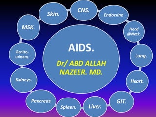
- 1. CNS. AIDS. Endocrine Dr/ ABD ALLAH NAZEER. MD. Head @Neck. Lung. Heart. GIT. Spleen. Liver. MSK. Kidneys. Pancreas Genito-urinary. Skin.
- 2. AIDS in the CNS 10-20% of pts with AIDS present with neurologic disease 40% of AIDS pts will have neurologic involvement in course of disease CNS symptoms may indicate overall deterioration HIV is the most common viral infection of the CNS and PNS. Up to 50% of HIV patients have clinical apparent neurological disease. Types of CNS Disease HIV itself Neoplasm, primary CNS lymphoma Metastases Opportunistic infection parasitic: toxoplasmosis (toxo), amebiasis mycobacterial: tuberculosis (TB), mycobacterium avium complex (MAC) viral: JC virus (JCV), herpes simplex virus (HSV), cytomegalovirus (CMV) fungal: cryptococcus, aspergillus, candida
- 11. Diffuse cerebral cortical atrophy with AIDS patient.
- 14. Toxoplasmic encephalitis in a 36-year-old patient with AIDS.
- 15. Toxoplasmosis
- 19. HIV Encephalitis Vs PML
- 20. Brain Lymphoma.
- 22. Two cases of Lymphoma: periventricular spread at CT case.
- 23. Contrast-enhancing lesions on CT scans (A–D) in 4 patients with AIDS-related PCNSL. Note irregularly enhancing lesions in the right parietal lobe (A), right occipital lobe (B), and right periventricular white matter (C and D); most of the lesions show ring enhancement (A, B, and C).
- 24. Solitary lesions on a noncontrast CT scan (A), contrast-enhanced CT scans (B and C), and an MR image (D) in 3 patients with non-AIDS PCNSL. Note a hyperattenuated lesion in the frontal lobe on the noncontrast CT scan (A) with marked enhancement on the contrast series (B) and focal contrast-enhancing lesions in the left basal ganglia (C) and temporal lobe (D). One lesion has ring enhancement (D).
- 25. Contrast-enhanced axial (B and D) and coronal (A and C) MR images in 4 patients with non-AIDS PCNSL. Note lesions in the basal ganglia (A), ventricles (B), frontal lobes (C), and cerebellar lobes (D).
- 29. HIV associated with cord neuropathy.
- 30. HIV associated with cord neuropathy.
- 32. Infection by a diverse array of organisms, as well as HIV-associated malignancies (i.e., Kaposi's sarcoma and lymphoma), have been detected in the pituitary and adrenal glands . Such occurrences were far more common prior to the widespread introduction of potent ART, although they may still be observed in patients not receiving ART or who have antiretroviral drug resistant infection. Tissue is generally required for a definitive diagnosis. When technically feasible, fine needle aspiration (FNA) biopsy of the adrenal gland provides a less invasive alternative to open biopsy. Pheochromocytoma must always be excluded before FNA biopsy of the adrenal gland is performed. Standard functional testing should also be performed since clinically significant endocrine dysfunction may accompany glandular infection or infiltration of the pituitary or adrenal glands.
- 33. Hypopituitarism secondary to HIV infection.
- 34. Acute hypophysitis and hypopituitarism in early syphilitic meningitis in a HIV-infected patient: at diagnosis and after treatment.
- 36. Chagas’ disease presenting with a suprasellar mass and panhypopituitarism secondary to HIV infection.
- 37. Hypoadrenalism secondary to HIV infection.
- 38. Infectious Process of the Neck Related to HIV In the HIV-infected population, however, Mycobacterium avium complex (MAC) infection is the most common mycobacterial infection. Fungal infections, including cryptococcosis, histoplasmosis, and coccidioidomycosis, can manifest as a cervical mass in the HIV-infected patient. Cryptococcus neoformans is the most prevalent cause of deep-seated fungal infections in the HIV-infected population, occurring in 5 to 10% of these patients and commonly involves the lungs and the meninges Candidiasis (Oral Thrush), Oral candidiasis is by far the most common oral condition in HIV /AIDS patients. Herpes labialis most commonly presents as crops of fever blisters on the palate, gingiva, or other oral mucosal surfaces. Otitis Media and externa. The most common otologic problems reported in HIV-infected patients are serous otitis media and recurrent acute otitis media and externa. The prevalence of rhinosinusitis ranges from 20 to 70% in patients with AIDS. Causative organisms include atypical opportunistic and common organisms responsible for sinusitis in hosts without AIDS.
- 39. Neoplastic disease in the head and neck of patients with AIDS. Immunosuppression increases the risk of developing malignancies. In immunosuppression due to human immunodeficiency virus (HIV) disease the common head and neck tumors are Kaposi's sarcoma and non- Hodgkin's lymphoma. Squamous cell carcinoma has also been reported. Kaposi's sarcoma is the commonest neoplastic disease in AIDS. The incidence of lymphoma is rapidly increasing. This article reviews the incidence, clinical presentation and management of these diseases in the head and neck in AIDS patients.
- 40. Invasive Aspergillosis Presenting as a Neck Mass in a Person With HIV/AIDS HIV with chronic bilateral bacterial maxillary sinusitis.
- 41. HIV two cases neck abscess caused by Mycobacterium avium complex.
- 42. Kaposi,s sarcoma of the oropharynx in AIDS patient.
- 43. Two HIV -related Kaposi’s sarcoma.
- 44. HIV -related Kaposi’s sarcoma of the oropharynx:
- 45. Disseminated AIDS-related KS in a 36-year-old man with involvement of the tongue base and soft and hard palates.
- 46. Neck lymphoma with AIDS patient.
- 47. HIV AND THE RESPIRATORY SYSTEM Lung is a major target organ for HIV infection that has been shown to be present in T and B lymphocytes, pulmonary fibroblasts, macrophages, Natural Killer cells, eosinophils, monocytes and dendritic cells. As a consequence, progressive quantitative and functional depression within the CD4 lymphocytes and other immunological subsets occur and render the patient more prone to a wide array of infectious and non-infectious complications. Over 98% of respiratory complications were infectious and the most frequent complications were acute bronchitis, bacterial pneumonia and PCP. The mortality and morbidity of HIV-infected patients have dramatically improved as a result of the introduction of highly active antiretroviral therapy (HAART). An analysis of the Centers for Disease Control and Prevention's HIV.
- 48. The most frequent respiratory diagnoses in the HIV-infected patients are upper respiratory tract infection, acute bronchitis, and acute sinusitis. They occur at all strata of CD4 cell counts and have higher rates compared with HIV-negative control. Recurrent bacterial pneumonia and pulmonary tuberculosis (PTB) occur more frequently in patients with CD4 cell counts less than 400 cells/μL; Pneumocystis pneumonia (PCP) and disseminated TB usually diagnose when CD4 cell counts drop below 200 cells/μL. Disseminated MAC, fungal pneumonia, and cytomegalovirus pneumonitis occur in patients with the most severe immunosuppression (CD4 cell counts less than 100 cells/μL). Bacterial pneumonia occurs more frequently in HIV-infected patients than in the general population. Pneumonia occurs at any CD4 cell count but is especially common as HIV infection progresses. Rate of pneumonia is higher in intravenous drug users than other transmission categories. The spectrum of bacterial pathogens is similar to that of community-acquired pneumonia in the general population and Streptococcus pneumoniae remains the most common pathogen. Staphylococcus aureus and gram-negative organisms in particular Pseudomonas aerugionsa are seen more frequently in advanced disease. The incidence of bacteraemia is increased too. Influenza and pneumococcal vaccination is indicated in HIV-infected patients.
- 49. Non-infectious pulmonary disease Pulmonary involvement is present in up to one third of patients with known Kaposi's sarcoma (KS). It usually follows the appearance of cutaneous disease. Intrathoracic involvement by KS may include parenchymal disease, endobronchial lesions, pleural disease, and adenopathy. The prognosis of pulmonary KS is poor with median survival of 2 to 10 months. However, there is significant reduction in mortality after the introduction of HAART and newer combination chemotherapy. The incidence of intrathoracic manifestations of AIDS-associated lymphoma ranges from 6% to 31%. Lung involvement is usually seen in association with other sites of disease but occasionally it can be the initial or predominant site of disease. The median CD4 cell count has been noted to be lower in patients with pulmonary involvement than in those without. Chest radiographs may show effusions, multi-nodular infiltrates, consolidation, mass lesions, focal or diffuse interstitial infiltrates, and hilar adenopathy. Epidemiological studies have suggested that lung cancer occurs more frequently in HIV-infected patients, but is often linked to the increased smoking rates. Adenocarcinoma has been the most common histology. Survival is significantly shorter for HIV-infected patients compared with HIV-negative subjects and outcomes of these patients remain poor despite HAART.
- 52. Chest radiograph of an HIV positive individual with a CD4 cell count above 200 cells/mm3, revealing right upper lobe consolidation. Sputum and blood cultures were positive for Streptococcus pneumoniae.
- 53. Two chest radiograph of an HIV positive individual with a CD4 cell count above 200 cells/mm3, revealing right upper and lower lobes consolidation with areas of cavitation at the upper lobe consolidation.
- 54. Chest high-resolution computed tomography (HRCT) scan of an HIV positive person with a CD4 cell count below 200 cells/mm3, whose chest radiograph was normal. Chest HRCT scan revealed the characteristic patchy ground-glass opacities of PCP. Induced sputum microscopic examination revealed Pneumocystis cysts and trophic forms.
- 56. Pneumocystis carinii pneumonia. These chest radiographs are of two patients. Both show -ground glass appearance. The left chest X-ray (CXR) shows a much more subtle ground-glass appearance while the right CXR shows a much more gross ground-glass appearance mimicking pulmonary edema.
- 57. Pneumocystis carinii pneumonia. Computed tomography (CT) in a subacute phase showing foci of consolidation and interlobular septal thickening due to organized inflammatory infiltrate on high-resolution CT
- 58. Pneumocystis carinii pneumonia (PCP). High-resolution computed tomography showing the hallmark of PCP in a clinical setting of immune compromise. Note the ground-glass attenuation with a geographic or mosaic distribution
- 59. Pneumocystis carinii pneumonia. Chest X-ray and computed tomography show a left-sided ground-glass pattern and a right-sided large tension pneumothorax. Note the mediastinal shift.
- 60. Bronchiolitis obliterans with or without organizing pneumonia in the absence of infection can be a feature of acquired immunodeficiency syndrome (AIDS). This is an infrequent imaging diagnosis, although focal air trapping on expiratory computed tomography, consistent with bronchiolitis obliterans, has been demonstrated in two-thirds of human immunodeficiency virus-positive patients without AIDS, the severity increasing with the duration of infection.
- 61. Mycobacterium xenopi in a human immunodeficiency virus, 36-year old, male patient with a CD4 count of 80 with four positive sputum samples and a bronchoalveolar lavage for acid-fast bacilli. The chest X-ray and computed tomography scans show cavitating consolidation, loss of volume, traction bronchiectasis and ground-glass appearances in the right apical region superimposed on bullous disease of the lungs. There is no associated lymphadenopathy
- 62. A 26-year-old human immunodeficiency virus-positive female presented with shortness of breath. The chest X-ray shows a large left-sided pleural effusion and loss of height and erosion of the articular plates between the 9th and 10th vertebral bodies associated with soft tissue swelling. A sagittal T2-weigted magnetic resonance scan of the dorsal spine shows complete obliteration of the disc between the 9th and 10th dorsal vertebral bodies associated with fluid collection anterior to the spine representing pus. Acid-fast bacilli were identified in the aspirated pus
- 63. CT Chest with lung windowing of Pulmonary Kaposi’s Sarcoma in an adult demonstrates multiple poorly circumscribed pulmonary nodules and ground glass opacification.
- 64. Kaposi’s Sarcoma with peri-hilar opacification, pleural effusion and lymphadenopathy.
- 65. Pulmonary KS in a 45-year-old man.
- 66. Kaposi's Sarcoma with peri-hilar opacification, pleural effusion and lymphadenopathy.
- 67. Lymphoma. Chest X-ray (CXR) on a human immunodeficiency virus patient that presented with multiple lung masses, which grew rapidly mimicking infection. Note that there is no associated lymphadenopathy. Well-defined solitary or multiple parenchymal nodules CXR are common. A percutaneous biopsy revealed a non-Hodgkin’s lymphoma
- 68. Pulmonary parenchymal lymphoma in a 41-year-old HIV-positive man.
- 69. NHL in a 23-year-old human immunodeficiency virus female. The chest radiograph shows multiple well-defined lung nodules within the left lung associated with mediastinal lymphadenopathy. Lymphadenopathy is a less common feature in acquired immunodeficiency disease-related NHL and nodes are rarely significant according to size criteria unlike as in the case shown here, where there is significant lymphadenopathy as confirmed by computed tomography (right upper frame). Magnetic resonance imaging is the imaging of choice to detect vascular encasement
- 70. Cardiovascular Manifestations of HIV Infection: Myocarditis Myocarditis and HIV-1 myocardial infection are still the most studied causes of dilated cardiomyopathy in HIV disease. HIV-1 virions appear to infect myocardial cells in patchy distributions without a clear direct association between HIV-1 and cardiac myocyte dysfunction Autoimmunity Cardiac-specific auto antibodies (anti-α myosin auto antibodies) have been reported in up to 30% of patients with HIV-associated cardiomyopathy. The finding supports the theory that cardiac autoimmunity plays a role in the pathogenesis of HIV-related heart disease and suggests that cardiac auto antibodies may be markers of left ventricular dysfunction in HIV-positive patients with previously normal echocardiographic findings. Dilated Cardiomyopathy HIV disease is recognized as an important cause of dilated rdiomyopathy, with an estimated annual incidence of 15.9 in 1000 before the introduction of HAART. The importance of cardiac dysfunction is demonstrated by its effect on survival in acquired immunodeficiency syndrome (AIDS).
- 71. Pericardial Effusion The prevalence of pericardial effusion in asymptomatic AIDS patients has been estimated at 11% before the introduction of HAART. HIV infection should be included in the differential diagnosis of unexplained pericardial effusion or tamponade. Pericardial effusion in HIV disease may be related to opportunistic infections or to malignancy, but most often a clear pathology is not found. The effusion may be part of a generalized serous effusive process also involving pleural and peritoneal surfaces. Endocarditis The prevalence of infective endocarditis in HIV-infected patients is similar to that in patients of other risk groups, such as intravenous drug users.6 Estimates of endocarditis prevalence vary from 6.3% to 34% of HIV-infected patients who use intravenous drugs independently of HAART regimens.6 Right-sided valves are predominantly affected, and the most frequent agents are Staphylococcus aureus (>75% of cases), Streptococcus pneumoniae, Haemophilus influenzae, Candida albicans, Aspergillus fumigatus, and Cryptococcus neoformans. Patients with HIV generally have presentations and survival from infective endocarditis similar to those without HIV (85% versus 93%).
- 72. HIV-Associated Pulmonary Hypertension The pathogenesis of primary pulmonary hypertension in HIV infection is multifactorial and poorly understood. Primary pulmonary hypertension has been found in hemophiliacs receiving lipophilized factor VIII, intravenous drug users, and patients with left ventricular dysfunction, obscuring any relationship with HIV-1. HIV-1 is frequently identified in alveolar macrophages on histology. Vasculitis and Coronary Artery Disease A wide range of inflammatory vascular diseases, including polyarteritis nodosa, Henoch-Schönlein purpura, and drug-induced hypersensitivity vasculitis, may develop in HIV-infected individuals. Kawasaki-like syndrome and Takayasu’s arteritis have been also described. Hypertension and Coagulative Disorders The prevalence of hypertension in HIV disease has been estimated to have been about 20% to 25% before the introduction of HAART. Recent reports indicate that elevated blood pressure may be related to protease inhibitor-induced lipodystrophy and metabolic disorders, especially fasting triglyceride, with a prevalence of hypertension in up to 74% of patients with HAART-related metabolic syndrome.
- 73. Chest radiograph showing cardiomeagly in a five-year-old girl with HIV infection, cardiomyopathy, and congestive heart failure.
- 75. HIV with dilated cardiomyopathy.
- 76. HIV with pericardial and pleural effusion.
- 77. This human immunodeficiency virus patient suffered from gram negative septicemia, which was successfully treated. However, routine physical examination revealed an audible bruit on thoracic auscultation. The chest X-ray shows a prominent hump over the proximal descending aorta due to mycotic aortic aneurysm. The axial computed tomography (CT) and coronal CT reconstruction elegantly demonstrate the abnormality
- 78. The gastrointestinal (GI) tract is a major site of disease in HIV infection: almost half of HIV-infected patients present with GI symptoms, and almost all patients develop GI complications. GI symptoms such as anorexia, weight loss, dysphagia, odynophagia, abdominal pain, and diarrhea are frequent and usually nonspecific among these patients. Endoscopy is the diagnostic test of choice for most HIV-associated GI diseases, as endoscopic and histopathologic evaluation can render diagnoses in patients with non-specific symptoms. In the past three decades, studies have elucidated a variety of HIV-associated inflammatory, infectious, and neoplastic GI diseases, often with specific predilection for various sites. HIV-associated esophageal disease, for example, commonly includes candidiasis, cytomegalovirus (CMV) and herpes simplex virus (HSV) infection, Kaposi's sarcoma (KS), and idiopathic ulceration. Gastric disease, though less common than esophageal disease, frequently involves CMV, Mycobacterium avium-intracellulare (MAI), and neoplasia (KS, lymphoma).
- 79. Double contrast barium esophagography shows innumerable pseudo membranes and plaques (arrows) "shaggy esophagus“ in a patient with AIDS. Candida Esophagitis.
- 81. CMV esophagitis.
- 82. Primary non-Hodgkin B-cell Lymphoma of the esophagus.
- 83. lymphoma of the duodenum.
- 84. AIDS with gastric lymphoma with circumferential wall thickening.
- 85. AIDS patient with Pneumatosis intestinalis and mesenteric air are present.
- 86. AIDS with non-Hodgkin Burkett's lymphoma of the terminal ileum.
- 87. Upper GI series demonstrating abnormal appearance of small bowel with diffuse thickening of mucosa (arrows) in an AIDS patient with CMV.
- 88. Diffuse large cell lymphoma in the mesentery with infiltration of the jejunum.
- 89. AIDS with Small bowel lymphoma
- 90. AIDS patient with Cytomegalovirus colitis
- 91. AIDS patient with Cytomegalovirus colitis.
- 92. KS in a 44-year-old man with AIDS who presented with fever and diarrhea. Abdominal CT scan shows circumferential wall thickening of the cecum (arrows) that is not associated with enlarged lymph nodes or adjacent fat stranding.
- 93. Burkett lymphoma of the thyroid, in a 26 year old pregnant woman, with involvement of the right colon a) T2 weighted MRI of the neck show homogeneous enlargement of the thyroid left lobe (orange arrow). (b) Axial, (c and d) coronal and (e) sagittal T2 weighted MRI, reveals a mass in the right colon (yellow arrow), hyperintense comparing to the liver, and the fetus (purple arrow).
- 94. CT of the abdomen showing an inflammatory matted mass from extensive gastrointestinal involvement with KS.
- 95. AIDS patient with eccentric thickening of the colon. Note the severe pericolonic stranding around the transverse colon.
- 96. An irregular thickening of rectal wall. Kaposi sarcoma was performed at the biopsy
- 97. Coronal reconstructions CT show large masses and bulky masses in a patient HIV-positive. AIDS-related lymphoma.
- 98. Pathology of AIDS-related liver disease. Hepatomegally and abnormalities of serum liver tests are common problems in patients with acquired immune deficiency syndrome. Opportunist infections (Mycobacterium avium-intracellulare and cytomegalovirus) and neoplasms (lymphoma, Kaposi's sarcoma) are among the most prevalent hepatic lesions in AIDS. Although Kupffer cells and endothelial cells are potential sites of human immunodeficiency virus 1 (HIV-1) infection, current studies do not indicate that the liver is a major reservoir for this virus. Drug hepatotoxicity, multimicrobial infections of the biliary tree resembling sclerosing cholangitis and a variety of nonspecific hepatic changes should be considered in evaluating AIDS patients or HIV-1-infected patients with evidence of liver dysfunction
- 99. Hepatobiliary System. Infections of the liver and biliary system with any of a number of organisms may result in solitary or multiple hepatic abscesses or different forms of biliary involvement, including papillary stenosis and sclerosing cholangitis. Papillary stenosis will cause bile duct dilatation and delayed emptying of contrast into the duodenum; sclerosing cholangitis presents with focal strictures and dilatations of the intra- and extrahepatic bile ducts. Both forms of biliary disease may be seen at the same time, and occasionally long biliary strictures are noted. Liver disease may be diagnosed with CT or ultrasound; biliary disease may be diagnosed with CT, ultrasound, or endoscopic retrograde cholangiopancreatography (ERCP).
- 101. PRIMARY LIVER AIDS-RELATED LYMPHOMA
- 102. An 35-years-old man with 1 year of history fever, weight loss and abdominal pain. Ultrasound detected a large hypoechoic mass into the right lobe liver. HIV-positive. Biopsy confirm that is primary extranodal lymphoma
- 103. AIDS with non-Hodgkin's lymphoma.
- 105. Disseminated AIDS-related KS with liver involvement, para-aortic and inguinal LN.
- 106. Ultrasound of liver demonstrating intrahepatic duct dilation (arrows) of AIDS patient.
- 107. ERCP demonstrating abnormal dilation and beaded appearance of extrahepatic and intrahepatic biliary system consistent with cholangitis. (1) Common hepatic duct. (2) Dilated branch of left intrahepatic duct of AIDS patient.
- 108. HIV in the spleen include amyloid-like protein deposition, perivascular hyalinization, infarcts, necrosis, hemosiderin pigment deposits, plasma cell infiltrates, neutrophilic microabscesses, spindle cell infiltrates, extramedullary hematopoiesis, and granulomatous reaction. Some of these changes have been shown to be associated with specific infections. Our aim in this study was to compare AIDS splenic pathology in a more recent group of patients with splenic pathology in AIDS before the use of multiagent therapy.
- 109. AIDS with splenic Pneumocystosis: An Atypical Presentation of Extra-pulmonary Pneumocystis Infection .
- 110. Disseminated AIDS-related KS in a 41-year-old man with abdominal compromise.
- 111. Splenic KS in a 50-year-old HIV-positive man. Abdominal CT scan shows multiple subcentimeter hypoattenuating nodules in the spleen
- 112. AIDS patient showing focal low- density lesions due to non-Hodgkin's lymphoma within an enlarged spleen.
- 113. Contrast-enhanced CT scan of an immunocompromised patient shows multiple rounded areas of decreased attenuation scattered throughout the spleen and liver
- 114. Pancreatic disease in AIDS--a review. Patients with the acquired immunodeficiency syndrome (AIDS) can develop pancreatic disease from causes unrelated to AIDS as well as AIDS-specific lesions. AIDS-specific causes include opportunistic infection, AIDS-associated neoplasia, and medications used to treat complications of AIDS. Reported pancreatic opportunistic pathogens include Mycobacterium tuberculosis, Mycobacterium avium intracellulare, Cryptococcus neoformans, Candida, Aspergillus, Toxoplasma gondii, Pneumocystis carinii, cytomegalovirus, herpes simplex, cryptosporidium, and microsporidium. Although cytomegaloviral pancreatic infection can occur without clinically evident pancreatic disease, cytomegalovirus can cause pancreatitis. Mycobacterial infection can produce a pancreatic abscess. Hepatobiliary or pancreatic duct infection by cytomegalovirus, cryptosporidium, and microsporidium causes irregular ductular narrowing and dilatation. This cholangiographic abnormality resembles the pattern found in idiopathic sclerosing cholangitis. Reported AIDS-associated pancreatic neoplasms include Kaposi's sarcoma and lymphoma. Pancreatic involvement is usually part of widely disseminated tumor and rarely produces clinical symptoms.
- 115. Two cases of pancreatitis of AIDS.
- 116. Diffuse Pancreatic Lesion Mimicking Autoimmune Pancreatitis in an HIV-Infected Patient
- 117. Primary pancreatic lymphoma in an HIV patient.
- 118. AIDS patient with pancreatic lymphoma.
- 119. AIDS patient with Kaposi sarcoma of the pancreas.
- 120. The HIV-associated renal diseases. The HIV-associated renal diseases: Since the description of a new renal syndrome in patients with the acquired immunodeficiency syndrome (AIDS) in the middle 1980s, much has been learned regarding the association of human immunodeficiency virus (HIV) infection and renal disease. The HIV-associated renal diseases represent a spectrum of clinical and histopathologic conditions. In this review, epidemiologic and clinical aspects of HIV-associated renal diseases are presented. Particular attention is placed on the pathologic and pathophysiologic mechanisms involved in HIV-associated focal glomerulosclerosis, immune complex–mediated disease, and thrombotic microangiopathies. Pharmaceutical treatment options, including the use of glucocorticoids, angiotensin-converting enzyme (ACE) inhibitors, and highly active antiretroviral therapy, are discussed. The therapeutic option of renal transplantation is presented, with insight into new clinical and basic research supporting a possible role of immunosuppressive therapy in this already immunocompromised patient population.
- 121. NEPHROPATHY HIV disease is associated with a variety of renal syndromes. Mild to moderate proteinuria occurs in 38 to82% of HIV-seropositive patients while nephrotic-like proteinuria is seen in approximately 10%. Fluid and electrolyte changes, including hyponatremia in 12 to 30% (from volume depletion) and hypokalemia (from diarrhea, vomiting) are seen and often worsen with progression of HIV disease. Prompt attention to electrolyte abnormalities may prevent the development of acute tubular necrosis and subsequent renal failure. HIV associated nephropathy (HIVAN), first reported in 1984, is seen in 5 to 10% of HIV-infected patients and is most commonly seen in black males with a history of intravenous drug abuse. It is not usually associated with hypertension. Histopathologic features include diffuse, global sclerosis, epithelial cell hypertrophy, severe tubulo-interstitial inflammation, edema and dilatation of tubules. On ultrasound, large, echogenic kidneys are seen, and pelvicalyceal thickening can occur. There is usually progression to end stage renal disease (ESRD) within 3 to 6 months. Patients can be treated by either peritoneal or hemodialysis65, but hemodialysis does not seem to prolong life in patients with AIDS as there is a 95% mortality rate within 6 months.
- 122. AIDS patient with acute renal failure with diffuse renal enlargement.
- 123. Tubercular renal Abscesses of AIDS patient.
- 124. Tubercular renal Abscesses of AIDS patient.
- 125. Burkett lymphoma in a 4 year-old boy infiltrating both kidneys, liver and spleen. a) Axial contrast enhanced CT reveals a homogeneous hypodense nodular lesion in the left kidney (white arrow) and another one that almost replaces the right kidney (yellow arrow). b) Two months later, after chemotherapy, the left kidney lesion was unapparent and the lesion in the right shrunken significantly (yellow arrow).
- 126. Kaposi sarcoma of the left kidney
- 127. The genitourinary system. The genitourinary system is both a primary site of HIV infection as well as a site for its complications, the genitourinary tract is generally protected. According to Miles and associates, urinary tract infections occur in 17% of patients, while there was a 16% incidence of urologic related symptoms. Symptoms of urinary tract infections include dysuria, frequency, urgency, and hematuria, but many patients are relatively asymptomatic. Escherichia coli, which accounts for up to 80% of urinary tract infections in the general population, only accounts for 25% of urinary tract infections in HIV-positive patients. Pseudomonas aeruginosa is found in up to 33%. Bacterial infections are more common in AIDS patients with CD4 counts of less than 200/ul. In patients with low CD4 counts, neurological symptoms may also occur. Bladder areflexia and hyporeflexia is a common neurologic complication, which leads to urinary stasis, and ultimately infection.
- 128. Prostatitis is a result of urinary stasis due to either a dysfunctional or obstructed urinary system. The prostate gland usually has its own mechanism of defense for resisting bacterial infections (including spermine, spermidine, and prostatic antibacterial factor), but in the HIV-infected patient, it is suspected that local immunodeficiency of the prostate fluid allows bacterial invasion. Typically, prostatic abscesses are found in patients with pre-disposing factors such as diabetes mellitus, previous bladder catheterizations or instrumentations, or urinary obstruction; but, prostatic abscesses are clearly an emerging problem in the AIDS population. VOIDING DYSFUNCTION There is some controversy as to the degree of urinary symptoms suffered by patients with HIV. Gyrtrup and colleagues, in a prospective study, concluded that symptoms are fairly modest and that neurological bladder dysfunction only occurs rarely in the late stage AIDS patient. Gonococcal urethritis, chlamydial urethritis, and nongonococcal, non-chlamydial urethritis might facilitate HIV transmission.
- 129. TESTICULAR ATROPHY: Atrophy of the testicles is usually related to advanced age, alcohol/cirrhosis, and cigarette smoking. In patients with AIDS, atrophy is the most prevalent AIDS-associated testicular disorder and is related to chronic illness, prolonged fever, malnutrition and cachexia ERECTILE DYSFUNCTION: Erectile and ejaculatory dysfunction also occurs in HIV-infected patients. Erectile dysfunction is often caused by psychologic and neurogenic factors. Patients with AIDS may suffer from fatigue and depression; this leading to decreased libido. Neurogenic factors include infections (viral myelitis/myelopathies), malignancy, and AIDS dementia. Dobs and associates reported 33% impotence and 66% decreased libido. Testicular neoplasms are the third most common AIDS-associated malignancy, following KS and non-Hodgkins lymphoma.44 The incidence of testicular malignancy in the non-HIV populations is 0.004% while it is 0.2% in those with HIV (especially black and hispanic patients).
- 130. Pelvic Inflammatory Disease (PID) An infection of the upper female genital tract affecting the uterus, fallopian tubes, and ovaries. It is usually caused by the bacteria responsible for two common sexually transmitted diseases (STDs), gonorrhea and chlamydia. If left untreated, PID can cause severe pain, tubal pregnancy, and infertility. Vaginal candidiasis is an overgrowth of that yeast in the vulva and vagina. Many things can disrupt the natural balance of the vaginal environment and cause a yeast infection. They can include birth control pills, steroids, pregnancy, obesity, diabetes and poor hygiene. For HIV-positive women, yeast infections are the most common first symptom of HIV Genital herpes is a sexually transmitted infection, most commonly caused by herpes simplex virus 2 (HSV-2). Its close relative, HSV-1, causes herpes of the mouth, lips and skin, like cold sores. Genital herpes recur and there is no cure. Symptoms include single or multiple small blisters that open and become sores after a few days. Other symptoms include swelling of the vulva, fever and enlarged and tender lymph nodes in the stomach and groin area (abdomen).
- 131. MENSTRUAL CHANGES Changes in periods are common, for both HIV-positive and negative women. Many of these changes in HIV-positive women include irregular, heavier or lighter periods; worsening of symptoms from pre-menstrual syndrome (PMS); darkening of menstrual blood; and no periods for more than 90 days (amenorrhea). HIV-positive women with changes in menstrual bleeding should seek medical attention to determine its cause. Heavy bleeding or painful periods can be associated with PID. They may also be explained by low platelets (the part of the blood involved in clotting and immune response) from HIV infection. Menopause, women usually experience menopause between the ages of 38- 58, and most enter it around the age of 50. There's some evidence that women with HIV may experience menopause earlier. This may be due to many factors such as anemia, lower hormone production, chronic illness, weight loss, anti-HIV drugs, street drugs and smoking. However, the symptoms of menopause appear to be the same for both HIV-positive and -negative women. They include heavier, irregular or missed periods; hot flashes; vaginal dryness; and other changes of the vagina.
- 132. Acute prostatitis Chronic prostatitis
- 133. 3 cases with orchitis of AIDS patient.
- 134. Hemorrhagic necrosis of right testicle with early atrophy of AIDS patient.
- 135. Primary diffuse large B-cell lymphoma of left testicle in AIDS patient.
- 136. Kaposi sarcoma of the right testis
- 137. Kaposi’s sarcoma of the penis of AIDS patient.
- 138. Prostatic abscess of AIDS patient.
- 139. Prostatic abscess of AIDS patient.
- 140. HIV patient with Pelvic inflammatory disease with pyosalpinx on MR.
- 141. Two cases with cervicitis with HIV infection.
- 142. HIV with cancer cervix.
- 143. Two cervical cancer with +ve HIV patients.
- 144. The musculoskeletal system can be affected by a number of conditions in HIV and the prevalence of these complaints is high with a reported incidence between 5.5% and 11%. Compared to CNS, respiratory and gastrointestinal manifestations of HIV, musculoskeletal disorders have been less well documented and are arguably less well understood. • The underlying mechanisms leading to these complications are complex and thought to be multifactorial, involving not only the immunosuppressed status of the patient, but also the virus itself, as well as complex immunologic, environmental, and genetic interactions. • HIV infection diminishes the body's defence mechanisms by impairing T lymphocyte response which predisposes the patient to a wide variety of opportunistic infections, immune-related neoplasms, and inflammatory disorders. Indeed musculoskeletal manifestations may be reactive in nature and secondary to the HIV virus itself, or secondary to its treatment with highly active antiretroviral therapy (HAART). • Musculoskeletal pathology can be broadly divided into 4 groups; myopathies, arthropathies, infections and neoplasms.
- 145. HIV positive man with a pre-existing diagnosis of rheumatoid arthritis represented with a worsening of his symptoms. Radiographs of the feet show multiple subluxations of the MTP joints with osteopenia, periarticular erosions and secondary osteoarthritis typical of RA. Note the lack of periostitis and new bone formation more typical of HIV associated symmetrical polyarthritis.
- 146. 46 male with HIV presented with acute onset pain in ankle. Sagittal STIR MRI image demonstrated an ankle joint effusion with no erosive changes or bone marrow edema typical of HIV associated arthropathies. tenosynovitis of the posterior tibial tendon is also noted.
- 147. HIV + male presenting with left hip and buttock pain and fevers for 6 weeks. STIR sequence MRI demonstrates a confirmed case of TB septic arthritis showing a joint effusion, joint space narrowing, bone marrow oedema and cortical destruction.
- 148. KS in a 42-year-old man with AIDS who presented with a painful soft-tissue mass of the left hip
- 149. HIV positive patient presented with weight loss and pelvic pain at night. CT shows a mixed lytic and sclerotic process involving the left iliac bone and sacrum with an associated soft tissue mass around the iliopsoas muscle. Note the lack of periostitis, a feature that is typical of NHL.
- 150. AIDS patient with the unusual finding of multiple low-density lymph nodes (arrows). There is infiltration of the left psoas muscle (curved arrow) and ascites (open arrow).
- 151. Disseminated AIDS-related KS in a 35-year-old man with a history of the disease.
- 152. Disseminated AIDS-related KS in a 41-year-old man with chronic back pain.
- 153. Disseminated AIDS-related KS in a 45-year-old man who presented with diffuse swelling of the left lower extremity.
- 154. HIV positive patient with multiple skin lesions typical of Kaposi sarcoma. Axial CT spine shows typical erosive, destructive process with some periostitis. Biopsy confirmed Kaposi sarcoma
- 155. Avascular Necrosis (AVN) Death of bone (osteonecrosis) caused by a loss of blood supply to the bone tissue. AVN has occurred in the hip bones of some people with HIV, but it is not clear if bone death occurs because of HIV infection itself or as a side effect of the medications used to treat HIV. Symptoms include pain in the affected area of the body, limited range of motion, joint stiffness, limping, and muscle spasms. If untreated, AVN can cause progressive bone damage leading to bone collapse.
- 156. AIDS patient with bilateral femoral heads and necks with collapse.
- 157. High Prevalence of Osteonecrosis of the Femoral Head in HIV-Infected Adults
- 158. High Prevalence of Osteonecrosis of the Femoral Head in HIV-Infected Adults
- 159. Muscle Atrophy Abnormal weight loss is a common characteristic of patients with HIV/AIDS, which often includes skeletal muscle wasting. The NIAMS supports research on many molecular mechanisms of muscle degeneration that may be related to this condition in HIV/AIDS patients, such as inflammation, metabolic changes, and muscle disuse and damage. The Institute funds numerous studies on treatments and approaches to block muscle degeneration, as well as regeneration of these tissues. NIAMS-supported research on fundamental molecular, cellular, and physiological processes, and development and testing of drug and non-pharmacologic interventions, such as exercise and nutrition, have important implications for the maintenance of muscle strength and physical activity in the HIV/AIDS patient population. Subcutaneous Adipose Tissue (SAT) A type of adipose (fat) tissue found directly under the skin. Both loss (lipoatrophy) and gain lipohypertrophy) of this fat tissue can occur as a side effect of HIV infection and some of the drugs used to treat HIV infection, especially PIs and NRTIs.
- 160. Acquired immune deficiency syndrome-related Kaposi sarcoma (KS). The axial image at the level of the midthoracic region demonstrates abnormal thickening of the skin in the anterior chest wall with subcutaneous nodules and an infiltrative mass of the left pectoralis muscle. Abnormally enlarged hilar lymph nodes and bilateral pleural fluid collections also are noted.
- 161. Pyomyositis, much more extensive, in a patient with AIDS who had a loculated abscess
- 162. HIV myositis
- 163. Non-necrotizing fasciitis with associated cellulitis
- 164. AP radiography showing a lobulated soft-tissue mass in the medial aspect of the right thigh (arrow), proved Kaposi sarcoma of patient HIV infection .
- 165. The same patient with Kaposi Sarcoma.
- 166. Acquired immune deficiency syndrome-related Kaposi sarcoma (KS) in a man aged 42 years who presented with a left gluteal and trochanteric mass. This patient had biopsy-proven, cutaneous KS. (Top) This coronal positron emission tomography (PET) image with 18-fluorodeoxyglucose demonstrates abnormal uptake of the gluteal mass, consistent with increased glucose metabolic activity. (Bottom) This contrast-enhanced axial computed tomography image shows a large, abnormal, low-density, soft tissue mass in the left gluteal region.
- 167. Primary intraosseous Kaposi’s sarcoma of the maxilla in AIDS patient.
- 168. Kaposi's Sarcoma. Multiple small subcutaneous nodules were palpated in this patient with AIDS.
- 169. Multiple plaques of Kaposi's sarcoma of AIDS patient.
- 170. Thank You.