SlideShare una empresa de Scribd logo
1 de 104
PRESENTER: Dr.Aditya
DNB RESIDENT
CARE HOSPITALS,
HYDERABAD
SCAPHOID FRACTURES AND
NON-UNION
ANATOMY
MECHANISM OF INJURY
PATHOPHYSIOLOGY
DIAGNOSIS AND CLINICAL EXAMINATION
CLASSIFICATIONS
TREATMENT
COMPLICATIONS
NONUNION OF SCAPHOID AND ITS MANAGEMENT
INTRODUCTION
Scaphoid is derived from Greek word SKAPHOS
meaning boat
It acts as a link between proximal and distal carpal rows
The scaphoid is the most commonly fractured carpal
bone.
AGE: 10-70 yrs. most common in young (20-30 yrs.)
Scaphoid fractures are uncommon in children because
the physis of distal radius fails first.
M:F- 4:1
About 5-12% of scaphoid fractures are associated with
other fractures
70-80% occur at the waist, 10-20% occurs at proximal
pole
• RULE OF 70’S FOR SCAPHOID
- 70% of all carpal bone fractures.
- 70% of blood supply is by the dorsal branch of the
radial artery.
- 70% of fractures occur at the waist of scaphoid.
- 70% of the scaphoid fractures unite .
ANATOMY
The scaphoid lies at the radial border of the proximal carpal
row
Scaphoid represents the floor of the anatomical snuff box
80% covered by articular cartilage. Its implications are that
1, articular cartilage may be damaged by screw insertion, 2,
Absence of periosteum results in minimal callus and 3, poor
blood supply predisposes to osteonecrosis.
Parts of scaphoid : 1.Tubercle
2.Distal pole
3.Waist
4.Proximal pole
ARTICULATIONS
The scaphoid articulates with
five bones: the radius,
trapezoid, trapezium, lunate
and capitate.
•proximal surface: radius
•distal surface: laterally with
the trapezoid and trapezium;
medially with the capitate
•ulnar surface: lunate
WRIST LIGAMENTS
The ligaments of the wrist include 
extrinsic ligaments: include volar and dorsal ligaments
bridge carpal bones to the radius or metacarpals
intrinsic ligaments
originate and insert on carpal bones
the most important intrinsic ligaments are the scapholunate
interosseous ligament and lunotriquetral interosseous ligament
Characteristics
1. volar ligaments are secondary stabilizers of the scaphoid
2. volar ligaments are stronger than dorsal ligaments
3. dorsal ligaments converge on the triquetrum
VOLAR LIGAMENTS
Volar radiocarpal ligaments:
1. Radial collateral ligament
2. Radioscaphocapitate ligament
3. Long radiolunate ligament
4. Short radiolunate ligament
5. Radioscapholunate ligament
Volar ulnocarpal ligaments:
1. Ulnotriquetral
2. Ulnolunate
3. Ulnocapitate
DORSAL LIGAMENTS
Radiotriquetral ligament
Dorsal intercarpal ligament
Radiolunate
radioscaphoid
INTRINSIC (INTEROSSEOUS)
LIGAMENTS
Proximal row
1. Scapholunate ligament
disruption leads to lunate extension
when the scaphoid flexes creating
DISI deformity
2.lunotriquetral ligament
disruption leads to lunate flexion
when the scaphoid is normally
aligned creating VISI deformity (in
combination with rupture of dorsal
Radiotriquetral rupture)
• Distal row
1. trapeziotrapezoidal lig
2. trapeziocapitate
3. capitohamate lig
Palmar midcarpal
1. scaphotrapeziotrapezoid   
2. scaphocapitate
3. triquetralcapitate
Blood supply to the scaphoid is primarily
through the radial artery.
The branches of the artery enter the
scaphoid through the foramina at the
dorsal ridge at the level of the waist of the
scaphoid. ( 80% of blood supply to
scaphoid)
Subsequently, these vessels divide and run
proximally and palmarly to supply blood
to the proximal pole of the scaphoid.
BLOOD SUPPLY TO THE
SCAPHOID
Other branches provide 20– 30% of
the blood flow and appear from the
distal palmar area of the scaphoid,
arising either directly from the radial
artery or from the superficial palmar
branch.
The proximal pole, therefore, is
dependent entirely on intraosseous
blood flow.
MECHANISM OF INJURY
It is caused by fall on the outstretched
hand, resulting in severe hyperextension
and slight radial deviation of the wrist
The scaphoid usually fractures in tension
with the wrist extended, concentrating
the load on the radial-palmar side.
The proximal pole locks in the scaphoid
fossa of the radius, and the distal pole
moves excessively dorsal resulting in
fracture.
PATHOPHYSIOLOGY
■ Essentially fractures of scaphoid have been explained as a failure
of bone caused by compressive or tension load
■ Compression, as explained by Cobey and White, against concave
surface by head of capitate
■ Position of radial and ulnar deviation thought to determine
where it breaks
■ Fryman subjected cadaver wrists to loading and observed that:
– extension of 35 degrees of less resulted in distal
forearm fractures
– >90degrees resulted in carpal fractures
■Combination of radial deviation and wrist extension locks
scaphoid within the scaphoid fossa
DIAGNOSISAND CLINICAL
EXAMINATION
A strong index of suspicion is the key to early diagnosis
Diagnosis should be based on
1.history
2.clinical examination
3. radiographic evaluation
CLINICAL EXAMINATION
SYMPTOMS:
1.Pain along the radial side of wrist
2.Inability or difficulty to move the wrist
SIGNS:
1.Swelling present in anatomical snuff box
2.Tenderness in anatomical snuffbox
3.Tenderness with axial compression of thumb towards the
snuff box
4.Radial & ulnar deviation results in pain on radial
side of wrist
TESTS
TAPPING THE SCAPHOID: by
placing one thumb in snuff box
and other on distal tubercle
produces pain or crepitus
SCAPHOID LIFT TEST:
reproduction of pain by dorsal
and volar shifting of scaphoid.
WATSON TEST: painful dorsal
scaphoid displacement as the
wrist is moved from ulnar to
radial deviation with
compression of tuberosity.
CLASSIFICATIONS
There are several classification systems available for fractures
of scaphoid. These include the following
RUSSE’S classification
AO classification
HERBERT’S classification
MAYO classification
RUSSE CLASSIFICATION
Russe classified scaphoid
fractures into 3 type
according to direction of
fracture
–Horizontal oblique #
–Transverse #
–Vertical oblique #
(unstable)
MAYO’SCLASSIFICATION
It divides scaphoid fractures into
three basic types according to
anatomic location of the fracture
line.
1.proximal third (10%)
2.middle third (70%)
3.distal third (20%)
Fracture of the distal third are
further divided according to
involvement of the distal articular
surface or the distal tubercle.
HERBERT’S
CLASSIFICATION• Herbert devised an alpha-numeric
system that combined fracture
anatomy, stability and chronicity of
injury.
 Type A (stable acute fractures)
– A1: fracture of tubercle
– A2: incomplete fracture
 Type B (unstable acute fractures)
– B1: distal oblique
– B2: complete fracture through waist
– B3: proximal pole fracture
– B4: trans-scaphoid perilunate fracture
dislocation of carpus
 Type C(delayed union)
 Type D (established non-union)
– D1: fibrous union
– D2: pseudarthrosis
– D3: sclerotic pseudoarthrosis
– D4: avascular necrosis
IMAGING
XRAYS: 4 essential views
PA view
Lateral view identify majority
Supinated oblique of fractures
Pronated oblique
SCAPHOID VIEW: is a PA radiograph with the wrist
extended 30° and deviated ulnarly 20°. This view helps to
stretch out the scaphoid and is also used for assessing the
degree of scaphoid fracture angulation
A clenched-fist radiograph has also been useful for
visualization of the scaphoid waist.
X-RAYS
Scaphoid axis
The true axis of the scaphoid is the line through the
midpoints of its proximal and distal poles. Since the
midpoint of the proximal pole is often difficult to
appreciate, an almost parallel line can be used that is
traced along the most ventral points of the proximal
and distal poles of the bone
The scapholunate angle is the angle between the long axis of the
scaphoid and the mid axis of the lunate on the sagittal imaging of the
wrist. In a normal situation it should be between 30o and 60o in the
resting (neutral) position
The capitate axis joins the midportion of the proximal convexity of
the third metacarpal and that of the proximal surface of the capitate.
Capitolunate angle
Normal: 0-30deg
Abnormal: > 30?.This indicates instability of the wrist.
DISI or dorsiflexion instability
DISI is short for dorsal intercalated segmental instability. 
The intercalated segment is the proximal carpal row identified by the lunate.
The term 'intercalated segment' refers to the part in between the proximal
segment of the wrist consisting of the radius and the ulna and the distal
segment, represented by the distal carpal row and the metacarpals. 
So all this means is that in DISI or dorsiflexion instability the lunate is angulated
dorsally. 
VISI or volar flexion instability
Volar intercalated segmental instability or palmar flexion
instability is when the lunate is tilted palmarly too much. 
While most DISI is abnormal, in many cases VISI is a normal
variant, especially if the wrist is very lax
CT SCAN
CT permits accurate anatomic assessment of the fracture.
Bone contusions are not evaluated with CT, but true fractures can
be excluded
Most sensitive and specific
Multiplanar and 3D-reconstructions are possible.
MRI
T1-weighted images obtained in a single plane (coronal) are
typically sufficient to determine the presence of a scaphoid
fracture.
Gaebler prospectively performed MRI on 32 patients, at
average of 2.8 days post injury
– 100% sensitivity and specificity
In recent study Dorsay has shown that immediate MRI
provides cost benefit when compared to splintage and repeat
x-ray
• Determine preop vascularity in a diagnosed scaphoid
fracture.
• Acute fractures- Normal or decreased T1 intensity or
increased T2 intensity.
• Low T1 and T2 marrow signal intensity indicates poor
vascularity.
BONE SCAN
Sensitive, but less specific
Increased osteocyte activity due to trauma will show as a
focal hot spot.
Fractures are seen in around 95% of non-osteoporotic
patients within 24 hrs.
Negative bone scan excludes any scaphoid fracture.
Teil-van studied cost effectiveness and concluded that initial
x-ray followed by bone scan at 2 weeks if patient is still
symptomatic is most effective management option
J Hand Surg Am. 2008 Jul-Aug; 33(6): 988–
997.
TREATMENT
Treatment of scaphoid fractures are determined by
displacement and stability of fracture.

nondisplaced,stable fractures displaced, unstable fractures
Non operative treatment operative treatment
ACUTE SCAPHOID FRACTURES
SCAPHOID CAST
• Forearm cast below the elbow proximally to the base of
thumbnail and the proximal palmar crease distally.
• Wrist in slight radial deviation and in neutral flexion.
• Thumb is maintained in functional position and the fingers are
free to move from MCP joints distally.
• 90-95% union in 10-12 weeks . During this time fracture is
observed radiographically for healing.
• If collapse or angulation of fractured fragments occurs, surgical
treatment is required.
Management of waist fractures
■ Most common type of fracture
■ Operative vs non-operative: Controversial
■ High rate of delayed and non-union
■ Most stable fractures - with below elbow thumb spica cast.
■ Unstable fractures best treated with compression screw
fixation
– >1mm displacement
– Fragment angulation
– Abnormal carpal alignment
■ With advent of percutaneous techniques of cannulated screws
under fluoroscopic control trend towards operative
management
Management of distal polefractures
 Distal Pole
– Are infrequent
– Usually extra-
articular with good
blood supply
– Best treated with
short arm thumb
spica for 3-6 weeks
• Fractures at and distal to the scaphoid waist heal sooner than the
fractures in proximal pole
• Immobilization for 6 weeks by using long arm thumb spica cast
is justified in case of proximal third fractures or those in which
diagnosis is delayed.
• Healing occurs by creeping substitution.
Management of proximal pole
fractures
Displaced, unstablefractures
 Following are the criteria to label it as unstable
1. Fragments are offset more than 1mm in the AP
or oblique view.
2. Lunocapitate angulation > 15 deg.
3. Scapholunate angulation is > 45 deg in lateral view.
Other criteria for evaluating displacement include:
1. Lateral intrascaphoid angle >45 deg
2. AP intrascaphoid angle <35 deg.
3. Height to length ratio of 0.65 or more.
OPERATIVE TREATMENT
INDICATIONS OF SURGERY:
1. Displaced unstable fractures
2. Scaphoid fracture associated with perilunate # or dislocation
3. Ligamentous injury
4. Non displaced fractures of proximal pole
5. Non displaced fractures if the patient will not tolerate
prolonged immobilization ( athletes and manual laborers)
The choice of surgical procedure
depends on surgeons preference and experience, the type of
fracture, pt’s age,
METHODSOF FIXATION
SURGICAL APPROACHES
VOLAR
APPROACH:
Indications
Best exposure for
scaphoid fractures at
and distal to the waist.
Comminuted fractures
ORIF of fractures
Bone grafting for
nonunion scaphoid
Angled skin incision
The landmarks for this incision are:
1.The scaphoid tubercle
2. The flexor carpi radialis (FCR) tendon
INCISION : The incision line can be marked on the skin, in
line with the FCR tendon, starting at the scaphoid tubercle,
and running proximally for about 2 cm. Distal of the scaphoid
tubercle, the incision angles towards the base of the thumb,
over the scaphotrapezial joint.
Ligate superficial palmar
branch of radial artery
The superficial palmar branch
of the radial artery passes
towards the palm, running close
to the scaphoid tubercle. If
necessary, it can be ligated and
divided.
Open the FCR sheath
The FCR sheath is opened as far distally as possible, and the tendon
retracted towards the ulnar side.
Exposure of the wrist capsule
The capsule is then incised obliquely from the tubercle
distally towards the palmar rim of the radius proximally.
As determined by the fracture configuration, preserve as
much of the palmar ligament complex as possible, as it helps
to contain the proximal pole and prevent palmar tilt of the
scaphoid.
Expose the
scaphoid
Retract the divided
radioscaphocapitate
ligament to expose the
scaphoid.
If it is necessary to expose the
proximal part of the scaphoid,
divide the long radiolunate
ligament, proximally as far as
the palmar rim of the radius.
Exposure of scaphotrapezial
joint
The scaphotrapezial joint must be
exposed to allow optimal
positioning of a screw.
The incision is deepened distally,
dividing the origin of the thenar
muscles in line with their fibres.
The scaphotrapezial joint is
identified, the scaphotrapezial
ligament divided in the line of its
fibers, and the joint capsule
opened.
WOUND CLOSURE:
The divided palmar ligaments
(radioscaphocapitate/long
radiolunate) must be repaired
with fine interrupted sutures in
order to prevent secondary
carpal instability.
Approximate the soft tissues
over the scaphotrapezial joint.
Test the integrity of the soft-
tissue repair by passive wrist
motion.
Finally, the FCR tendon sheath is
repaired and covered with
subcutaneous tissue.
Indications
This approach is used for the
following injuries:
•ORIF of Proximal pole
fractures
•Excision of the proximal
fragment of a nonunion scaphoid
•Bone grafting for nonunion
DORSAL APPROACH
Straight skin incision
Make a straight dorsal skin incision starting over
Lister’s tubercle and extending for about 4 cm
distally.
Identify the radial nerve
Identify and preserve the dorsal
superficial branch of the radial
nerve, which runs in the radial skin
flap of the wound.
Incise the retinaculum
Incise the extensor retinaculum over the
extensor pollicis longus (EPL) tendon
opening the distal part of the third
extensor compartment.
Retraction of the tendons
The EPL tendon is then retracted
radially together with the tendons
of the second extensor
compartment.
The fourth extensor compartment,
containing the extensor digitorum
and extensor indicis, is located on
the ulnar side.
Opening the capsule
Make a longitudinal, or inverted T-shaped, incision, starting at the
dorsal rim of the distal radius, extending to the dorsal intercarpal
ligament.
Take care to preserve the vessels to the dorsal ridge of the scaphoid.
The capsule is not stripped from this area.
Expose the scaphoid
To expose the proximal pole of the scaphoid, it is necessary to flex
the wrist.
The scaphoid now comes into view. Identify the SL ligament.
Wound closure
Close the capsule with interrupted
sutures.
Close the third extensor
compartment, avoiding any tension
over the EPL tendon, which must
glide smoothly. If this is not
possible, the EPL tendon is best left
superficial to the retinaculum, in the
subcutaneous tissue.
COMPLICATIONS
DELAYED UNION
MALUNION
NONUNION
AVASCULAR NECROSIS
OA OF RADIOCARPAL AND INTERCARPAL JOINTS
NON UNION OF SCAPHOID
A non union of scaphoid fractures are influenced by
1.Delayed diagnosis
2.Gross displacement
3.Associated injuries of carpus and
4.Impaired blood supply
40% of scaphoid fractures are undiagnosed at the time of
injury
Displaced scaphoid # - nonunion rate is 92%
The incidence of osteonecrosis is 30%- 40%, occurring most
frequently in proximal third fractures.
Non union of proximal pole fractures depends on
1.blood supply to proximal pole
2. size of the fragments
Non union of proximal pole fractures
Good blood supply poor blood supply
Non vascularized bone grafts vascularized bone grafts
Blood supply is determined preoperatively by gadolinum
enhanced MRI and by intraoperative assessment of bone
bleeding.
If small, avascular, ununited fragments, excision of proximal
pole is done
Electrical and ultrasound stimulations found to be of variable
effectiveness.
Bone grafting should be considered a better option than
PEMF for scaphoid nonunions.
2. The clinical and radiological outcome of pulsed electromagnetic field treatment
for acute scaphoid fractures: A randomised double-blind placebo-controlled
multicentre trial
J Bone Joint Surg Br. 2012 Oct;94(10):1403-8
 The use of PEMF therapy, as a treatment for acute scaphoid fractures, did not
provide any significant improvements in clinical or radiological measures of
union compared to a placebo control. There were also no improvements in
range of motion or grip strength.
1. Delayed Union of Scaphoid Fracture and Effectiveness of Pulsed
Electromagnetic Fields: A Case Report and Review of the Literature
Middle East J Rehabil Health Stud. 2018 January; 5(1):e63850
 PEMF is a safe technique that has shown a promising therapeutic effectiveness in the
healing of delayed union of scaphoid fracture. PEMF could recover delayed
nonunion of scaphoid fracture and decreased pain as evaluated clinically and
radiologically in our case report.
Degenerative arthritis is seen after many years in scaphoid
nonunions
Radiographic features of arthritis are:
1. Radioscaphoid narrowing
2. Capitolunate narrowing
3. Cyst formation
4. Pronounced dorsal intercalated segment instability
This is so called scaphoid nonunion advanced
collapse pattern
STAGES OF SCAPHOID NONUNION ADVANCECD
COLLAPSE ARE:
STAGE I: arthritis at radial styloid
STAGE II: scaphoid fossa arthritis
STAGE III: capitolunate arthritis
STAGE IV: diffuse arthritis of carpus
SLADE AND GIESSLER
CLASSIFICATION FOR SCAPHOID
NONUNION
Type I injury : are the result of a delayed presentation (4 to 12
weeks after injury).
 Type II injuries: a fibrous union is present.
 Type III injuries: minimal sclerosis is seen at the fracture site.
Sclerosis < 1 mm.
 Type IV injuries: cystic formation is present.
 Type V injuries: cystic changes > 5 mm in diameter, rotation of
the lunate has occurred, resulting in a humpback deformity as
seen with plain radiography or CT.
 Type VI injuries: secondary degenerative changes are present,
(i.e., scaphoid nonunion advanced collapse [SNAC]).
GOALSOF MANAGEMENT
1. relieve symptoms,
2. correct the carpal deformity,
3. achieve union,
4.delay the onset of wrist arthrosis
The major principles to follow
are the following:
1. Make an early diagnosis
2. Perform a complete resection of the
nonunion
3. Correct the deformity secondary to
carpal collapse and carpal instability
4. Preserve the blood supply
throughout
KNOLL AND TRUMBLE ALGORITHM FOR
MANAGEMENT OF SCAPHOID NON UNION
(Adapted from Knoll VD, Trumble TE:
Scaphoid fractures and nonunions, in Trumble TE [ed]: Hand Surgery
Update 3.Rosemont, IL: American Academy of Orthopaedic Surgeons,
2003, pp 161-173.)
Jupiter et al observed that ununited fractures of the scaphoid
fall into 3 groups depending upon the extent of arthrosis
1. Established nonunions without arthrosis
2. Nonunions with radiocarpal arthrosis
3. Nonunions with advanced radiocarpal and
intercarpal arthrosis
Bone healing is needed for nonunions without arthrosis,
additional salvage procedures may be required in patients
with extensive arthrosis.
Following operations can be useful for nonunions of
scaphoid.
1.Traditional bone grafting
2. Vascularized bone grafting
3. Excision of proximal fragment,distal
Fragment or entire scaphoid.
4. Radial styloidectomy salvage
5. Proximal row carpectomy procedures
6. Partial or total arthrodesis of wrist.
GRAFTING OPERATIONS
Cancellous bone grafting for scaphoid nonunion, as first
described by matti and modified by russe.
Produces bony union in 80-97%
This technique is most useful for ununited fractures that do
not have associated shortening or angulation.
MATTI-RUSSE TECHNIQUE
VOLAR APPROACH
Volar incision over FCR tendon ending distally over the scaphoid
tuberosity.
Identify the scaphoid bone and expose the nonunion by
dorsiflexion and ulnar deviation of wrist.
Freshen the sclerotic ends and form a cavity that extends
well into each adjacent fragments.
From the iliac crest obtain a piece of cancellous bone and fit
into the cavity and stabilize the two fragments with k-wires.
Malpositioned nonunion of scaphoid
fractures (Humpback deformity)
• Resorption/communition at fracture site.
• The deformity includes extension of the proximal pole of the scaphoid,
resulting extension of the lunate, and a form of dorsal intercalated
instability pattern seen on lateral plain radiographs
• Techniques-
– Fernandez et al
– Tomaino et al
– Stark et al
FERNANDEZ TECHNIQUE
Calculate the amount of resection, size of graft, and deformity
on tracing paper by x-rays.
Volar approach is used and care must be taken to preserve the
vascularity of fragments
Interpositional grafting
Distract the osteotomy site to correct flexion deformity and
shortening along with dorsal rotation of lunate
Shape the graft from iliac crest. cortical part of the graft should
be palmar
Scaphoid fixed with 1.2mm k-wires (one or two)
TOMAINO ET AL:
Approach between FCR and radial artery
Incise the capsule, RSC lig longitudinally and expose the
proximal trapezium and scaphotrapezial joint
Correct lunate extension, fix it with 1.1mm k-wire
Open up nonunion site and resect it
Obtain a tricorticocancellous graft from the iliac crest
Fit the graft into nonunion site stabilize with k-wire
Using c-arm insert herbert whipple screw
Assess wrist flexion, extension, radial and ulnar deviation if
graft impinges perform radial styloidectomy
Repair the capsule; RSC ligament and sheath of FCR.
TOMAINO ET AL:
Tomaino technique
VASCULARIZED BONE
GRAFTS
INDICATION: Nonunion and avascular necrosis and if previous
iliac grafting has failed.
SOURCES:
pronator quadratus pedicle graft from the distal radius
iliac crest free flap
a vascularized bone graft from the distal dorsolateral radius
pedicle bone grafts based on the 1,2 intercompartmental
supraretinacular artery.
TECHNIQUES:
• KAWAI AND YAMAMOTO
• ZAIDEMBERG ET AL.
PEDICLE BONE GRAFT BY
KAWAI AND YAMAMOTO
Volar approach bone exposed, sclerotic ends excised.
Large oval cavity 10-20mm long created, pronator quadratus
identified and block of bone graft 11-20mm outlined at its
distal insertion on the distal radius close to the abductor
pollicis longus tendon
Outline margin of the graft with k-wire holes separate with
fine osteotomy, dissect the muscle towards ulna to secure a
pedicle 20mm thick.
Align the fracture fragments and insert into the cavity and
introduce 2 k-wires from the tuberosity.
PEDICLE BONE GRAFT BY KAWAI
AND YAMAMOTO
Non union scaphoid fracture
Pronator quadratus
ZAIDEMBERG ET AL:
• Oblique Incision on the dorsoradial side of the wrist- centered on
the radiocarpal joint.
• On distal radius identify the longitudinal course of ascending
irrigating branch of the radial artery
• Design a bone graft with longitudinal vessel at its center and use a
small gauge to harvest a graft beneath the periosteal vessel and
transpose it in the long trough created in the scaphoid and stabilize
it with k-wires
RADIAL STYLOIDECTOMY
 It is a kind of early salvage procedure which aims to reduce
pain arising from radial styloid impingement that may occur
in scaphoid nonunion or scapholunate instability.
Indicated along with grafting of scaphoid or excision of its
ulnar fragment when arthritic changes involve the scaphoid
fossa.
Technique : Stewart
He recommended resecting enough of the styloid to remove
entire articulation with the scaphoid
To avoid ulnar translocation of the carpus, it is important to
preserve palmar radiocarpal ligaments
EXCISION OF THE PROXIMAL
FRAGMENT
•Excising both fragments of the scaphoid as the only procedure is unwise;
although the immediate result may be satisfactory, eventual derangement
of the wrist is likely.
•Soto-Hall and Haldeman reported gradual migration of the capitate into
the space previously occupied by the scaphoid.
•If excision of both fragments is considered, it is preferable to add some
other procedure to stabilize the capitolunate joint (e.g., capitolunate or
capital-lunate-triquetral-hamate fusions).
•Excising the proximal scaphoid fragment usually is satisfactory; the loss
of one fourth or less of the scaphoid usually causes minimal impairment of
wrist motion. Because postoperative immobilization is brief, function
usually returns rapidly.
Indications for excising the proximal fragment
of a scaphoid nonunion:
1.The fragment is one fourth or less of the scaphoid.
2.The fragment is one fourth or less of the scaphoid and is
sclerotic, comminuted, or severely displaced.
3.The fragment is one fourth or less of the scaphoid, and
grafting has failed.
4.Arthritic changes are present in the region of the radial
styloid.
EXCISION OF DISTAL
SCAPHOID
Satisfactory results have been reported with distal scaphoid
resection for the treatment of scaphoid nonunions with
radioscaphoid arthritis treated with distal scaphoid resection.
If capitolunate arthritis is present, an additional procedure (e.g.,
limited intercarpal arthrodesis) should be added to distal scaphoid
excision.
PROXIMAL ROW
CARPECTOMY
• Proximal row carpectomy is used as a reconstructive procedure for
posttraumatic degenerative conditions in the wrist, especially
conditions involving the scaphoid and lunate.
• alternative to arthrodesis.
• is considered to be a satisfactory procedure in patients who have
limited requirements, desire some wrist mobility, and accept the
possibility of minimal persistent pain
• When proximal row carpectomy is done for degenerative changes,
healthy articular surfaces should be present in the lunate fossa of the
radius and the proximal articular surface of the capitate to allow for
satisfactory articulation between these surfaces.
Excision of the triquetrum, lunate, and entire scaphoid
usually is recommended.
The distal pole of the scaphoid at its articulation with the
trapezium can be left, however, to provide a more stable
base for the thumb.( in addition, radial styloidectomy should
be done to avoid impingement of the distal scaphoid pole and
trapezium on the radial styloid)
After proximal carpectomy
ARTHROSCOPIC PROXIMAL
ROW CARPECTOMY by WEISS et.al
ARTHRODESIS
If patient wants painless wrist or
in cases of nonunion or Malunion
A/W radiolunate traumatic
arthritis with fracture of distal
end of radius.
1.LIMITED INTERCARPAL
ARTHRODESIS: when
degenerative changes involve the
midcarpal joint.
Four corner fusion : if
radiolunate joint is not involved
2. TOTAL ARTHROSESIS:
Involvement of radiolunate joint
The wrist is fused in 10-20 deg of extension with the long
axis of third metacarpal shaft aligned with long axis of the radial
shaft.
REFERENCES
1. CAMPBELL’S ORTHOPAEDICS-13TH
EDT
2. ROCKWOOD AND GREEN’S FRACTURE IN ADULT-
8TH
EDT
3. SURGICAL EXPOSURES IN ORTHOPAEDICS
4. GREEN’S OPERATIVE HAND SURGERY
5. JOURNALS: JHS,AAOS,
THANK
YOU

Más contenido relacionado

La actualidad más candente

Bennetts Fracture
Bennetts FractureBennetts Fracture
Bennetts Fracturejfreshour
 
ULNAR NERVE PALSY AND TENDON TRANSFERS
ULNAR NERVE PALSY AND TENDON TRANSFERSULNAR NERVE PALSY AND TENDON TRANSFERS
ULNAR NERVE PALSY AND TENDON TRANSFERSBenthungo Tungoe
 
Principles of deformity correction
Principles of deformity correctionPrinciples of deformity correction
Principles of deformity correctionAbdulla Kamal
 
Galleazi fracture dislocation
Galleazi fracture dislocationGalleazi fracture dislocation
Galleazi fracture dislocationFawas Muhammad
 
Fractures of distal end radius
Fractures of distal end radiusFractures of distal end radius
Fractures of distal end radiusMahak Jain
 
supracondylar fracture humerus in children
supracondylar fracture humerus in childrensupracondylar fracture humerus in children
supracondylar fracture humerus in childrenHardik Pawar
 
Radial head fracture
Radial head fractureRadial head fracture
Radial head fractureKrunal Patel
 
Intramedullary nailing seminar by dr ashwani panchal
Intramedullary nailing seminar by dr ashwani panchalIntramedullary nailing seminar by dr ashwani panchal
Intramedullary nailing seminar by dr ashwani panchalDr ashwani panchal
 
Bennett , rolando , tendon injuries
Bennett , rolando , tendon injuriesBennett , rolando , tendon injuries
Bennett , rolando , tendon injuriesSagar Savsani
 
Perilunate dislocations
Perilunate dislocationsPerilunate dislocations
Perilunate dislocationsRashik Ismail
 
Dr.S.Senthil Sailesh-functional cast bracing,PTBcast,sarmiento principle
Dr.S.Senthil Sailesh-functional cast bracing,PTBcast,sarmiento principleDr.S.Senthil Sailesh-functional cast bracing,PTBcast,sarmiento principle
Dr.S.Senthil Sailesh-functional cast bracing,PTBcast,sarmiento principleSenthil sailesh
 
Hoffa's Fracture: Diagnosis, management & New Classification System by BAGARI...
Hoffa's Fracture: Diagnosis, management & New Classification System by BAGARI...Hoffa's Fracture: Diagnosis, management & New Classification System by BAGARI...
Hoffa's Fracture: Diagnosis, management & New Classification System by BAGARI...Vaibhav Bagaria
 
Meniscal injury
Meniscal injuryMeniscal injury
Meniscal injurymanoj das
 
Shoulder instability (anatomy,types, management )
Shoulder instability (anatomy,types, management )Shoulder instability (anatomy,types, management )
Shoulder instability (anatomy,types, management )DrHarpreet Bhatia
 
Carpal instability
Carpal instabilityCarpal instability
Carpal instabilityazhanrubeesh
 
Osteotomies around hip by dr rohit kumar
Osteotomies around hip by dr rohit kumarOsteotomies around hip by dr rohit kumar
Osteotomies around hip by dr rohit kumarDr Rohit Kumar
 

La actualidad más candente (20)

Bennetts Fracture
Bennetts FractureBennetts Fracture
Bennetts Fracture
 
Monteggia
MonteggiaMonteggia
Monteggia
 
ULNAR NERVE PALSY AND TENDON TRANSFERS
ULNAR NERVE PALSY AND TENDON TRANSFERSULNAR NERVE PALSY AND TENDON TRANSFERS
ULNAR NERVE PALSY AND TENDON TRANSFERS
 
Principles of deformity correction
Principles of deformity correctionPrinciples of deformity correction
Principles of deformity correction
 
Cubitus varus deformity
Cubitus varus deformityCubitus varus deformity
Cubitus varus deformity
 
Galleazi fracture dislocation
Galleazi fracture dislocationGalleazi fracture dislocation
Galleazi fracture dislocation
 
Fractures of distal end radius
Fractures of distal end radiusFractures of distal end radius
Fractures of distal end radius
 
supracondylar fracture humerus in children
supracondylar fracture humerus in childrensupracondylar fracture humerus in children
supracondylar fracture humerus in children
 
Radial head fracture
Radial head fractureRadial head fracture
Radial head fracture
 
Intramedullary nailing seminar by dr ashwani panchal
Intramedullary nailing seminar by dr ashwani panchalIntramedullary nailing seminar by dr ashwani panchal
Intramedullary nailing seminar by dr ashwani panchal
 
Bennett , rolando , tendon injuries
Bennett , rolando , tendon injuriesBennett , rolando , tendon injuries
Bennett , rolando , tendon injuries
 
Perilunate dislocations
Perilunate dislocationsPerilunate dislocations
Perilunate dislocations
 
Tb hip
Tb hipTb hip
Tb hip
 
Dr.S.Senthil Sailesh-functional cast bracing,PTBcast,sarmiento principle
Dr.S.Senthil Sailesh-functional cast bracing,PTBcast,sarmiento principleDr.S.Senthil Sailesh-functional cast bracing,PTBcast,sarmiento principle
Dr.S.Senthil Sailesh-functional cast bracing,PTBcast,sarmiento principle
 
Hoffa's Fracture: Diagnosis, management & New Classification System by BAGARI...
Hoffa's Fracture: Diagnosis, management & New Classification System by BAGARI...Hoffa's Fracture: Diagnosis, management & New Classification System by BAGARI...
Hoffa's Fracture: Diagnosis, management & New Classification System by BAGARI...
 
Meniscal injury
Meniscal injuryMeniscal injury
Meniscal injury
 
Shoulder instability (anatomy,types, management )
Shoulder instability (anatomy,types, management )Shoulder instability (anatomy,types, management )
Shoulder instability (anatomy,types, management )
 
Carpal instability
Carpal instabilityCarpal instability
Carpal instability
 
Supracondylar Fractures
Supracondylar FracturesSupracondylar Fractures
Supracondylar Fractures
 
Osteotomies around hip by dr rohit kumar
Osteotomies around hip by dr rohit kumarOsteotomies around hip by dr rohit kumar
Osteotomies around hip by dr rohit kumar
 

Similar a Dr. Aditya Discusses Scaphoid Fractures and Non-Union Treatment

Scaphoid fracture and non union
Scaphoid fracture and non unionScaphoid fracture and non union
Scaphoid fracture and non unionratish mishra
 
Scaphoid fracturesw
Scaphoid fractureswScaphoid fracturesw
Scaphoid fractureswdrmoradisyd
 
Lunate Fracture & Perilunate dislocation of carpals by Dr.@rpan Chaudhary (Re...
Lunate Fracture & Perilunate dislocation of carpals by Dr.@rpan Chaudhary (Re...Lunate Fracture & Perilunate dislocation of carpals by Dr.@rpan Chaudhary (Re...
Lunate Fracture & Perilunate dislocation of carpals by Dr.@rpan Chaudhary (Re...CHAUDHARY ARPAN
 
Scaphoid fractures
Scaphoid fracturesScaphoid fractures
Scaphoid fracturesKaushik Ys
 
Fractures Of The Distal Radius
Fractures Of The Distal RadiusFractures Of The Distal Radius
Fractures Of The Distal Radiusnavigator13
 
Kienbock,s diseases
Kienbock,s diseasesKienbock,s diseases
Kienbock,s diseasesdrjumarasekh
 
Lateral condyle of humerus fracture in children
Lateral condyle of humerus fracture in childrenLateral condyle of humerus fracture in children
Lateral condyle of humerus fracture in childrenAnilKC5
 
Presentation1.pptx, ultrasound examination of the wrist joint.
Presentation1.pptx, ultrasound examination of the wrist joint.Presentation1.pptx, ultrasound examination of the wrist joint.
Presentation1.pptx, ultrasound examination of the wrist joint.Abdellah Nazeer
 
Blood supply & fractures of scaphoid
Blood supply & fractures of scaphoidBlood supply & fractures of scaphoid
Blood supply & fractures of scaphoidorthoprince
 
Hand and fore arm radiology truma girish gunar
Hand and fore arm radiology truma  girish gunarHand and fore arm radiology truma  girish gunar
Hand and fore arm radiology truma girish gunarDr-Girish Gunari
 
Anatomy of wrist joint
Anatomy of wrist jointAnatomy of wrist joint
Anatomy of wrist jointBipulBorthakur
 
usg shoulder by dr. kanishka.pptx
usg shoulder by dr. kanishka.pptxusg shoulder by dr. kanishka.pptx
usg shoulder by dr. kanishka.pptxRajVaghasia
 
Scaphoid fracture and perilunate dislocation
Scaphoid fracture and perilunate dislocation Scaphoid fracture and perilunate dislocation
Scaphoid fracture and perilunate dislocation Thiyagarajan G
 

Similar a Dr. Aditya Discusses Scaphoid Fractures and Non-Union Treatment (20)

Scaphoid fracture and non union
Scaphoid fracture and non unionScaphoid fracture and non union
Scaphoid fracture and non union
 
Scaphoid fractures
Scaphoid fracturesScaphoid fractures
Scaphoid fractures
 
Scaphoid fracturesw
Scaphoid fractureswScaphoid fracturesw
Scaphoid fracturesw
 
Lunate Fracture & Perilunate dislocation of carpals by Dr.@rpan Chaudhary (Re...
Lunate Fracture & Perilunate dislocation of carpals by Dr.@rpan Chaudhary (Re...Lunate Fracture & Perilunate dislocation of carpals by Dr.@rpan Chaudhary (Re...
Lunate Fracture & Perilunate dislocation of carpals by Dr.@rpan Chaudhary (Re...
 
scaphoid and lunate fractures
scaphoid and lunate fracturesscaphoid and lunate fractures
scaphoid and lunate fractures
 
Scaphoid fractures
Scaphoid fracturesScaphoid fractures
Scaphoid fractures
 
Scaphoid fractures
Scaphoid fracturesScaphoid fractures
Scaphoid fractures
 
Fractures Of The Distal Radius
Fractures Of The Distal RadiusFractures Of The Distal Radius
Fractures Of The Distal Radius
 
Kienbock,s diseases
Kienbock,s diseasesKienbock,s diseases
Kienbock,s diseases
 
Druj and tfcc injuries f
Druj and tfcc injuries fDruj and tfcc injuries f
Druj and tfcc injuries f
 
Scaphoid fractures
Scaphoid fracturesScaphoid fractures
Scaphoid fractures
 
Lateral condyle of humerus fracture in children
Lateral condyle of humerus fracture in childrenLateral condyle of humerus fracture in children
Lateral condyle of humerus fracture in children
 
Presentation1.pptx, ultrasound examination of the wrist joint.
Presentation1.pptx, ultrasound examination of the wrist joint.Presentation1.pptx, ultrasound examination of the wrist joint.
Presentation1.pptx, ultrasound examination of the wrist joint.
 
Blood supply & fractures of scaphoid
Blood supply & fractures of scaphoidBlood supply & fractures of scaphoid
Blood supply & fractures of scaphoid
 
Hand and fore arm radiology truma girish gunar
Hand and fore arm radiology truma  girish gunarHand and fore arm radiology truma  girish gunar
Hand and fore arm radiology truma girish gunar
 
DER #
DER #DER #
DER #
 
Anatomy of wrist joint
Anatomy of wrist jointAnatomy of wrist joint
Anatomy of wrist joint
 
usg shoulder by dr. kanishka.pptx
usg shoulder by dr. kanishka.pptxusg shoulder by dr. kanishka.pptx
usg shoulder by dr. kanishka.pptx
 
Scaphoid fracture and perilunate dislocation
Scaphoid fracture and perilunate dislocation Scaphoid fracture and perilunate dislocation
Scaphoid fracture and perilunate dislocation
 
Galeazzi fracture..23
Galeazzi fracture..23Galeazzi fracture..23
Galeazzi fracture..23
 

Último

Russian Call Girls Chickpet - 7001305949 Booking and charges genuine rate for...
Russian Call Girls Chickpet - 7001305949 Booking and charges genuine rate for...Russian Call Girls Chickpet - 7001305949 Booking and charges genuine rate for...
Russian Call Girls Chickpet - 7001305949 Booking and charges genuine rate for...narwatsonia7
 
Call Girl Service Bidadi - For 7001305949 Cheap & Best with original Photos
Call Girl Service Bidadi - For 7001305949 Cheap & Best with original PhotosCall Girl Service Bidadi - For 7001305949 Cheap & Best with original Photos
Call Girl Service Bidadi - For 7001305949 Cheap & Best with original Photosnarwatsonia7
 
Call Girls Kanakapura Road Just Call 7001305949 Top Class Call Girl Service A...
Call Girls Kanakapura Road Just Call 7001305949 Top Class Call Girl Service A...Call Girls Kanakapura Road Just Call 7001305949 Top Class Call Girl Service A...
Call Girls Kanakapura Road Just Call 7001305949 Top Class Call Girl Service A...narwatsonia7
 
Hemostasis Physiology and Clinical correlations by Dr Faiza.pdf
Hemostasis Physiology and Clinical correlations by Dr Faiza.pdfHemostasis Physiology and Clinical correlations by Dr Faiza.pdf
Hemostasis Physiology and Clinical correlations by Dr Faiza.pdfMedicoseAcademics
 
Call Girls Frazer Town Just Call 7001305949 Top Class Call Girl Service Avail...
Call Girls Frazer Town Just Call 7001305949 Top Class Call Girl Service Avail...Call Girls Frazer Town Just Call 7001305949 Top Class Call Girl Service Avail...
Call Girls Frazer Town Just Call 7001305949 Top Class Call Girl Service Avail...narwatsonia7
 
Call Girls Hosur Just Call 7001305949 Top Class Call Girl Service Available
Call Girls Hosur Just Call 7001305949 Top Class Call Girl Service AvailableCall Girls Hosur Just Call 7001305949 Top Class Call Girl Service Available
Call Girls Hosur Just Call 7001305949 Top Class Call Girl Service Availablenarwatsonia7
 
Glomerular Filtration and determinants of glomerular filtration .pptx
Glomerular Filtration and  determinants of glomerular filtration .pptxGlomerular Filtration and  determinants of glomerular filtration .pptx
Glomerular Filtration and determinants of glomerular filtration .pptxDr.Nusrat Tariq
 
High Profile Call Girls Kodigehalli - 7001305949 Escorts Service with Real Ph...
High Profile Call Girls Kodigehalli - 7001305949 Escorts Service with Real Ph...High Profile Call Girls Kodigehalli - 7001305949 Escorts Service with Real Ph...
High Profile Call Girls Kodigehalli - 7001305949 Escorts Service with Real Ph...narwatsonia7
 
call girls in Connaught Place DELHI 🔝 >༒9540349809 🔝 genuine Escort Service ...
call girls in Connaught Place  DELHI 🔝 >༒9540349809 🔝 genuine Escort Service ...call girls in Connaught Place  DELHI 🔝 >༒9540349809 🔝 genuine Escort Service ...
call girls in Connaught Place DELHI 🔝 >༒9540349809 🔝 genuine Escort Service ...saminamagar
 
97111 47426 Call Girls In Delhi MUNIRKAA
97111 47426 Call Girls In Delhi MUNIRKAA97111 47426 Call Girls In Delhi MUNIRKAA
97111 47426 Call Girls In Delhi MUNIRKAAjennyeacort
 
High Profile Call Girls Jaipur Vani 8445551418 Independent Escort Service Jaipur
High Profile Call Girls Jaipur Vani 8445551418 Independent Escort Service JaipurHigh Profile Call Girls Jaipur Vani 8445551418 Independent Escort Service Jaipur
High Profile Call Girls Jaipur Vani 8445551418 Independent Escort Service Jaipurparulsinha
 
call girls in paharganj DELHI 🔝 >༒9540349809 🔝 genuine Escort Service 🔝✔️✔️
call girls in paharganj DELHI 🔝 >༒9540349809 🔝 genuine Escort Service 🔝✔️✔️call girls in paharganj DELHI 🔝 >༒9540349809 🔝 genuine Escort Service 🔝✔️✔️
call girls in paharganj DELHI 🔝 >༒9540349809 🔝 genuine Escort Service 🔝✔️✔️saminamagar
 
Kolkata Call Girls Services 9907093804 @24x7 High Class Babes Here Call Now
Kolkata Call Girls Services 9907093804 @24x7 High Class Babes Here Call NowKolkata Call Girls Services 9907093804 @24x7 High Class Babes Here Call Now
Kolkata Call Girls Services 9907093804 @24x7 High Class Babes Here Call NowNehru place Escorts
 
9873777170 Full Enjoy @24/7 Call Girls In North Avenue Delhi Ncr
9873777170 Full Enjoy @24/7 Call Girls In North Avenue Delhi Ncr9873777170 Full Enjoy @24/7 Call Girls In North Avenue Delhi Ncr
9873777170 Full Enjoy @24/7 Call Girls In North Avenue Delhi NcrDelhi Call Girls
 
Book Call Girls in Yelahanka - For 7001305949 Cheap & Best with original Photos
Book Call Girls in Yelahanka - For 7001305949 Cheap & Best with original PhotosBook Call Girls in Yelahanka - For 7001305949 Cheap & Best with original Photos
Book Call Girls in Yelahanka - For 7001305949 Cheap & Best with original Photosnarwatsonia7
 
call girls in munirka DELHI 🔝 >༒9540349809 🔝 genuine Escort Service 🔝✔️✔️
call girls in munirka  DELHI 🔝 >༒9540349809 🔝 genuine Escort Service 🔝✔️✔️call girls in munirka  DELHI 🔝 >༒9540349809 🔝 genuine Escort Service 🔝✔️✔️
call girls in munirka DELHI 🔝 >༒9540349809 🔝 genuine Escort Service 🔝✔️✔️saminamagar
 
Russian Call Girl Brookfield - 7001305949 Escorts Service 50% Off with Cash O...
Russian Call Girl Brookfield - 7001305949 Escorts Service 50% Off with Cash O...Russian Call Girl Brookfield - 7001305949 Escorts Service 50% Off with Cash O...
Russian Call Girl Brookfield - 7001305949 Escorts Service 50% Off with Cash O...narwatsonia7
 
Call Girls Service in Virugambakkam - 7001305949 | 24x7 Service Available Nea...
Call Girls Service in Virugambakkam - 7001305949 | 24x7 Service Available Nea...Call Girls Service in Virugambakkam - 7001305949 | 24x7 Service Available Nea...
Call Girls Service in Virugambakkam - 7001305949 | 24x7 Service Available Nea...Nehru place Escorts
 
Glomerular Filtration rate and its determinants.pptx
Glomerular Filtration rate and its determinants.pptxGlomerular Filtration rate and its determinants.pptx
Glomerular Filtration rate and its determinants.pptxDr.Nusrat Tariq
 
Pharmaceutical Marketting: Unit-5, Pricing
Pharmaceutical Marketting: Unit-5, PricingPharmaceutical Marketting: Unit-5, Pricing
Pharmaceutical Marketting: Unit-5, PricingArunagarwal328757
 

Último (20)

Russian Call Girls Chickpet - 7001305949 Booking and charges genuine rate for...
Russian Call Girls Chickpet - 7001305949 Booking and charges genuine rate for...Russian Call Girls Chickpet - 7001305949 Booking and charges genuine rate for...
Russian Call Girls Chickpet - 7001305949 Booking and charges genuine rate for...
 
Call Girl Service Bidadi - For 7001305949 Cheap & Best with original Photos
Call Girl Service Bidadi - For 7001305949 Cheap & Best with original PhotosCall Girl Service Bidadi - For 7001305949 Cheap & Best with original Photos
Call Girl Service Bidadi - For 7001305949 Cheap & Best with original Photos
 
Call Girls Kanakapura Road Just Call 7001305949 Top Class Call Girl Service A...
Call Girls Kanakapura Road Just Call 7001305949 Top Class Call Girl Service A...Call Girls Kanakapura Road Just Call 7001305949 Top Class Call Girl Service A...
Call Girls Kanakapura Road Just Call 7001305949 Top Class Call Girl Service A...
 
Hemostasis Physiology and Clinical correlations by Dr Faiza.pdf
Hemostasis Physiology and Clinical correlations by Dr Faiza.pdfHemostasis Physiology and Clinical correlations by Dr Faiza.pdf
Hemostasis Physiology and Clinical correlations by Dr Faiza.pdf
 
Call Girls Frazer Town Just Call 7001305949 Top Class Call Girl Service Avail...
Call Girls Frazer Town Just Call 7001305949 Top Class Call Girl Service Avail...Call Girls Frazer Town Just Call 7001305949 Top Class Call Girl Service Avail...
Call Girls Frazer Town Just Call 7001305949 Top Class Call Girl Service Avail...
 
Call Girls Hosur Just Call 7001305949 Top Class Call Girl Service Available
Call Girls Hosur Just Call 7001305949 Top Class Call Girl Service AvailableCall Girls Hosur Just Call 7001305949 Top Class Call Girl Service Available
Call Girls Hosur Just Call 7001305949 Top Class Call Girl Service Available
 
Glomerular Filtration and determinants of glomerular filtration .pptx
Glomerular Filtration and  determinants of glomerular filtration .pptxGlomerular Filtration and  determinants of glomerular filtration .pptx
Glomerular Filtration and determinants of glomerular filtration .pptx
 
High Profile Call Girls Kodigehalli - 7001305949 Escorts Service with Real Ph...
High Profile Call Girls Kodigehalli - 7001305949 Escorts Service with Real Ph...High Profile Call Girls Kodigehalli - 7001305949 Escorts Service with Real Ph...
High Profile Call Girls Kodigehalli - 7001305949 Escorts Service with Real Ph...
 
call girls in Connaught Place DELHI 🔝 >༒9540349809 🔝 genuine Escort Service ...
call girls in Connaught Place  DELHI 🔝 >༒9540349809 🔝 genuine Escort Service ...call girls in Connaught Place  DELHI 🔝 >༒9540349809 🔝 genuine Escort Service ...
call girls in Connaught Place DELHI 🔝 >༒9540349809 🔝 genuine Escort Service ...
 
97111 47426 Call Girls In Delhi MUNIRKAA
97111 47426 Call Girls In Delhi MUNIRKAA97111 47426 Call Girls In Delhi MUNIRKAA
97111 47426 Call Girls In Delhi MUNIRKAA
 
High Profile Call Girls Jaipur Vani 8445551418 Independent Escort Service Jaipur
High Profile Call Girls Jaipur Vani 8445551418 Independent Escort Service JaipurHigh Profile Call Girls Jaipur Vani 8445551418 Independent Escort Service Jaipur
High Profile Call Girls Jaipur Vani 8445551418 Independent Escort Service Jaipur
 
call girls in paharganj DELHI 🔝 >༒9540349809 🔝 genuine Escort Service 🔝✔️✔️
call girls in paharganj DELHI 🔝 >༒9540349809 🔝 genuine Escort Service 🔝✔️✔️call girls in paharganj DELHI 🔝 >༒9540349809 🔝 genuine Escort Service 🔝✔️✔️
call girls in paharganj DELHI 🔝 >༒9540349809 🔝 genuine Escort Service 🔝✔️✔️
 
Kolkata Call Girls Services 9907093804 @24x7 High Class Babes Here Call Now
Kolkata Call Girls Services 9907093804 @24x7 High Class Babes Here Call NowKolkata Call Girls Services 9907093804 @24x7 High Class Babes Here Call Now
Kolkata Call Girls Services 9907093804 @24x7 High Class Babes Here Call Now
 
9873777170 Full Enjoy @24/7 Call Girls In North Avenue Delhi Ncr
9873777170 Full Enjoy @24/7 Call Girls In North Avenue Delhi Ncr9873777170 Full Enjoy @24/7 Call Girls In North Avenue Delhi Ncr
9873777170 Full Enjoy @24/7 Call Girls In North Avenue Delhi Ncr
 
Book Call Girls in Yelahanka - For 7001305949 Cheap & Best with original Photos
Book Call Girls in Yelahanka - For 7001305949 Cheap & Best with original PhotosBook Call Girls in Yelahanka - For 7001305949 Cheap & Best with original Photos
Book Call Girls in Yelahanka - For 7001305949 Cheap & Best with original Photos
 
call girls in munirka DELHI 🔝 >༒9540349809 🔝 genuine Escort Service 🔝✔️✔️
call girls in munirka  DELHI 🔝 >༒9540349809 🔝 genuine Escort Service 🔝✔️✔️call girls in munirka  DELHI 🔝 >༒9540349809 🔝 genuine Escort Service 🔝✔️✔️
call girls in munirka DELHI 🔝 >༒9540349809 🔝 genuine Escort Service 🔝✔️✔️
 
Russian Call Girl Brookfield - 7001305949 Escorts Service 50% Off with Cash O...
Russian Call Girl Brookfield - 7001305949 Escorts Service 50% Off with Cash O...Russian Call Girl Brookfield - 7001305949 Escorts Service 50% Off with Cash O...
Russian Call Girl Brookfield - 7001305949 Escorts Service 50% Off with Cash O...
 
Call Girls Service in Virugambakkam - 7001305949 | 24x7 Service Available Nea...
Call Girls Service in Virugambakkam - 7001305949 | 24x7 Service Available Nea...Call Girls Service in Virugambakkam - 7001305949 | 24x7 Service Available Nea...
Call Girls Service in Virugambakkam - 7001305949 | 24x7 Service Available Nea...
 
Glomerular Filtration rate and its determinants.pptx
Glomerular Filtration rate and its determinants.pptxGlomerular Filtration rate and its determinants.pptx
Glomerular Filtration rate and its determinants.pptx
 
Pharmaceutical Marketting: Unit-5, Pricing
Pharmaceutical Marketting: Unit-5, PricingPharmaceutical Marketting: Unit-5, Pricing
Pharmaceutical Marketting: Unit-5, Pricing
 

Dr. Aditya Discusses Scaphoid Fractures and Non-Union Treatment

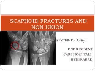
  • 1. PRESENTER: Dr.Aditya DNB RESIDENT CARE HOSPITALS, HYDERABAD SCAPHOID FRACTURES AND NON-UNION
  • 2. ANATOMY MECHANISM OF INJURY PATHOPHYSIOLOGY DIAGNOSIS AND CLINICAL EXAMINATION CLASSIFICATIONS TREATMENT COMPLICATIONS NONUNION OF SCAPHOID AND ITS MANAGEMENT
  • 3. INTRODUCTION Scaphoid is derived from Greek word SKAPHOS meaning boat It acts as a link between proximal and distal carpal rows The scaphoid is the most commonly fractured carpal bone. AGE: 10-70 yrs. most common in young (20-30 yrs.) Scaphoid fractures are uncommon in children because the physis of distal radius fails first.
  • 4. M:F- 4:1 About 5-12% of scaphoid fractures are associated with other fractures 70-80% occur at the waist, 10-20% occurs at proximal pole • RULE OF 70’S FOR SCAPHOID - 70% of all carpal bone fractures. - 70% of blood supply is by the dorsal branch of the radial artery. - 70% of fractures occur at the waist of scaphoid. - 70% of the scaphoid fractures unite .
  • 5. ANATOMY The scaphoid lies at the radial border of the proximal carpal row Scaphoid represents the floor of the anatomical snuff box 80% covered by articular cartilage. Its implications are that 1, articular cartilage may be damaged by screw insertion, 2, Absence of periosteum results in minimal callus and 3, poor blood supply predisposes to osteonecrosis. Parts of scaphoid : 1.Tubercle 2.Distal pole 3.Waist 4.Proximal pole
  • 6. ARTICULATIONS The scaphoid articulates with five bones: the radius, trapezoid, trapezium, lunate and capitate. •proximal surface: radius •distal surface: laterally with the trapezoid and trapezium; medially with the capitate •ulnar surface: lunate
  • 7. WRIST LIGAMENTS The ligaments of the wrist include  extrinsic ligaments: include volar and dorsal ligaments bridge carpal bones to the radius or metacarpals intrinsic ligaments originate and insert on carpal bones the most important intrinsic ligaments are the scapholunate interosseous ligament and lunotriquetral interosseous ligament Characteristics 1. volar ligaments are secondary stabilizers of the scaphoid 2. volar ligaments are stronger than dorsal ligaments 3. dorsal ligaments converge on the triquetrum
  • 8. VOLAR LIGAMENTS Volar radiocarpal ligaments: 1. Radial collateral ligament 2. Radioscaphocapitate ligament 3. Long radiolunate ligament 4. Short radiolunate ligament 5. Radioscapholunate ligament Volar ulnocarpal ligaments: 1. Ulnotriquetral 2. Ulnolunate 3. Ulnocapitate
  • 9. DORSAL LIGAMENTS Radiotriquetral ligament Dorsal intercarpal ligament Radiolunate radioscaphoid
  • 10. INTRINSIC (INTEROSSEOUS) LIGAMENTS Proximal row 1. Scapholunate ligament disruption leads to lunate extension when the scaphoid flexes creating DISI deformity 2.lunotriquetral ligament disruption leads to lunate flexion when the scaphoid is normally aligned creating VISI deformity (in combination with rupture of dorsal Radiotriquetral rupture)
  • 11. • Distal row 1. trapeziotrapezoidal lig 2. trapeziocapitate 3. capitohamate lig Palmar midcarpal 1. scaphotrapeziotrapezoid    2. scaphocapitate 3. triquetralcapitate
  • 12. Blood supply to the scaphoid is primarily through the radial artery. The branches of the artery enter the scaphoid through the foramina at the dorsal ridge at the level of the waist of the scaphoid. ( 80% of blood supply to scaphoid) Subsequently, these vessels divide and run proximally and palmarly to supply blood to the proximal pole of the scaphoid. BLOOD SUPPLY TO THE SCAPHOID
  • 13. Other branches provide 20– 30% of the blood flow and appear from the distal palmar area of the scaphoid, arising either directly from the radial artery or from the superficial palmar branch. The proximal pole, therefore, is dependent entirely on intraosseous blood flow.
  • 14. MECHANISM OF INJURY It is caused by fall on the outstretched hand, resulting in severe hyperextension and slight radial deviation of the wrist The scaphoid usually fractures in tension with the wrist extended, concentrating the load on the radial-palmar side. The proximal pole locks in the scaphoid fossa of the radius, and the distal pole moves excessively dorsal resulting in fracture.
  • 15. PATHOPHYSIOLOGY ■ Essentially fractures of scaphoid have been explained as a failure of bone caused by compressive or tension load ■ Compression, as explained by Cobey and White, against concave surface by head of capitate ■ Position of radial and ulnar deviation thought to determine where it breaks ■ Fryman subjected cadaver wrists to loading and observed that: – extension of 35 degrees of less resulted in distal forearm fractures – >90degrees resulted in carpal fractures ■Combination of radial deviation and wrist extension locks scaphoid within the scaphoid fossa
  • 16. DIAGNOSISAND CLINICAL EXAMINATION A strong index of suspicion is the key to early diagnosis Diagnosis should be based on 1.history 2.clinical examination 3. radiographic evaluation
  • 17. CLINICAL EXAMINATION SYMPTOMS: 1.Pain along the radial side of wrist 2.Inability or difficulty to move the wrist SIGNS: 1.Swelling present in anatomical snuff box 2.Tenderness in anatomical snuffbox 3.Tenderness with axial compression of thumb towards the snuff box 4.Radial & ulnar deviation results in pain on radial side of wrist
  • 18. TESTS TAPPING THE SCAPHOID: by placing one thumb in snuff box and other on distal tubercle produces pain or crepitus SCAPHOID LIFT TEST: reproduction of pain by dorsal and volar shifting of scaphoid. WATSON TEST: painful dorsal scaphoid displacement as the wrist is moved from ulnar to radial deviation with compression of tuberosity.
  • 19. CLASSIFICATIONS There are several classification systems available for fractures of scaphoid. These include the following RUSSE’S classification AO classification HERBERT’S classification MAYO classification
  • 20. RUSSE CLASSIFICATION Russe classified scaphoid fractures into 3 type according to direction of fracture –Horizontal oblique # –Transverse # –Vertical oblique # (unstable)
  • 21. MAYO’SCLASSIFICATION It divides scaphoid fractures into three basic types according to anatomic location of the fracture line. 1.proximal third (10%) 2.middle third (70%) 3.distal third (20%) Fracture of the distal third are further divided according to involvement of the distal articular surface or the distal tubercle.
  • 22. HERBERT’S CLASSIFICATION• Herbert devised an alpha-numeric system that combined fracture anatomy, stability and chronicity of injury.  Type A (stable acute fractures) – A1: fracture of tubercle – A2: incomplete fracture  Type B (unstable acute fractures) – B1: distal oblique – B2: complete fracture through waist – B3: proximal pole fracture – B4: trans-scaphoid perilunate fracture dislocation of carpus
  • 23.  Type C(delayed union)  Type D (established non-union) – D1: fibrous union – D2: pseudarthrosis – D3: sclerotic pseudoarthrosis – D4: avascular necrosis
  • 24.
  • 25. IMAGING XRAYS: 4 essential views PA view Lateral view identify majority Supinated oblique of fractures Pronated oblique SCAPHOID VIEW: is a PA radiograph with the wrist extended 30° and deviated ulnarly 20°. This view helps to stretch out the scaphoid and is also used for assessing the degree of scaphoid fracture angulation A clenched-fist radiograph has also been useful for visualization of the scaphoid waist.
  • 27.
  • 28. Scaphoid axis The true axis of the scaphoid is the line through the midpoints of its proximal and distal poles. Since the midpoint of the proximal pole is often difficult to appreciate, an almost parallel line can be used that is traced along the most ventral points of the proximal and distal poles of the bone
  • 29. The scapholunate angle is the angle between the long axis of the scaphoid and the mid axis of the lunate on the sagittal imaging of the wrist. In a normal situation it should be between 30o and 60o in the resting (neutral) position
  • 30. The capitate axis joins the midportion of the proximal convexity of the third metacarpal and that of the proximal surface of the capitate. Capitolunate angle Normal: 0-30deg Abnormal: > 30?.This indicates instability of the wrist.
  • 31. DISI or dorsiflexion instability DISI is short for dorsal intercalated segmental instability.  The intercalated segment is the proximal carpal row identified by the lunate. The term 'intercalated segment' refers to the part in between the proximal segment of the wrist consisting of the radius and the ulna and the distal segment, represented by the distal carpal row and the metacarpals.  So all this means is that in DISI or dorsiflexion instability the lunate is angulated dorsally. 
  • 32. VISI or volar flexion instability Volar intercalated segmental instability or palmar flexion instability is when the lunate is tilted palmarly too much.  While most DISI is abnormal, in many cases VISI is a normal variant, especially if the wrist is very lax
  • 33. CT SCAN CT permits accurate anatomic assessment of the fracture. Bone contusions are not evaluated with CT, but true fractures can be excluded Most sensitive and specific Multiplanar and 3D-reconstructions are possible.
  • 34. MRI T1-weighted images obtained in a single plane (coronal) are typically sufficient to determine the presence of a scaphoid fracture. Gaebler prospectively performed MRI on 32 patients, at average of 2.8 days post injury – 100% sensitivity and specificity In recent study Dorsay has shown that immediate MRI provides cost benefit when compared to splintage and repeat x-ray
  • 35. • Determine preop vascularity in a diagnosed scaphoid fracture. • Acute fractures- Normal or decreased T1 intensity or increased T2 intensity. • Low T1 and T2 marrow signal intensity indicates poor vascularity.
  • 36.
  • 37. BONE SCAN Sensitive, but less specific Increased osteocyte activity due to trauma will show as a focal hot spot. Fractures are seen in around 95% of non-osteoporotic patients within 24 hrs. Negative bone scan excludes any scaphoid fracture. Teil-van studied cost effectiveness and concluded that initial x-ray followed by bone scan at 2 weeks if patient is still symptomatic is most effective management option
  • 38. J Hand Surg Am. 2008 Jul-Aug; 33(6): 988– 997.
  • 39. TREATMENT Treatment of scaphoid fractures are determined by displacement and stability of fracture.  nondisplaced,stable fractures displaced, unstable fractures Non operative treatment operative treatment ACUTE SCAPHOID FRACTURES
  • 40. SCAPHOID CAST • Forearm cast below the elbow proximally to the base of thumbnail and the proximal palmar crease distally. • Wrist in slight radial deviation and in neutral flexion. • Thumb is maintained in functional position and the fingers are free to move from MCP joints distally. • 90-95% union in 10-12 weeks . During this time fracture is observed radiographically for healing. • If collapse or angulation of fractured fragments occurs, surgical treatment is required.
  • 41.
  • 42. Management of waist fractures ■ Most common type of fracture ■ Operative vs non-operative: Controversial ■ High rate of delayed and non-union ■ Most stable fractures - with below elbow thumb spica cast. ■ Unstable fractures best treated with compression screw fixation – >1mm displacement – Fragment angulation – Abnormal carpal alignment ■ With advent of percutaneous techniques of cannulated screws under fluoroscopic control trend towards operative management
  • 43. Management of distal polefractures  Distal Pole – Are infrequent – Usually extra- articular with good blood supply – Best treated with short arm thumb spica for 3-6 weeks
  • 44. • Fractures at and distal to the scaphoid waist heal sooner than the fractures in proximal pole • Immobilization for 6 weeks by using long arm thumb spica cast is justified in case of proximal third fractures or those in which diagnosis is delayed. • Healing occurs by creeping substitution. Management of proximal pole fractures
  • 45. Displaced, unstablefractures  Following are the criteria to label it as unstable 1. Fragments are offset more than 1mm in the AP or oblique view. 2. Lunocapitate angulation > 15 deg. 3. Scapholunate angulation is > 45 deg in lateral view. Other criteria for evaluating displacement include: 1. Lateral intrascaphoid angle >45 deg 2. AP intrascaphoid angle <35 deg. 3. Height to length ratio of 0.65 or more.
  • 46.
  • 47. OPERATIVE TREATMENT INDICATIONS OF SURGERY: 1. Displaced unstable fractures 2. Scaphoid fracture associated with perilunate # or dislocation 3. Ligamentous injury 4. Non displaced fractures of proximal pole 5. Non displaced fractures if the patient will not tolerate prolonged immobilization ( athletes and manual laborers) The choice of surgical procedure depends on surgeons preference and experience, the type of fracture, pt’s age,
  • 49. SURGICAL APPROACHES VOLAR APPROACH: Indications Best exposure for scaphoid fractures at and distal to the waist. Comminuted fractures ORIF of fractures Bone grafting for nonunion scaphoid
  • 50. Angled skin incision The landmarks for this incision are: 1.The scaphoid tubercle 2. The flexor carpi radialis (FCR) tendon
  • 51. INCISION : The incision line can be marked on the skin, in line with the FCR tendon, starting at the scaphoid tubercle, and running proximally for about 2 cm. Distal of the scaphoid tubercle, the incision angles towards the base of the thumb, over the scaphotrapezial joint. Ligate superficial palmar branch of radial artery The superficial palmar branch of the radial artery passes towards the palm, running close to the scaphoid tubercle. If necessary, it can be ligated and divided.
  • 52. Open the FCR sheath The FCR sheath is opened as far distally as possible, and the tendon retracted towards the ulnar side.
  • 53. Exposure of the wrist capsule The capsule is then incised obliquely from the tubercle distally towards the palmar rim of the radius proximally. As determined by the fracture configuration, preserve as much of the palmar ligament complex as possible, as it helps to contain the proximal pole and prevent palmar tilt of the scaphoid.
  • 54. Expose the scaphoid Retract the divided radioscaphocapitate ligament to expose the scaphoid. If it is necessary to expose the proximal part of the scaphoid, divide the long radiolunate ligament, proximally as far as the palmar rim of the radius.
  • 55. Exposure of scaphotrapezial joint The scaphotrapezial joint must be exposed to allow optimal positioning of a screw. The incision is deepened distally, dividing the origin of the thenar muscles in line with their fibres. The scaphotrapezial joint is identified, the scaphotrapezial ligament divided in the line of its fibers, and the joint capsule opened.
  • 56. WOUND CLOSURE: The divided palmar ligaments (radioscaphocapitate/long radiolunate) must be repaired with fine interrupted sutures in order to prevent secondary carpal instability. Approximate the soft tissues over the scaphotrapezial joint. Test the integrity of the soft- tissue repair by passive wrist motion. Finally, the FCR tendon sheath is repaired and covered with subcutaneous tissue.
  • 57. Indications This approach is used for the following injuries: •ORIF of Proximal pole fractures •Excision of the proximal fragment of a nonunion scaphoid •Bone grafting for nonunion DORSAL APPROACH
  • 58. Straight skin incision Make a straight dorsal skin incision starting over Lister’s tubercle and extending for about 4 cm distally.
  • 59. Identify the radial nerve Identify and preserve the dorsal superficial branch of the radial nerve, which runs in the radial skin flap of the wound.
  • 60. Incise the retinaculum Incise the extensor retinaculum over the extensor pollicis longus (EPL) tendon opening the distal part of the third extensor compartment.
  • 61. Retraction of the tendons The EPL tendon is then retracted radially together with the tendons of the second extensor compartment. The fourth extensor compartment, containing the extensor digitorum and extensor indicis, is located on the ulnar side.
  • 62. Opening the capsule Make a longitudinal, or inverted T-shaped, incision, starting at the dorsal rim of the distal radius, extending to the dorsal intercarpal ligament.
  • 63. Take care to preserve the vessels to the dorsal ridge of the scaphoid. The capsule is not stripped from this area.
  • 64. Expose the scaphoid To expose the proximal pole of the scaphoid, it is necessary to flex the wrist. The scaphoid now comes into view. Identify the SL ligament.
  • 65. Wound closure Close the capsule with interrupted sutures. Close the third extensor compartment, avoiding any tension over the EPL tendon, which must glide smoothly. If this is not possible, the EPL tendon is best left superficial to the retinaculum, in the subcutaneous tissue.
  • 67.
  • 68. NON UNION OF SCAPHOID A non union of scaphoid fractures are influenced by 1.Delayed diagnosis 2.Gross displacement 3.Associated injuries of carpus and 4.Impaired blood supply 40% of scaphoid fractures are undiagnosed at the time of injury Displaced scaphoid # - nonunion rate is 92% The incidence of osteonecrosis is 30%- 40%, occurring most frequently in proximal third fractures.
  • 69. Non union of proximal pole fractures depends on 1.blood supply to proximal pole 2. size of the fragments Non union of proximal pole fractures Good blood supply poor blood supply Non vascularized bone grafts vascularized bone grafts
  • 70. Blood supply is determined preoperatively by gadolinum enhanced MRI and by intraoperative assessment of bone bleeding. If small, avascular, ununited fragments, excision of proximal pole is done Electrical and ultrasound stimulations found to be of variable effectiveness. Bone grafting should be considered a better option than PEMF for scaphoid nonunions.
  • 71. 2. The clinical and radiological outcome of pulsed electromagnetic field treatment for acute scaphoid fractures: A randomised double-blind placebo-controlled multicentre trial J Bone Joint Surg Br. 2012 Oct;94(10):1403-8  The use of PEMF therapy, as a treatment for acute scaphoid fractures, did not provide any significant improvements in clinical or radiological measures of union compared to a placebo control. There were also no improvements in range of motion or grip strength. 1. Delayed Union of Scaphoid Fracture and Effectiveness of Pulsed Electromagnetic Fields: A Case Report and Review of the Literature Middle East J Rehabil Health Stud. 2018 January; 5(1):e63850  PEMF is a safe technique that has shown a promising therapeutic effectiveness in the healing of delayed union of scaphoid fracture. PEMF could recover delayed nonunion of scaphoid fracture and decreased pain as evaluated clinically and radiologically in our case report.
  • 72. Degenerative arthritis is seen after many years in scaphoid nonunions Radiographic features of arthritis are: 1. Radioscaphoid narrowing 2. Capitolunate narrowing 3. Cyst formation 4. Pronounced dorsal intercalated segment instability This is so called scaphoid nonunion advanced collapse pattern
  • 73. STAGES OF SCAPHOID NONUNION ADVANCECD COLLAPSE ARE: STAGE I: arthritis at radial styloid STAGE II: scaphoid fossa arthritis STAGE III: capitolunate arthritis STAGE IV: diffuse arthritis of carpus
  • 74. SLADE AND GIESSLER CLASSIFICATION FOR SCAPHOID NONUNION Type I injury : are the result of a delayed presentation (4 to 12 weeks after injury).  Type II injuries: a fibrous union is present.  Type III injuries: minimal sclerosis is seen at the fracture site. Sclerosis < 1 mm.  Type IV injuries: cystic formation is present.  Type V injuries: cystic changes > 5 mm in diameter, rotation of the lunate has occurred, resulting in a humpback deformity as seen with plain radiography or CT.  Type VI injuries: secondary degenerative changes are present, (i.e., scaphoid nonunion advanced collapse [SNAC]).
  • 75. GOALSOF MANAGEMENT 1. relieve symptoms, 2. correct the carpal deformity, 3. achieve union, 4.delay the onset of wrist arthrosis The major principles to follow are the following: 1. Make an early diagnosis 2. Perform a complete resection of the nonunion 3. Correct the deformity secondary to carpal collapse and carpal instability 4. Preserve the blood supply throughout
  • 76. KNOLL AND TRUMBLE ALGORITHM FOR MANAGEMENT OF SCAPHOID NON UNION (Adapted from Knoll VD, Trumble TE: Scaphoid fractures and nonunions, in Trumble TE [ed]: Hand Surgery Update 3.Rosemont, IL: American Academy of Orthopaedic Surgeons, 2003, pp 161-173.)
  • 77. Jupiter et al observed that ununited fractures of the scaphoid fall into 3 groups depending upon the extent of arthrosis 1. Established nonunions without arthrosis 2. Nonunions with radiocarpal arthrosis 3. Nonunions with advanced radiocarpal and intercarpal arthrosis Bone healing is needed for nonunions without arthrosis, additional salvage procedures may be required in patients with extensive arthrosis.
  • 78. Following operations can be useful for nonunions of scaphoid. 1.Traditional bone grafting 2. Vascularized bone grafting 3. Excision of proximal fragment,distal Fragment or entire scaphoid. 4. Radial styloidectomy salvage 5. Proximal row carpectomy procedures 6. Partial or total arthrodesis of wrist.
  • 79. GRAFTING OPERATIONS Cancellous bone grafting for scaphoid nonunion, as first described by matti and modified by russe. Produces bony union in 80-97% This technique is most useful for ununited fractures that do not have associated shortening or angulation.
  • 80. MATTI-RUSSE TECHNIQUE VOLAR APPROACH Volar incision over FCR tendon ending distally over the scaphoid tuberosity. Identify the scaphoid bone and expose the nonunion by dorsiflexion and ulnar deviation of wrist. Freshen the sclerotic ends and form a cavity that extends well into each adjacent fragments. From the iliac crest obtain a piece of cancellous bone and fit into the cavity and stabilize the two fragments with k-wires.
  • 81.
  • 82. Malpositioned nonunion of scaphoid fractures (Humpback deformity) • Resorption/communition at fracture site. • The deformity includes extension of the proximal pole of the scaphoid, resulting extension of the lunate, and a form of dorsal intercalated instability pattern seen on lateral plain radiographs • Techniques- – Fernandez et al – Tomaino et al – Stark et al
  • 83. FERNANDEZ TECHNIQUE Calculate the amount of resection, size of graft, and deformity on tracing paper by x-rays. Volar approach is used and care must be taken to preserve the vascularity of fragments Interpositional grafting Distract the osteotomy site to correct flexion deformity and shortening along with dorsal rotation of lunate Shape the graft from iliac crest. cortical part of the graft should be palmar Scaphoid fixed with 1.2mm k-wires (one or two)
  • 84.
  • 85. TOMAINO ET AL: Approach between FCR and radial artery Incise the capsule, RSC lig longitudinally and expose the proximal trapezium and scaphotrapezial joint Correct lunate extension, fix it with 1.1mm k-wire Open up nonunion site and resect it Obtain a tricorticocancellous graft from the iliac crest Fit the graft into nonunion site stabilize with k-wire Using c-arm insert herbert whipple screw Assess wrist flexion, extension, radial and ulnar deviation if graft impinges perform radial styloidectomy Repair the capsule; RSC ligament and sheath of FCR.
  • 88. VASCULARIZED BONE GRAFTS INDICATION: Nonunion and avascular necrosis and if previous iliac grafting has failed. SOURCES: pronator quadratus pedicle graft from the distal radius iliac crest free flap a vascularized bone graft from the distal dorsolateral radius pedicle bone grafts based on the 1,2 intercompartmental supraretinacular artery. TECHNIQUES: • KAWAI AND YAMAMOTO • ZAIDEMBERG ET AL.
  • 89. PEDICLE BONE GRAFT BY KAWAI AND YAMAMOTO Volar approach bone exposed, sclerotic ends excised. Large oval cavity 10-20mm long created, pronator quadratus identified and block of bone graft 11-20mm outlined at its distal insertion on the distal radius close to the abductor pollicis longus tendon Outline margin of the graft with k-wire holes separate with fine osteotomy, dissect the muscle towards ulna to secure a pedicle 20mm thick. Align the fracture fragments and insert into the cavity and introduce 2 k-wires from the tuberosity.
  • 90. PEDICLE BONE GRAFT BY KAWAI AND YAMAMOTO Non union scaphoid fracture Pronator quadratus
  • 91. ZAIDEMBERG ET AL: • Oblique Incision on the dorsoradial side of the wrist- centered on the radiocarpal joint. • On distal radius identify the longitudinal course of ascending irrigating branch of the radial artery • Design a bone graft with longitudinal vessel at its center and use a small gauge to harvest a graft beneath the periosteal vessel and transpose it in the long trough created in the scaphoid and stabilize it with k-wires
  • 92.
  • 93. RADIAL STYLOIDECTOMY  It is a kind of early salvage procedure which aims to reduce pain arising from radial styloid impingement that may occur in scaphoid nonunion or scapholunate instability. Indicated along with grafting of scaphoid or excision of its ulnar fragment when arthritic changes involve the scaphoid fossa. Technique : Stewart He recommended resecting enough of the styloid to remove entire articulation with the scaphoid To avoid ulnar translocation of the carpus, it is important to preserve palmar radiocarpal ligaments
  • 94.
  • 95. EXCISION OF THE PROXIMAL FRAGMENT •Excising both fragments of the scaphoid as the only procedure is unwise; although the immediate result may be satisfactory, eventual derangement of the wrist is likely. •Soto-Hall and Haldeman reported gradual migration of the capitate into the space previously occupied by the scaphoid. •If excision of both fragments is considered, it is preferable to add some other procedure to stabilize the capitolunate joint (e.g., capitolunate or capital-lunate-triquetral-hamate fusions). •Excising the proximal scaphoid fragment usually is satisfactory; the loss of one fourth or less of the scaphoid usually causes minimal impairment of wrist motion. Because postoperative immobilization is brief, function usually returns rapidly.
  • 96. Indications for excising the proximal fragment of a scaphoid nonunion: 1.The fragment is one fourth or less of the scaphoid. 2.The fragment is one fourth or less of the scaphoid and is sclerotic, comminuted, or severely displaced. 3.The fragment is one fourth or less of the scaphoid, and grafting has failed. 4.Arthritic changes are present in the region of the radial styloid.
  • 97. EXCISION OF DISTAL SCAPHOID Satisfactory results have been reported with distal scaphoid resection for the treatment of scaphoid nonunions with radioscaphoid arthritis treated with distal scaphoid resection. If capitolunate arthritis is present, an additional procedure (e.g., limited intercarpal arthrodesis) should be added to distal scaphoid excision.
  • 98. PROXIMAL ROW CARPECTOMY • Proximal row carpectomy is used as a reconstructive procedure for posttraumatic degenerative conditions in the wrist, especially conditions involving the scaphoid and lunate. • alternative to arthrodesis. • is considered to be a satisfactory procedure in patients who have limited requirements, desire some wrist mobility, and accept the possibility of minimal persistent pain • When proximal row carpectomy is done for degenerative changes, healthy articular surfaces should be present in the lunate fossa of the radius and the proximal articular surface of the capitate to allow for satisfactory articulation between these surfaces.
  • 99. Excision of the triquetrum, lunate, and entire scaphoid usually is recommended. The distal pole of the scaphoid at its articulation with the trapezium can be left, however, to provide a more stable base for the thumb.( in addition, radial styloidectomy should be done to avoid impingement of the distal scaphoid pole and trapezium on the radial styloid)
  • 102. ARTHRODESIS If patient wants painless wrist or in cases of nonunion or Malunion A/W radiolunate traumatic arthritis with fracture of distal end of radius. 1.LIMITED INTERCARPAL ARTHRODESIS: when degenerative changes involve the midcarpal joint. Four corner fusion : if radiolunate joint is not involved
  • 103. 2. TOTAL ARTHROSESIS: Involvement of radiolunate joint The wrist is fused in 10-20 deg of extension with the long axis of third metacarpal shaft aligned with long axis of the radial shaft.
  • 104. REFERENCES 1. CAMPBELL’S ORTHOPAEDICS-13TH EDT 2. ROCKWOOD AND GREEN’S FRACTURE IN ADULT- 8TH EDT 3. SURGICAL EXPOSURES IN ORTHOPAEDICS 4. GREEN’S OPERATIVE HAND SURGERY 5. JOURNALS: JHS,AAOS, THANK YOU