MNPS System Presentation. 2020
•
0 recomendaciones•98 vistas

MNPS is a stereotactic planning and post-op evaluation system developed by Mevis Informatica Médica LTDA. Support for biopsy, functional neurosurgery, DBS, VTA, X-ray stereotactic localization, 2D-3D registration, image fusion, tractography, sEEG, and much more. Versions for a lot of frames manufactures and models; FiMe, Micromar, Bramsys, Leksell, CRW, BRW, ZD, Macom, Riechert-Mundiguer, Adeor-Zeppelin, EstereoFlex. DBS support: Medtronic, St.Jude-Abbott, Boston Sc. and SceneRay. Isotropic and directional DBS

Recomendados
Recomendados
Más contenido relacionado
La actualidad más candente
La actualidad más candente (7)
Similar a MNPS System Presentation. 2020
Similar a MNPS System Presentation. 2020 (20)
Más de Armando Alaminos Bouza
Más de Armando Alaminos Bouza (20)
Último
Último (20)
MNPS System Presentation. 2020
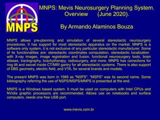
- 1. MNPS: Mevis Neurosurgery Planning System. Overview (June 2020). By Armando Alaminos Bouza MNPS allows pre-planning and simulation of several stereotactic neurosurgery procedures. It has support for most stereotactic apparatus on the market. MNPS is a software only system, it is not exclusive of any particular stereotactic manufacturer. Some of its functionalities are: stereotactic coordinates computation, stereotactic localization with X-ray images, image registration and fusion, functional neurosurgery tools, brain atlases, tractography, brachytherapy, radiosurgery, and more. MNPS has corrections for ring tilt and swivel inside CT/MRI gantry for all stereotactic systems. There is also support of DBS geometry, electric field, and VTA, for several brands and models. The present MNPS was born in 1989 as "NSPS". "MSPS" was its second name. Some bibliography referring the use of NSPS/MSPS/MNPS is presented at the end. MNPS is a Windows based system. It must be used on computers with Intel CPUs and NVidia graphic processors are recommended. Allows use on notebooks and surface computers, needs one free USB port. www.mevis.com.br
- 2. MNPS has a long history. The first lines of its code were written in 1989, for a Riechert- Mundinger system. The right image shows a banner from a meeting in Brazil, 2010.
- 3. Support for DICOM, NIfTI and some proprietary image formats. Allows use of CT, MRI, PET and SPECT, but for 99% of the cases only CT and MRI are used. MNPS : Importing patient's images
- 4. MNPS : Automatic fiducials detection for any supported stereotactic device, even those systems with markers embedded in plastic plates, resulting in lower contrast localizers. In the rare case where MNPS fails to find the fiducials, the operator can set the fiducials manually for that tomogram.
- 5. 2D planning window. Stereotactic coordinates of the red cursor are continuously displayed. Reformatted images at the left are co-planar with red cross at main window. Reformatted images are rendered in real time following cursor movement.
- 6. Main window with reformatted image. Screenshot of planning over a reformatted sagittal image. MNPS accepts images with tilt and swivel. Stereotactic coordinates from images with tilt/swivel are corrected. CT calibration bias in stereotactic images are also corrected based on fiducials markers positions.
- 7. MNPS 3D window. Isodensity solid surface at skin level.
- 8. MNPS. Detail from 3D window with projection of principal planes and rendering the surface of a Region of Interest (ROI) drawn by the operator (red surface). There are several tools for image segmentation.
- 9. Non-stereotactic MRI images can be registered into stereotactic CT space. MRI images can be distorted by some stereotactic frame parts inside the magnetic field, so it is advisable to use CT instead of MRI with the frame attached. MNPS: Multimodality image registration and fusion.
- 10. Reformatted images rendering MRI. Coordinates are stereotactic, even if cursor is over MRI. MNPS allows image registration with axial, coronal or sagittal MRI sets. MNPS: Multimodality image registration and fusion.
- 11. MRI rendered in false color. In the main window a high-band filter was used removing MRI voxels with low signal response. MNPS : Stereotactic CT and non-stereotactic MRI.
- 12. Stereotactic coordinates and trajectory projection. MNPS shows the stereotactic coordinates as a table at the left bottom side of the main window. For a Riechert-Mundinger system (case of the image), four angles an the needle depth are shown. For isocentric systems, only two angles are presented.
- 13. Most functional resources depends on the positions of three landmarks: AC (anterior commissure), PC (posterior commissure) and IHP (a point on the mid sagittal plane). The registration of all maps to the brain are based on those points. AC, PC and IHP definition is the operator's responsibility, there is not automatic method so far. Functional neurosurgery tools on MNPS.
- 14. Maps overlayed on patient's brain. The maps were digitalized as vectors. Vectorial representation of maps has the advantage of allowing deformations for a better fit between the model's brain and the patient's brain. The name of the nucleus is also stored, so that the operator can query MNPS about the name of any structure.
- 15. MNPS : Other tools for functional surgery planning Vectorial maps allows some deformations and scaling operations to try a better fit into patient's topography. This is a detail of the maps setting window. Maps allows query for the name of each structure as the cursor is moved
- 16. MNPS : Other tools for functional surgery planning Nuclei with 3D representation Some common targets can be suggested by MNPS based on AC, PC and IHP location. Once the user marks AC, PC and IHP, MNPS stars reporting commissural coordinates. The commissural Cartesian system in MNPS has origin at the inter-comissural point.
- 17. MNPS : 3D volumetric rendering of some functional nuclei with projected trajectories for a DBS or electrode.
- 18. Precise geometrical model and rendering of several DBS by brand and model. DBS Selection Menu Boston Scientific Medtronic Saint Jude
- 19. 3D Rendering of bilateral DBS entering the STn, as part of pre-planning for a functional procedure
- 20. MNPS showing DBS over registered MRI image with overlayed functional maps. This is a DBS with 1.5 mm electrode size with 1.5 mm spacing.
- 21. Implementation of a Electric Field model (FEM solution) allowing presentation of Electric Field Modulus around a set of DBS leads. Brain tissue is treated as homogeneous and isotropic (conductivity and permittivity are constant for all tissue).
- 22. Finite Element Model (FEM) of the electric field allows simulation of arbitrary shaped contact. Note the eccentricity of the field near the contact, specially for the tip of a Saint Jude lead. Also note the influence of several active contacts on the resulting field.
- 23. VTA shown with 50% transparency. Conductivity and permittivity of tissue are assumed isotropic. Correlation between electric field and VTA are taken from literature. Implemented only as voltage-controlled so far. MNPS rendering VTA on the 3D window.
- 25. MNPS: Support to SceneRay DBS models Three models with the same electric parameters resulting in slightly different electric fields and VTA, due to contact spacing. SceneRay 1200 SceneRay 1211 SceneRay 1210
- 26. Kinesthetic points rendering. All the following points were collected by Dr. Mark Sedrak with software tools developed by him. By courtesy of Dr. Sedrak the tools are available at the following link https://app.box.com/s/4zxysoc3bltxvvt4cwcplpbkntoi9cll Points collected following trajectories to STn. Each color represent a different location of the response.
- 27. Less square regression line of kinesthetic points collected in STn explorations. Courtesy of Dr. Mark Sedrak
- 28. Probabilistic ellipsoid of kinesthetic points, with main axis as two root mean square of the point cloud measure from the points center of mass. Lateral axis equals two standard deviations measured as distance to main axis. Courtesy of Dr. Mark Sedrak
- 29. Registration and fusion after skull stripping of MRI images
- 30. Registration and fusion after skull stripping of MRI images. 3D rendering
- 31. “Virtual Fiducials Mode” allows non-stereotactic images directly into MNPS. WARNING : Coordinates in Virtual Fiducials Mode are not stereotactic Do not use on surgery ! On the other hand, comissural coordinates are useful for pre- and post-planning.
- 32. Virtual Fiducials Mode is convenient for pre- planning and also for post- planning. In Virtual Fiducials Mode all the tools available for Stereotactic image sequences can be used on non-stereotactic image sets
- 33. MNPS imports DTI tractography from .OBJ or .TRK files. The user can process the original DTI volume with its preferred DTI analysis system and export the fiber tracks for use on MNPS. We have successfully imported fiber tracks from DTI-Studio, FSL, and 3D-Slicer.
- 34. Fiber tracks can be overlaid to functional map nuclei, ROI volumes, instrument tracks, etc, rendered in 2D and 3D. The user can select fibers from a region and hide fibers not crossing that region. Sample images showing fibers crossing the region of STn and Red Nuclei.
- 35. Fiber tracks can be imported do stereo-CT via multi-modal image registration with MRI volumes.
- 36. Fiber tracks imported into stereo-CT space via image registration. Border of a surgery target segmented and rendered as solid 3D object (in red). Also, use image registration to import tracks from DTI volume into T1 or T2 volumes. Care should be taken with significant image distortion, common on DTI volumes. Non-rigid registration can be handy to deal with some distortion.
- 37. Digitally Reconstructed Radiograph (DRR) generation.
- 38. 2D-3D registration for intra-op verification of DBS or any probe position with X-rays
- 39. “Portal Check”: New tool for comparing DRR, including a virtual probe, with an X-ray taken intra-op. It helps for verifying the placement of a DBS or any other instrument.
- 40. Checking a DBS position by matching a DRR and its corresponding X-ray Wrong registration, MI = 1.1089 Best registration MI = 1.1770
- 41. MNPS - DRR and X-rays, registration using intrinsic landmarks.
- 42. MNPS : Stereotactic Radiosurgery (SRS) tools. It is LINAC based SRS, with circular collimators. The dose should be delivered by non-coplanar radiations arcs. Up to 64 arcs are available. Isodose maps are rendered in 2D and 3D surfaces. For SRS planning the stereo-CT is mandatory, all dosimetry is based on Houndfield units, but MRIs could be used via registration and fusion with stereo-CT.
- 43. MNPS and SRS: Acoustic neuroma.
- 44. MNPS SRS, Treating multiple isocenters MNPS SRS: Automatic optimization tools and conformity evaluation report. Some inverse planning capabilities, optimize isocenter positions and arc weights
- 45. Stereotactic target localization based of orthogonal X-ray pairs. Mainly used for DSA localization and treatment with SRS. Cursor in X-rays correlated to position on CT or MRI continuously. The orthogonal setup is not mandatory, only need to “see” the 8 markers on each X-ray view. The image below shows a phantom for quality assurance with a ZD system attached.
- 46. Sample AVM treatment plan with MNPS, (plan by Physicist Dra. S. Gusman, Lima, Peru)
- 47. MNPS and SRS planning: Brain fibers crossing an isodose volume 80% Isodose volume rendered with all fibers Only those fibers crossing 80% isodose
- 48. MNPS: Support for stereotactic brachytherapy with Iodine 125 seeds. Up to 256 sources per plan. Dose computation for temporary as well as permanent implants. Isodose overlays in 2D images and isodose surfaces on 3D window.
- 49. MNPS is a Windows based application, but if you want to run it on a Mac-OS there are to ways: Use a virtual machine (tested so far with Parallels and VMware Fusion) or create a Bootcamp.
- 50. - Bramsys - BrainLAB - BRW (Brown-Roberts-Wells) - CRW (Cosman-Roberts-Wells) - EstereoFlex - FiMe - Leksell - G - Macom - Micromar - Riechert-Mundinger - ZD (Zamorano-Dujovny) - Zeppelin Note: One MNPS license supports only one device model. If the user has several device models, more than one license will be necessary. Supported devices until January 2018 :
- 51. Some bibliography with references to “NSPS” or “MSPS” or “MNPS”: Alaminos Bouza A., Ortega, Molina, Valladares, Quiñones; “A Stereotactic Surgical Planning System for the IBM 386/486 PC family”. Stereotact Funct Neurosurg. 1994; 63: 35-37. Quiñones-Molina R, Molina H, Ohye C, Macías R, Alaminos Bouza A, Alvarez L, Teijeiro J, Muñoz J, Ortega I, Piedra J, Torres A, Morales F, Soler W: “CT-oriented microrecording guided selective thalamotomy”. Stereotact Funct Neurosurg 1994;62:200-203. Quiñones-Molina R, Alaminos Bouza A, Molina H, Muñoz J, López G, Alvarez L, Ortega I, Piedra J, Soler W, Torres A: “Computer-assisted CT-guided stereotactic biopsy and brachytherapy of brain tumors”. Stereotact Funct Neurosurg. 1994;63(1-4):52-5. Teijeiro J, Ohye C, Macías RJ, Ortega I, Alaminos Bouza A, Alvarez L: “Deep recording and digital processing system for brain electrical activity evaluation” (abstract). Stereotact Funct Neurosurg 1994; 62:198. José Augusto Nasser, Asdrubal Falavigna, Armando Alaminos Bouza, Antônio Bonatelli, Fernando Ferraz: "ESTIMULAÇÃO CEREBRAL PROFUNDA NO NÚCLEO SUBTALÂMICO PARA DOENÇA DE PARKINSON" . Arq. Neuro-Psiquiatr. vol.60 no.1 São Paulo Mar. 2002 . Ferraz Fernando, Aguiar PM, Ferraz H.B., Bidó J., Alaminos Bouza .A, Franco de Andrade L.A. "Talamotomia e Palidotommia estereotáxica com planejamento computadorizado no tratamento da doença de Parkinson" . Arq. Neuropsiquiatria 1998; 56(4): 789-797. Nasser JA, Falavigna A, Alaminos A, Bonatelli Ade P, Ferraz F. : “Deep brain stimulation of thalamus for tremor control”, Arq Neuropsiquiatr. 2002 Jun;60(2-B):429-34. Franca da Silva C.A., Lanes Vieira S., Campbell Penna A.B., Silva F., Guizzardi M.,F., Pereira Pedro P., Bardella Lúcia H., “Radiocirurgia estereotáxica com acelerador linear para tratamento da malformação arteriovenosa cerebral”. J.Bras. Neurocirurg. 15 (2), 53-58, 2004. Jacobsen Teixeira M., Fonof Erich T., Montenegro M.C.,”Dorsal Root Entry Zone Lesions for Treatment of Pain-Related to Radiation-Induced Plexotomy”. Spine, VOL 32, No.10, 2007.
- 52. Renato Ros, “Fusão de imagens médicas para aplicação em sistemas de planejamento de tratamento em Radioterapia”. Tese de Doutorado, São Paulo, 2006. Biblioteca digital de teses USP. Jacobsen Teixeira M., Talamoni Fonoff E., Mandel M., Leite Alves H. “Stereotactic biopsies of brain lesions”. Arq. Neuro-Psiquiatria. Vol.67, no.1, 2009. Teixeira MJ., Lepski G, Aguiar PH, Cescato VA., Rogano L., Alaminos Bouza A., “Bulbar trigeminal stereotactic nucleotractotomy for treatment of facial pain”. Stereotact Funct Neurosurg. 2003; 81(1-4): 37-42. Fonoff Erich T, et al., “Long-term outcome of atlas-based lesion of posterior zona incerta in secondary hemidystonia, Parkinsonism and Related Disorders”. 2011, Sep;17(8):649-50. doi: 10.1016/j.parkreldis.2011.05.005. Epub 2011 Jun 8. Teixeira Jacobsen M., Almeida F.F., Oliveira Y.S., Talamoni Fonof E., “Microendoscopic stereotactic-guided percutaneous radiofrequency trigeminal nucleotractotomy”. J. Neurosurgery 116:331-335, 2012. Alaminos Bouza Armando L., “Imaging, Stereotactic Space and Targeting in Functional Neurosurgery”. Chapter of book: “Functional Neurosurgery”, editor Arthur Cukiert. Alaúde Editorial 2014. pages 67-79. Contreras Ricardo, Hernandez Erick. “Planning Experience of Radiosurgery using a LINAC in Guatemala City”. Conference paper. Int. Conf. on Physics in Medicine & Clin. Neuroelectrophysiology. 19-20 February 2015. https://www.researchgate.net/publication/274314189_Planning_Experience_of_Radiosurgery_using_a_LINAC_in _Guatemala_City Ghilardi Maria Gabriela S., Cury R.G., Ângelos J.S, Barbosa E.R., Jacobsen Teixeira M., Fonoff E.T., “Long-Term improvement of tremor and ataxia after bilateral DBS of VoP/Zona Incerta in FXTAS. Neurology 84, May 5, 2015. William O Contreras Lopez, Azevedo A., Alencar C., Neville I, Reis, P.R., Navarro J, Monaco, B., Teixeira Manoel, Fonoff Erich. “Caudal Zona Incerta/VOP Radiofrequency Lesioning guided by combined Stereotactic MRI and Microelectrode recording for post-traumatic midbrain resting-kinetic tremor” World Neurosurgery, Sept. 8, 2015; http://www.worldneurosurgery.org/article/S1878-8750%2815%2901194-8/ abstract
- 53. Talamoni Fonoff, et.al. “Simultaneous bilateral stereotactic procedure for deep brain stimulation on implants: a significant step for reducing operating time” Journal of Neurosurgery. Dec. 2015. https://www.researchgate.net/publication/287483075_Simultaneous_bilateral_stereotactic_procedure_for_deep_ brain_stimulation_implants_a_significant_step_for_reducing_operation_time Erick Hernandez, Contreras R., Ortega M., Ureta L.; “Planning Experience of radiosurgery using LINAC in Guatemala city” , Int. Conf. On Physics In Medicine & Clin. Neurophysiology. Apr. 2015. https://www.researchgate.net/publication/274314189_Planning_Experience_of_Radiosurgery_using_a_LINAC_in _Guatemala_City Paul Rodrigo dos Reis, William O. Contrerars, Eduardo Alho, Armando Alaminos Bouza, Manoel Jacobsen Teixeira, Erich Talamoni Fonoff.; “3-D POSITION OF STN IN T2 WEIGHTED MRI IMAGES COMPARED TO STEREOTACTIC ATLASES FOR FUNCTIONAL NEUROSURGERY PLANNING”. Congresso da SBENF 2016. Brasil. Nevair Gallani, MD; Armando Alaminos Bouza, Sylvine Carrondo Cottin, Michel Prud’homme, Léo Cantin, “Simulation of Targeting two Simultaneous Nuclei With a Single 8-Contact DBS Lead for Movement Disorders”. NANS 20th Annual Meeting Neuromodulation: “From Frontier to Frontline”, Las Vegas Nevada, 2017. Online at ResearchGate: https://www.researchgate.net/publication/312591139_Simulation_of_Targeting_two_Simultaneous_Nuclei_with_a _single_8-contact_DBS_Lead_for_Movement_Disorders Ali Khedr, Armando L. Alaminos-Bouza, Russell A. Brown. “Use of the Brown-Roberts-Wells Stereotactic Frame in a Developing Country” . Cureus 10(1): e2126. doi:10.7759/cureus.2126. January 2018. https://www.cureus.com/articles/10590-use-of-the-brown-roberts-wells-stereotactic-frame-in-a-developing-country Maria Gabriela Dos Santos Ghilardi, Melisa Ibarra, Eduardo J.L. Alho, Paul Rodrigo Reis, William Omar Lopez Contreras, Clement Hamani, Erich Talamoni Fonoff. “Double target DBS for essential tremor: 8-contact lead for cZI and vim aligned in the same trajectory”, Neurology, February 07, 2018. Sedrak Mark, Sabelman E., Pezeshkian P., Duncan J., Bernstein I., Bruce D., Tse Vi., Khandhar S., Call E., Heit G., Alaminos-Bouza A., “Biplanar X-ray Methods for Stereotactic Intraoperative Localization in Deep Brain Stimulation Surgery”. Operative Neurosurgery. 2019. https://academic.oup.com/ons/advance-article/doi/10.1093/ons/opz397/5681720
- 54. For additional information, please visit us atFor additional information, please visit us at www.mevis.com.br.www.mevis.com.br. Or contact us by email: info@mevis.com.brOr contact us by email: info@mevis.com.br Thanks for your attention !Thanks for your attention !