

Recomendados
Más contenido relacionado
La actualidad más candente
La actualidad más candente (20)
Similar a Female rprdctive
Similar a Female rprdctive (20)
Más de MBBS IMS MSU
Más de MBBS IMS MSU (20)
Female rprdctive
- 1. Female Reproductive System Anatomy By Dr: Mohammed Faez
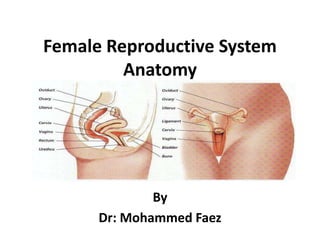
- 2. Female Reproductive System The reproductive tract in women is contained mainly in the pelvic cavity and perineum. It is divided to: External genitalia Internal genitalia
- 3. Female External Genitalia Vulva is the term given to the female external genitalia. The vulva includes: Mons pubis Labia majora Labia minora Clitoris Urethral opening Vaginal opening Perineum
- 5. MONS PUBIS The triangular mound of fatty tissue that covers the pubic bone It protects the pubic symphysis. During adolescence sex hormones trigger the growth of pubic hair on the monspubis. Hair varies in coarseness curliness, amount, color and thickness. 5
- 6. LABIA MAJORA The outer lip-like structure They have a darker pigmentation The Labia Majora: Protect the introitus and urethral openings Are covered with hair and sebaceous glands Become flaccid with age and after childbirth 6
- 7. LABIA MINORA Referred to as the “inner lips” Made up of erectile, connective tissue that darkens and swells during sexual arousal. Located inside the labia majora. They are more sensitive and responsive to touch than the labia majora. The labia minora tightens during intercourse. 7
- 8. CLITORIS The small penis-like structure. Highly sensitive organ composed of nerves, blood vessels, and erectile tissue Located under the prepuce It is made up of a shaft and a glans. Becomes engorged with blood during sexual stimulation. Key to sexual pleasure for most women. Urethral opening is located directly below clitoris 8
- 9. VAGINAL OPENINGINTROITUS A mucous membrane normally covers the opening into the vagina, called the hymen. Using the presence of an intact hymen for determining virginity is erroneous. Some women are born without hymens. Posterior to the vaginal opening are the two Bartholin’s glands. 9
- 10. PERINEUM The muscle and tissue located between the vaginal opening and anal canal. It supports and surrounds the lower parts of the urinary and digestive tracts. The perinium contains an abundance of nerve endings that make it sensitive to touch. 10
- 11. 11
- 12. Female Internal Genitalia The internal genitalia consists of the: Vagina Cervix Uterus Fallopian Tubes Ovaries
- 13. 13
- 15. VAGINA It is a distensible fibromuscular tube that extends from the perineum through the pelvic floor and into the pelvic cavity It measures approximately 8–12 cm in length. It extends from the vestibule to the uterus, and is situated behind the bladder and in front of the rectum. It is directed upward and backward. Its axis forming with that of the uterus an angle of over 90°, opening forward. 15
- 16. VAGINA The anterior wall of the vagina is related to the base of the bladder and to the urethra; in fact, the urethra is embedded in, or fused to, the anterior vaginal wall. Posteriorly, the vagina is related principally to the rectum.
- 17. VAGINA The vaginal fornix is the recess formed between the margin of the cervix and the vaginal wall. Based on position, the fornix is subdivided into a posterior fornix, an anterior fornix, and two lateral fornices. 17
- 18. VAGINA Blood Supply The blood supply to the vagina is from the vaginal artery (branch of the internal iliac artery) and the vaginal branch of the uterine artery. Lymphatic drainage Lymph from the upper vagina drains into the internal and external iliac nodes. Lymph from the lower vagina drains to the superficial inguinal nodes 18
- 19. Uterus The uterus is a thick-walled muscular organ in the midline between the bladder and rectum. It consists of a body and a cervix. Inferiorly it joins the vagina. Superiorly, uterine tubes project laterally from the uterus and open into the peritoneal cavity immediately adjacent to the ovaries. 19
- 20. Uterus The body of the uterus is flattened anteroposteriorlyand above the level of origin of the uterine tubes it has a rounded superior end (fundus of uterus). The cavity of the body of the uterus is a narrow slit, when viewed laterally. It is shaped like an inverted triangle, when viewed anteriorly.
- 21. Uterus Each of the superior corners of the cavity is continuous with the lumen of a uterine tube; the inferior corner is continuous with the central canal of the cervix The body of the uterus normally arches forward (anteflexed on the cervix) over the superior surface of the emptied bladder
- 22. Uterus The cervix forms the inferior part of the uterus and is shaped like a short, broad cylinder with a narrow central channel. The cervix is angled forward (anteverted) on the vagina so that the inferior end of the cervix projects into the upper anterior aspect of the vagina.
- 23. Relations of Uterine Anteriorly The uterus and cervix are related to the uterovesical pouch and superior surface of the bladder. Posteriorly The uterus is related to the recto-uterine pouch (of douglas), which extends down as far as the posterior fornix of the vagina. Laterally: the broad ligament
- 24. Uterus Blood supply The uterine artery (a branch of the internal iliac artery) Lymph drainage Lymphatics from the fundus accompany the ovarian artery and drain into the para-aortic nodes. Lymphatics from the body and cervix drain to the internal and external iliac lymph nodes.
- 25. Uterus
- 26. The ovary The ovaries are the sites of egg production (oogenesis). Like the testes in men. The ovaries lie adjacent to the lateral pelvic wall just inferior to the pelvic inlet. Each of the two almond-shaped ovaries is about 3 cm long and is suspended by a mesentery (the mesovarium) from the posterior aspect of the broad ligament.
- 27. The ovary Blood supply The ovarian artery (a branch of the abdominal aorta) Venous drainage To the inferior vena cava on the right and to the left renal vein on the left. Lymphatic drainage To the para-aortic nodes
- 28. The ovary