
Recommended
More Related Content
What's hot
What's hot (20)
Viewers also liked
Viewers also liked (20)
Similar to Postmortem
Similar to Postmortem (20)
Recently uploaded
Recently uploaded (20)
Postmortem
- 1. S A CHAPTER 3 SPECIFIC DISEASES OF CATTLE Diseases causedby viruses *Foot and mouth disease (FMD, Aphthous fever) FMD is an acute viral and extremely contagious diseaseof cloven footed animals such as cattle, sheep, goats, pigs and antelope. It is manifested by vesicles and erosions in the muzzle, nares, mouth, feet, teats, udder and pillar of the rumen. There are three main strains of viruses causing FMD, namely A, O and C. Three additional strains, SAT1, SAT 2 and SAT 3 have been isolated from Africaand a further strain ASIA-1 fromAsia and the Far East. Transmission: Directand indirect contact with infected animals and their secretions including saliva, blood, urine, feces, milk and semen, aerosol droplet dispersion, infected animal by-products, swillcontaining scraps of meat or other animal tissue and fomites and vaccines. Antemortemfindings: Before vesicle formation: 1. Incubation is 1 - 5 days or longer 2. Morbidity: Nearly 100 % 3. Mortality: variable depending on the strain of virus and its virulence and susceptibility of host; 50 % in young animals, 5 % in adults 4. Fever up to 41.7°C 5. Dullness 6. Lack of appetite
- 2. 7. Drastic drop in milk production. 8. Uneasiness and muscle tremors Vesicle formation: 9. Smacking and quivering of lips 10.Extensive salivation (Fig. 45) and drooling 11.Shaking of feet and lameness The vesicles and later erosions arecommonly found on the muzzle, tongue (Fig. 46), oral cavity, teat and on the skin between and above the hoofs of the feet. In morechronic cases in cattle the hoof become loose and the animal may walk with characteristic “clicking” sound (Slippering). Some strains of FMD, particularly in swine, sheep and goats cause erosions instead of vesicles. *Postmortemfindings : 1. Necrosis of heart muscle(tiger heart), usually only in young acutely infected animals. 2. Ulcerative lesions on tongue, palate, gums, pillars of the rumen and feet. Judgement : In countries or in zones within a country free or nearly free of FMD diseased or suspectanimals are prohibited to be admitted in an abattoir or slaughtered. If FMD is suspected on postmortem examination the carcass and viscera are condemned and appropriate action recommended by the regulatory authorities of the country must be taken. In countries where this disease is present, the judgement should be in accordance with the currentanimalhealth requirements, and consisted with effective public health protection. Particular attention should be paid to secondary bacterialinfections and general findings. Sanitary measures should be taken to comply with national animal health policy.
- 3. Remarks : Latent infections with Salmonella organisms werereported in animals affected with FMD. Differential diagnosisinbovine and ovine species : Vesicular stomatitis, allergic stomatitis, feedlot glossitis, photosensitization, bluetongue, rinderpest, infectious bovine rhinotracheitis, malignant catarrhal fever, bovine papular stomatitis, bovineviral diarrhoea, pseudocowpox, ovine pox, contagious ecthyma, footrot, mycotoxicosis and increased salt in concentrate. Discussion : In order to prevent the spread of the virus in the abattoir, the equipment and roomshould be disinfected with 2 % NaOH (caustic soda). In somecountries sodiumcarbonate (Na2CO3) is used. The vehicle conveying diseased animals should also be disinfected and abattoir personnelleaving the abattoir should pass through a footbath with 1 % solution of NaOH. The virus of FMD can survivein meat and meat products for a considerablelength of time. Outside the pH rangeof 6 – 9, viral infectivity is destroyed. A bovinecarcass matured at above +2°C produces a drop in the pH of muscle tissueto between 5.3 – 5.7 within 24 hours of slaughter. This is caused by the formation of sarcolactic acid. Quick freezing of the meat arrests acid production and consequently the virus remains infective for about 6 months. In salted meat at 4°C, the virus is still infective in bone marrow and lymph nodes for 6 months. In blood clots in large vessels of cattle and swine, the virus is infective for 2 months. The virus is inactivated by ultraviolet rays, acetic acid, 2 % lye and ethylene oxide. At high temperatures, the virus is only active for a shortperiod. 2 % NaOH solution inactivates the virus in 1 – 2 minutes. In dry refusein stalls, the virus remains infective for 14 days, 3 days on soil surfaces in summer compared to 39 days in fall. Itis also infective for 39 days in urine and for 20 weeks on hay dried at 22°C. The virus can be destroyed with 0.5 % citric or lactic acid, by cooking meat to an internal temperature of 69°Cand by pasteurization processes of milk.
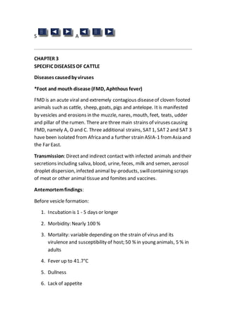
- 4. Fig. 45: Excessivesalivation in a cow affected with FMD.
- 5. Fig 46: FMD. Extensive areas of eroded epithelium on a bovinetongue.
- 6. Rinderpest (RP) Rinderpest is an acute, highly contagious, fatal viral diseaseof cattle, buffalo and wild ruminants manifested by inflammation, haemorrhage, erosions of the digestive tract, wasting and often bloody diarrhoea. Some swinespecies are also susceptible. Man is not susceptible to RP virus. Transmission : Direct contact with infected animals or their excretions and secretions and fomites. The virus appears in the blood and in secretions before the onset of clinical signs and this may causeinfection in abattoirs and stockyards. Antemortemfindings : 1. Incubation: 3 – 10 days or longer 2. Morbidity: Up to 100 % in a susceptible herd 3. Mortality: 50 % and may reach 90 – 95 % 4. High fever (41–42°C) 5. Nasal dischargeand excessive salivation 6. Punched out erosions in the mouth (Fig. 47) 7. Loss of appetite and depression 8. Abdominal pain (grunting, arched back) 9. Constipation followed by bloody diarrhoea and straining 10.Dehydration and rough hair coat 11.Marked debility 12.Abortion 13.The classical “milk fever position” in cattle Postmortemfindings : 1. Punched out erosions in the oesophagus
- 7. 2. Edema or emphysema of the lungs 3. Haemorrhagein the spleen, gallbladder and urinary bladder 4. Haemorrhagic or ulcerative lesions in the omasum 5. Congested abomasumfilled with bloody fluid. Ulcers may also be observed. 6. Severe congestion and haemorrhagein the intestine and enlarged and necrotic Peyer's patches (Fig. 48) 7. Last portion of the large intestine and rectum are haemorrhagic showing “tiger stripping” of longitudinal folds 8. Enlarged and edematous lymph nodes 9. Emaciated carcass Judgement : The carcass derived froma feverish and debilitated animal showing the sign of acute diseaseon antemortem examination should be condemned. In theareas free of RP and in zones wherefinal stages of eradication exist, the animals arealso condemned. In endemic zones, if acute symptoms of the diseaseare not present during clinical examination, the carcass may have limited distribution. In areas affected with outbreak which are protected by vaccination, heat treatmentof meat is suggested if economically worthwhile. The affected organs are condemned. Remarks : Rinderpest virus is sensitive to environmental changes and is destroyed by heat, drying and great number of disinfectants. Differential diagnosis: Bovine viraldiarrhoea, malignant catarrhal fever, infectious bovinerhinotracheitis, bluetongue, coccidiosis, foot and mouth disease, vesicular and necrotic stomatitis and bovine papular stomatitis. Vesicular diseases do not haveaccompanying haemorrhage and blisters should be differentiated fromerosions (ulcers) seen at RP.
- 8. Fig. 47: RinderpestErosions on the dental pad and the hard palate which resemble FMD.
- 9. Fig. 48: The mucosalsurfaceof Peyer's Patches showing necrosis and congestion. Vesicular stomatitis (VS) This is a viraldisease of ruminants, horses and swine characterized by vesicular lesions of the mouth, feet and teats. VS virus has two immunologically distinct serotypes, Indiana and New Jersey. Transmission: In susceptibleanimals, contamination of pre-existing abrasions with saliva or lesion material, by ingestion of contaminated pastureor during milking within dairy herds. Mechanical transmission by biting arthropods is also a possibility. The virus is isolated frommites, tropical sand flies and mosquitos. Antemortemfindings : 1. Fever 2. Mouth lesions in cattle and horses 3. Vesicles tend to disappear quickly and only papules may be seen in cattle outbreaks. 4. Marked weight loss and cessation of lactation in dairy cows. 5. Chewing movements and profusesalivation 6. Refuse food but eagerly accept water 7. Horses rub lips on edges of mangers 8. Foot lesions occur in about 50 % cases in cattle. 9. Lameness 10.Teat lesion may occur in all species. Postmortemfindings : 1. The skin and mucous membrane lesions resemble the lesions of other vesicular diseases.
- 10. 2. Secondary bacterial or fungal infections 3. Mastitis Judgement : The carcass of an animal affected with vesicular stomatitis is approved if the disease is not in the acute stage and secondary changes are not present. Parts of the affected carcass and organs are condemned. A carcass showing acutechanges and systemic lesions is condemned. If VS is notconfirmed by laboratory examination, the judgement will be the same as for the FMD. Differential diagnosis: Footand mouth disease, swinevesicular exanthema, vesicular disease, bovine papular stomatitis The mouth and muzzlelesions: Bovine viraldiarrhoea, rinderpest, mycotic stomatitis, photosensitization and Potomac valley fever in horses Teat lesions: Cowpox, pseudo-cowpox, pseudo-lumpyskin diseaseand bovine herpes mammillitis Fig. 49: Vesicular stomatitis. Tongue lesions.
- 11. Malignant catarrhal fever (MCF) An acute viral diseaseof cattle, deer, bison and buffalo characterized by inflammation of mucous membranes of the nose, eyes, cornealopacity, profusenasaldischargeand enlargement of lymph nodes. MCF is arbitrarily divided into peracute, intestinal, head-eyeand mild forms according to antemortem findings. Itis not communicable to man. Transmission: Closecontact between cattle and wildebeest (gnu, antelope), by common use of drinking troughs or by direct contact between cattle and newborn wildebeest and placenta of parturient dams. In American or European MCF, cattle are infected from sheep. Antemortemfindings : 1. Incubation: 9 – 44 days 2. Morbidity is low and mortality is high 3. Increased temperature 4. Bilateral ocular and nasaldischarges 5. Dyspnea and cyanosis 6. Loss of appetite 7. Encrustation of muzzleand eczema of the perineum, scrotumand udder 8. Erosions on the lips, tongue, gums, softand hard palate 9. Swollen reddened eyelids, corneal opacity and conjunctivitis (Fig. 50) 10.Photophobia associated with corneal opacity and blindness 11.Reluctance to swallow becauseof oesophageal erosions and drooling 12.Enlarged body lymph nodes 13.Rarely, uncoordinated movements and shivering
- 12. Postmortemfindings : 1. Lesions are not present in acute cases 2. Crater like erosions of the nose, mouth, conjunctiva, oesophagus and gastrointestinaltract 3. Lungs may be congested, swollen or emphysematous 4. White areas in the kidneys 5. Swollen and reddened abomasal folds 6. Intestinaledema and petechial haemorrhage 7. “Tiger striping” in the distal colon (Fig. 51) 8. Enlarged and reddened lymph nodes 9. Dehydrated and emaciated carcass Judgement :In the early stages of the disease, when fever, emaciation and systemic signs arelacking, the carcass of the affected animal may be approved asinferior meat. Otherwise, when fever, emaciation and systemic signs are present, the entire carcass and viscera are condemned. Thecondemned material may be used for rendering. Differential diagnosis: Bluetongue, rinderpest, bovineviral diarrhoea/mucosaldisease, footand mouth disease, vesicular stomatitis
- 13. Fig. 50: Malignant catarrhalfever Early stages of corneal opacity, conjunctivitis and the reddening of the eye lids.
- 14. Fig. 51: Malignant catarrhalfever. “Tiger striping” in the distal colon. Rift valley fever (RVF) (see Chapter 5) Rabies This is an acute infectious viraldisease of the central nervous systemin mammals. Transmission: Itis usually transmitted through the saliva by a bite from a rabid animal, commonly the dog or jackal. Man is infected the same way. Antemortemfindings : Furious form 1. Incubation from2 weeks to 6 months or longer 2. Restlessness 3. Aggressive, may attack other animals 4. Sexual excitement 5. Bellowing 6. Paralysis and death Paralytic form 7. Sagging and swaying of the hind quarters 8. Drooling and salivation 9. The tail is held to one side 10.Tenesmus or paralysis of the anus 11.Paralysis 12.The animal falls to the ground 13.Death after 48 hours of decubitus
- 15. Postmortemfindings: Possibleinflammation of gastrointestinalmucosa Judgement: In endemic areas carcasses may be approved if the animal was bitten eight days before slaughter and within 48 hours of slaughter. The bite area and surrounding tissuemustbe condemned, and prevention taken to prevent occupationalhazards. Differential diagnosis: Indigestion, milk fever or acetonemia when first seen, foreign body in the mouth, early infectious disease, poisoning Discussion: In a diseased animal, the virus is found in saliva, salivary gland and nervous tissue. Extreme caution should be instituted in abattoirs in order to prevent occupational hazards. Abattoir personnel can contract the disease through surfacecontactwith infected tissue. Infection does not occur by consumption of meat from a rabid animal. Slaughter may be prohibited during a quarantineperiod of 8 months following exposure to the disease. An animal suspected of having rabies should be placed under a “Held tag”. The warning sign should read “The animal is not to be handled”. Any person who was in touch with the animal should thoroughly wash his/her hands with strong soap and/or disinfectant. If possible, the wound should be opened to encourage bleeding in order to flush out the virus and expose the deeper area of the wound. Tinctureof iodine (up to 0.001 % aqueous solution of iodine or ethanol 43.70%) should befurther applied. Infectious bovine rhinotracheitis(IBR) IBRis a highly infectious viralrespiratory diseaseof cattle, goats and pigs manifested by inflammation of respiratory passages and pustular lesions on the male and female genital organs. Generally four forms of the diseaseare recognized; the respiratory form, the genital form, the enteric formand the encephalitic form. Transmission: Respiratory dropletand nasalexudate in the respiratory formof IBR. Obstetricaloperations, coitus and licking of genitalia of affected animals in the genital formof disease. Antemortemfindings:
- 16. Respiratory form 1. Incubation: 5 – 14 days 2. Fever 3. Nasal and ocular dischargeand red, swollen conjunctiva 4. Drop in milk yield 5. Breathing through the mouth and salivation (Fig. 56) 6. Hyperaemia of the nasalmucosa and necrotic areas on the nasal septum 7. Secondary bronchopneumonia 8. Abortion Genital form 9. Frequent urination and tail elevation 10.Edematous swelling of the vulva and pustule formation on reddened vaginal mucosa 11.Mucoid or mucopurulent exudate in the vagina Enteric form 12.Severe oraland stomach necrosis in new born animals 13.High mortality The encephalitic formin calves 14.Depression 15.Excitement 16.High mortality Postmortemfindings: 1. Acute inflammation of the larynx, trachea (Fig. 57) and bronchi
- 17. 2. Profusefibrino-purulentexudate in the upper respiratory tractin severecases 3. Chronic ulcerative gastro-enteritis in feedlot cattle 4. Lung emphysema 5. Secondary bronchopneumonia Judgement: Carcass of an animal affected with IBR is approved if signs of acute infection are not presentand the animal is in good body condition. Differential diagnosis: Pneumonic pasteurellosis, bovineviraldiarrhoea, malignant catarrhal fever and calf diphtheria Fig. 56: Breathing through the mouth and salivation in a bovine affected with IBR.
- 18. Fig. 57: Infectious bovine rhinotracheitis (IBR) . Acute inflammation of the larynxand trachea 1. Diseases causedby RickettsiaandMycoplasmaspp. Heartwater (Hydropericardium) “Black dung” whenaffecting Africancattle andbuffalo “0 fever”whenseeninsheep Heartwater is an acute, non contagious diseaseof cattle, sheep, goats, antelopesand wild ruminants. Itis caused by the rickettsial organism Cowdria (Rickettsia) ruminantium. Transmission: Heartwater is transmitted by various species of Amblyomma ticks. Transstadialtransmission of the organismoccur in vector ticks. Antemortemfindings : Peracute form
- 19. 1. Incubation 14 – 28 days 2. Fever 3. Diarrhoea 4. Convulsions and death Acute form 5. Fever up to 41.7°C 6. Rapid breathing 7. Lack of appetite, depression and listlessness Nervous signs include 8. Twitching of the eyelids 9. Protrusion of the tongue 10.Champing of the jaw 11.Walking in circles 12.Paddling with legs in recumbent animals 13.Opisthotonos and convulsions Postmortemfindings : 1. Hydropericardium 2. Hydrothorax 3. Pulmonary edema and ascites 4. Haemorrhagic gastroenteritis 5. Enlarged liver, spleen and lymph nodes 6. Haemorrhagein the abomasumand intestine 7. Edema and haemorrhageof the brain
- 20. Judgement : Carcass of an animal affected with heartwater is condemned in the acute stage of the disease. In a chronic case, the carcass may be approved if adequately bled and muscles are wholesome in colour and texture. The affected organs arecondemned. Differential diagnosis: Peracuteformof heartwater should be differentiated from anthrax. The acute nervous formof the diseaseis differentiated from tetanus, rabies, cerebral trypanosomiasis, strychnine poisoning, piroplasmosis, theileriosis, lead and organophosphate poisoning, parasitism, arsenicalpoisoning and poisoning with certain plants. Fig. 63 : Heartwater Cowdria ruminantiumin bovinebrain smear (arrow). Q fever (Queenslandfever, Ninemile fever, AmericanQ fever, AustralianQ fever) Q fever is a disease of cattle, sheep, goats, donkeys, camels, fowl, dogs, cats, pigeons and humans. Itis caused by Coxiella burnetii. Q fever is an
- 21. occupational diseaseof livestock personnel. farmers and laboratory personnel. Transmission : Ticks spread infection to cattle which develop mild disease. The faeces deposited on animal hide by ticks may be the source of infection for humans. Q fever is also transmitted by inhalation or dust contaminated with infected animal secreta or excreta. Healthy animals may serveas a carrier and shed the organismin milk, urine, faeces, placenta and fetal fluids. They harbour the infection and no clinical signs are observed. Contaminated meat and water are further means of infection read. field cases there are no clinical signs of this disease. In the disease produced by the inoculation of cows via the udder the clinical signs may include: 1. Acute mastitis 2. Loss of appetite and depression 3. Serous nasaland lacrimal discharge 4. Difficult breathing 5. Atony of the rumen 6. Abortion in pregnantcows No gross lesions arereported in cattle. Discussions : Coxiella burnetii is highly resistantand was isolated from farmsoil 6 months after the removalof animals. Itmay persistin the udder up to 3 years. The temperatures of milk pasteurisation (in bulk at 63°C for 30 minutes or the common method at 72°Cfor 15 seconds) kill this agent in milk. Vaccination will reduce shedding of organisms in milk. This diseasein humanshas a sudden onsetand is characterized by loss of appetite, weakness and generalized malaise lasting from 1 – 2 weeks. Pneumonia may also be present. Death may be caused by endocarditis in older people. Moresevere symptoms of Q fever are noticed.
- 22. Contagious bovine pleuropneumonia This is an acute, subacuteor chronic highly infectious disease of cattle caused by Mycoplasma mycoidesvar, mycoides. Transmission : Aerosoland droplet infection fromthe infected animals. The recovered animal called “lungers” act as carriers and shedders, especially under stress. Antemortemfindings : 1. Incubation: acute 10 – 14 days, chronic 3 – 6 months 2. Morbidity: 90 % in susceptible cattle 3. Mortality: 10 – 50 % 4. Fever 5. Depression 6. Lack of appetite and loss of weight 7. Coughing on exercise 8. Shallow rapid respiration, grunting and gurgling 9. Extended neck, lowered head and open mouth 10.Arched back and outward rotated elbow 11.Arthritis in young animals Postmortemfindings : 1. Fibrinous inflammation of the pleura (pleuritis) 2. Straw coloured fluid in the thorax (Fig. 64) 3. Lobar pneumonia with red hepatization, marbled appearance of lung lobules (Fig. 65) due to thickening of interlobular septae and interlobular pulmonary edema 4. Enlarged mediastinal lymph nodes
- 23. 5. Walled-off sequestra formation in chronic cases 6. Haemorrhagein the heart 7. Arthritis and tenosynovitis Judgement : Carcass of an animal affected with contagious bovine pleuropneumonia is condemned if the diseaseis associated with fever, inadequate bleeding of carcass, serous infiltration of the brisketand emaciation. Recovered animals showing no generalized signs of the diseaseare approved and the affected organs are condemned. Differential diagnosis: Shipping fever (Pasteurellosis). Eastcoastfever, foreign body pneumonia, IBR, tuberculosis, chlamidial infections and lungworms Fig. 64: Contagious bovine pleuropneumonia. Straw coloured fluid in the thorax and partial lung hepatization.
- 24. Fig. 65: Contagious bovine pleuropneumonia. Lobar pneumonia with red hepatization and marbled appearnceof lung lobules. Diseases causedby bacteria Malignant edema Malignant edema is a bacterial disease of cattle, sheep, goats, swine, horses and poultry. Itis caused by Clostridium septicum and is manifested by wound infection. The infection is commonly soil-borne. Deep wounds associated with trauma provideideal condition for the growth of this agent. Antemortemfindings : 1. Fever 41 – 42°C 2. Depression and weakness 3. Muscle tremor and lameness 4. Soft doughy swelling and erythema around the infection site
- 25. Postmortemfindings: 1. Gangreneof the skin in area of infection site 2. Foul putrid odour is frequently present 3. Gelatinous exudate in the subcutaneous and intramuscular connective tissue 4. Subserosalhaemorrhage 5. Accumulation of sero-sanguineousfluid in body cavities 6. Muscle tissueis dark-red buthas little or no gas Judgement : Carcasses of animals affected with malignant edema are condemned. Differential diagnosis: Blackleg. In malignantedema the muscle is not involved and the wound site is noted. Anthraxin pigs. Subcutaneous edema in the throat region is present. Tuberculosis Tuberculosis is a chronic diseaseof many animal species and poultry caused by bacteria of the genus Mycobacterium. Itis characterized by development of tubercles in the organs of mostspecies. Bovine tuberculosis is caused by Mycobacterium bovis. Itis a significant zoonotic disease. Transmission : An infected animal is the main sourceof transmission. The organisms areexcreted in the exhaled air and in all secretions and excretions. Inhalation is the chief mode of entry and for calves infected milk is an important sourceof infection. When infection has occurred tuberculosis may spread: a) by primary complex (lesion at point of entry and the local lymph node) and b) by dissemination from primary complex. Antemortemfindings: 1. Low grade fever
- 26. 2. Chronic intermittent hacking cough and associated pneumonia 3. Difficult breathing 4. Weakness and loss of appetite 5. Emaciation 6. Swelling superficialbody lymph nodes Postmortemfindings: 1. Tuberculous granuloma in the lymph nodes of the head, lungs (Fig. 68), intestine and carcass. Thesehave usually a well defined capsule enclosing a caseous mass with a calcified centre. They are usually yellow in colour in cattle, white in buffaloes and greyish white in other animals. 2. Active lesions may have a reddened periphery and caseous mass in the centre of a lymph node. 3. Inactivelesions may be calcified and encapsulated 4. Nodules on the pleura and peritoneum 5. Lesions in the lungs (Fig. 69), liver, spleen, kidney 6. Bronchopneumonia 7. Firmer and enlarged udder, particularly rear quarters 8. Lesions in the meninges, bone marrow and joints The diagnosis may be confirmed by making a smear of the lesion and with Ziehl-Neelsen. The TB bacterium is a very small red staining bacillus.
- 27. Fig. 68: Tuberculous granuloma in the mediastinal lymph nodes. M. bovis was isolated.
- 28. Fig. 69: Lesion of tuberculosis in the lungs. Discussion : Mycobacteria invade cattle by respiratory (90 – 95 %) and oral routes (5–10 %). Congenital infection in the bovine fetus occurs froman infected dam. Tuberculosis lesions can be classified as acute miliary, nodular lesions and chronic organ tuberculosis. Young calves are infected by ingestion of contaminated milk. The incidence of human tuberculosis caused by Mycobacterium bovis has markedly dropped with the pasteurization of milk. Italso has dropped in areas whereprograms of tuberculosis eradication are in place. Man is susceptibleto the bovine type. In cattle, lesions of tuberculosis caused by the avian type are commonly found in the mesenteric lymph nodes. Tuberculosis in small ruminants is rare. In pigs the diseasemay be caused by the bovine and avian types. Superinfection is specific in cattle. Judgement : Carcass of an animal affected with tuberculosis requires additional postmortemexamination of the lymph nodes, joints, bones and meninges. Itis suggested that the Codex Alimentarius judgement recommendations for cattle and buffalo carcasses befollowed.
- 29. Carcasses arecondemned i. wherean eradication schemehas terminated or in cases of residualinfection or re-infection ii. in final stages of eradication - naturalprevalence low iii. during early stages in high prevalence areas Carcass of a reactor animal without lesions may be approved for limited distribution. If the economic situation permits, this carcass should be condemned. Heattreatment of meat is suggested during early and final stages of an eradication programme: in low and high prevalence areas whereone or more organs areaffected, and wheremiliary lesions, signs of generalization or recent haematogenous spread are not observed. If the economical situation permits, then the carcass is condemned. In somecountries, the carcass is approved if inactive lesions (calcified and/or encapsulated) are observed in organs and without generalization in lymph nodes of carcass. Differential diagnosis: Lung and lymph node abscess, pleurisy. pericarditis, chronic contagious pleuropneumonia, actinobacillosis, mycotic and parasitic lesions, tumours, caseous lymphadenitis Johne's disease, adrenal gland tumour and lymphomatosis Johne's disease (Bovine paratuberculosis) Johne's disease is a chronic, infectious bacterial diseaseof adult wild and domestic ruminants such as cattle, sheep, and goats. It is characterized by the thickening and corrugation of the wall of the intestine, gradual weight loss and chronic diarrhoea and is caused by Mycobacterium paratuberculosis. Transmission : Ingestion of faeces harbouring Mycobacterium paratuberculosis 1. The agent is persistentin soil, pasture, manureand stagnant water for prolonged period.
- 30. 2. Carrier animals, so called “faecal shedders”, arethe most important sourceof infection. 3. Ingestion of organism. Calves may become infected froma nursing infected dam. 4. Transmission with semen and in-utero are minor sourceof infection Antemortemfindings : 1. Incubation period 2 - 3 years with range from6 months to 15 years. 2. Indifferentanimal which stops eating at the end of the disease 3. Gradualand chronic weight loss and emaciation 4. Rough hair coat and dry skin 5. Non responsivediarrhoea with watery fluid faeces 6. Submandibular edema (“bottle jaw”) 7. Reduced milk production 8. Mastitis and infertility 9. Debility and death Postmortemfindings : 1. Thickened and corrugated intestinal mucosa (Fig. 70) 2. Enlarged caecal lymph nodes Judgement : Carcass of an animal affected with Johne's diseaseis approved when generalized systemic signs of diseaseare not present. A poor, thin and slightly moist carcass should beheld in the chiller and assessed after 24 or 48 hours. If thedryness and setting of the carcass improves during this time it can be released. The carcass with associated edema and emaciation is condemned.
- 31. Differential diagnosis: Other causes of diarrhoea and weight loss, malnutrition, chronic salmonellosis, parasitism(e.g. Ostertagiasis), winter dysentery, BovineViral Diarrhoea (BVD), “hardware” disease, coccidiosis, liver abscesses, kidney disease, inflammation of the heart and its sac, toxic inflammation of the intestine caused by arsenic, plants and mycotoxicosis and neoplasm. Fig. 70 : Johne's disease. Thickened and corrugated intestinal mucosa. Leptospirosis Leptospirosis is an important and relatively common disease of domestic and wild animals and humans. In cattle, it is manifested by interstitial nephritis, anaemia and mastitis and abortion in mostspecies. Leptospira spp. are the causativeagents. Transmission : Animals contract the disease by eating and drinking leptospira-contaminated urine, water, or by direct contact of broken skin or mucous membranes with mud, vegetation or aborted fetuses of infected or carrier animals. Recovered animals and animals with
- 32. unapparent(subclinical) leptospirosis frequently excrete billions of leptospiras in their urine for severalmonths or years. Antemortemfindings : Acute and subacuteforms 1. Transient fever 2. Loss of appetite 3. Lactating cows may stop milking 4. Mastitis 5. Milk may be yellow, clotted and frequently blood stained Severely affected animals 6. Jaundice and anaemia 7. Pneumonia 8. Abortion with frequent retention of the placenta (afterbirth) Severe illness in young calves may be associated with yellowish discoloration of mucous membranes and reddish-brown urinebefore death. The chronic formhas mild clinical signs and only abortion may be observed. If meningitis occurs, the animal may show incoordination, salivation and muscular rigidity. Postmortemfindings : 1. Anaemia and jaundice 2. Subserosaland submucosalhaemormage 3. Ulcers and haemorrhages in the abomasal mucosa 4. Rarely pulmonary edema or emphysema 5. Interstitialnephritis (Fig. 71) 6. Septicaemia
- 33. Fig. 71 : Leptospirosis. Interstitialnephritis in a bovine. Judgement : Carcass of an animal affected with acute leptospirosis is condemned. A chronic and localized condition may warrantan approval of the carcass. Differential diagnosis: Acute and subacute forms to be differentiated frombabesiosis, anaplasmosis, rapeand kale poisoning, bacillary haemoglobinuria, post parturienthaemoglobinuria and acute haemolytic anaemia in calves. The presence of blood in the milk is a characteristic clinical sign which will differentiate leptospirosis fromother infectious diseases. Discussion : Leptospirosis is a zoonosis and is also an occupational hazard for farmers, veterinarians and butchers. Human infection may occur by contamination with infected urine and urine contents. The bacteria may be also found in milk in acute cases, however, it does not survivefor long period of time in milk. Pasteurization will also kill leptospiras. They can survivefor months in
- 34. moist and humid environments, particularly in swamps, ponds and streams or poorly drained pastures. Brucellosis(contagious abortion, Bang's disease) Brucellosis of cattle is an infectious, contagious disease caused by Brucella abortus and is characterized by abortion in late pregnancy and a high rate of infertility. B. melitensis affects goats, B. ovis sheep and B. suis swine. B. abortusmay occur in horses. Transmission : An uninfected animal may become infected with Brucella organisms by contaminated feed, pasture, water, milk, by an aborted fetus, fetal membranes and uterine fluid and discharges. Thedisease may also be spread by dogs, rats, flies, boots, vehicles, the milking machine and other equipment used in the barn. The Brucella organism may be occasionally shed in urine. Antemortemandpostmortemfindings : In cattle 1. Abortion in non vaccinated pregnant cows in the last 3 - 4 months of pregnancy 2. Occasionalinflammation of testes and epididymis 3. Swelling of scrotum(oneor both sacs) 4. Edematous placenta and fetus 5. Hygromas on the knees (Fig. 72), stifles, hock and angle of the haunch, and between the nuchalligament and the primary thoracic spines. In sheep 6. Fever, increased respiration and depression 7. Inferior quality of semen in rams 8. Edema and swelling of scrotum(seeFig. 163A in Chapter 5)
- 35. 9. In chronic stage enlarged and hard epididymis, thickened scrotal tunics and frequently atrophic testicles 10.Infertility in rams and abortion in ewes Judgement: Cattle and horsecarcasses affected with brucellosis are approved (after removalof affected parts), as Brucella bacteria remain viable for only a shortperiod in the muscles after slaughter. In acute abortive form(after the miscarriage), cattle carcasses are condemned. Pig, sheep, goat and buffalo carcasses require total condemnation. Heat treatmentmay be recommended in some areas for these species due to economic reasons. Affected part of the carcass, udder, genital organs and corresponding lymph nodes mustbe condemned. Reactor animals should be carefully handled during slaughter and dressing procedures. Gloves and goggles should be worn when known reactors are being slaughtered and hygroma lesions should be sprayed liberally with 1 % lactic acid at meat inspection. Differential diagnosis: Causes of abortion in cattle, IBR, vibriosis, leptospirosis, trichomoniasis, mycoplasma infections, mycosis, nutritional and physiologicalcauses. Discussion: Brucella organisms haveonly a shortlife in the muscles of slaughtered animals. They are destroyed by lactic acid. While slaughtering and dressing the reactors, a hook should be used in handling the uterus and udder. Employees in close contact with infected animals should wear gloves and avoid accidental cuts. In humans, brucellosis is called “Undulant Fever”. The general population is not at risk with this diseaseif high levels of hygiene and sanitation are practised. Pasteurized milk is brucella-free. Affected humans will suffer fromintermittent high fever, headache and generalized malaise. Brucellosis is an important zoonosis in particular in ruralareas in developing countries and is an important occupationalhazard for veterinarians, meat inspectors, farmers, animalhealth inspectors and butchers.
- 37. Fig. 72: Brucellosis, Hygromas on the knee joints. This condition may be a sequel to Brucella abortus infection. Anthrax Anthraxis a peracute disease of ruminants manifested with septicemia, sudden death and tarry blood fromthe body openings of the cadaver. It is caused by Bacillus anthracis. Transmission: Man may contractanthrax by inhalation, ingestion and through a wound in the skin. Biting flies havebeen shown to be transmitters. Antemortemfindings: The peracute and acute forms in cattle and sheep are without clinical signs. Death may follow in the acute formafter 1 – 2 hours of illness. The acute form lasts about 48 hours. In pigs and horses this disease is usually localized and chronic and is often characterized by swelling around the throat and head. Antemortem findings in pigs: 1. Incubation 1 – 2 weeks 2. Edematous swelling of the throatand neck 3. Swallowing and breathing difficulties 4. Death due to choking or toxaemia 5. Septicemia is not observed. Postmortemfindings: 1. Dark-tarry blood dischargefrombody orifices 2. Absenceof rigor mortis 3. Haemorrhageof the mucous and serous membranes, lymph nodes and subcutaneous tissue
- 38. 4. Enlarged spleen 5. Severe haemorrhagic enteritis 6. Degeneration of the liver and kidneys 7. Bloating and rapid decomposition of carcass 8. Localized lesions in the intestine of pigs (dysentery) Diagnosisof anthrax is carried out by direct microscopic examination of tissues and fluids (Fig. 73). Fig. 73: Anthrax. Toluidine blue stain. Bacillus anthracis in a bovine spleen. Anthraxbacilli in tissueseen in shortchains surrounded by a common capsule. Judgement: Condemnation of the carcass and its parts by burning or burial. If disposed by burial, the carcass should beburied at least 6 feet below ground. The site should be surrounded by a foot thick layer of quicklime.
- 39. Differential diagnosis: Peracuteblackquarter and septicaemic formof other diseases. In splenic enlargement as seen in babesiosis, anaplasmosis and leucosis, spleen consistency is firm. In anthrax, the spleen is softand upon incision the pulp exudes like thick blackish-red blood. Discussion: If an animalhas died froman unknown causein an abattoir's pen or in the stockyard, a blood smear fromthe tip of the ear should be examined to eliminate anthraxas a causeof death. All measures should be taken to preventfurther contact with the carcass. Theorifices of the nose, vulva and anus should be packed with cotton swabs to eliminate further spillage of discharge. The carcass mustnot be opened. Due to insufficient oxygen supply in the closed carcass, spores of B. anthracis will not be formed and the organismwill be killed. The spilled discharge is firstly removed by drying with sawdustand sand and is then destroyed together with the carcass. Thecarcass is wrapped in thick plastic sheets and destruction is performed under the supervision of an appropriate governmentofficial. An open carcass facilitates exposureof B. anthracis to air and consequently, spores areformed within a few hours. Anthraxspores are resistantto heat and disinfectants and may survivein a suitable environmentfor years. The abattoir's pen or stockyard area suspected of being in contact with an anthrax animal should be disinfected with 10 % NaOH or 5 % formaldehyde and cleaned. This cleaning should also include the cattle trucks or cars used for the transportation of infected animals. All personnelthat werein contact with anthraxor that handled contaminated material, are also subjected to decontamination. The arms and hands should be washed with liquid soap and hot water. After they have been rinsed, they should be immersed for about one minute, in an organic iodine solution or 1 p.p.m. solution of mercuric perchloride or other acceptable agents. This is followed by a potable water rinse. Clothing of the personnelinvolved should also be cleaned and thoroughly disinfected by boiling.
- 40. If the carcass is discovered on the killing floor, all operations mustcease. The carcass and its parts including hides, hooves, viscera and blood must be condemned and destroyed. The carcasses which havebeen dressed by the same abattoir employees prior to or after the affected carcass must also be condemned and destroyed. Thosecarcasses which had been dressed before the affected carcass may have a second option of being salvaged with sterilization. They mustbe boiled for a minimum of 3 hours if contamination occurred with blood splashes. If impractical, these carcasses may be used for “canned meat” for which heat treatment is recommended. Disinfection of equipment used for the dressing of a diseased carcass as well as the infected abattoir area, should be done with 5 % solution of sodium hydroxide (NaOH). This disinfectantis used because of its action on fat and greaseremoval. Heat in the formof a blowtorch can be used for disinfecting buildings. Salmonellosis inbovine Salmonellosis is a disease which occurs in all animals and humans. In animals, salmonellosis is characterized clinically by one of three syndromes: a) peracute septicemic form:, b) acute enteritis or c) chronic enteritis. The young, old, debilitated and stressed animals are at greater risk. More then 200 antigenically different serotypes of Salmonella have been identified and all of these possess pathogenic potential. The most frequently identified serotypes of the organisms which causethe disease in cattle are S. typhimurium, S. dublin, S. muenster and S. newport. Salmonellosis in stressed animals is frequently associated with inadequate diet, irregular feeding, water deprivation, overcrowding, parasitism, weather extremes, pregnancy, parturition, intercurrent diseases etc. The calving complications which may predisposethe diseaseinclude abortion or early termination of pregnancy, retained placenta, endometritis and post-parturientmetabolic conditions. Transmission: Ingestion of feed that have been contaminated by the faeces of infected animals, by drinking water in stagnantponds and by
- 41. the carrier animals. In housed animals, transmission is via contaminated feedstuff containing improperly sterilized animal by-products such as bone and meat meal and fish meal. Casualworkers, infected clothing and utensils, transportation trucks and birds may transmit the diseaseto the farm. Active carrier animals shed Salmonella organisms intermittently and without obvious stress factors. Latentcarriers with stress factors arealso identified in the transmission of salmonellosis. Human infection is transmitted via contaminated water, raw milk and meat. Compared to bovines, pigs and poultry are more significant sources of infection in humans (seeChapter 4 and 7). Antemortemfindings: Peracute septicemic form 1. Occurs mostfrequently in colostrumdeficient animals up to four months of age. 2. Increased temperature40.4°C – 41.5°C. 3. Depression 4. Diarrhoea and dehydration 5. Death within 24–48 hours Approximately four weeks after the onset of diarrhoea 6. Polyarthritis 7. Meningoencephalitis 8. Necrosis of distal limbs, tails and ear Acute enteritis 9. Common formin adult cattle in late pregnancy and early postpartum 10.High temperature of 40°C – 41°C 11.Depression and loss of appetite
- 42. 12.Watery, foul smelling diarrhoea and dehydration 13.Emaciation 14.Reduced milk production and abortion 15.Death Chronic enteritis - Preceded by acute enteric form 16.Further emaciation (poor doer), diarrhoea and dehydration 17.Fluctuating fever (35.5°C – 40.0°C) Postmortemfindings: Septicemic form 1. Absenceof gross lesions in animals 2. Submucosaland subserosalhaemorrhage Acute enteritis 3. Mucoenteritis to diffusehaemorrhagic enteritis 4. Severe necrotic enteritis of ileum and large intestine caused by S. typhimurium 5. Abomasitis in S. dublin infection 6. Enlarged, edematous and haemorrhagic lymph nodes 7. Thickened inflamed gall bladder wall 8. Fatty changeof the enlarged liver 9. Subserous and epicardial haemorrhage Chronic enteritis 10.Areas of necrosis in the wall of caecum and colon 11.Swollen mesenteric lymph nodes and spleen 12.Chronic pneumonia
- 43. In the septicemic and acute enteric forms, Salmonella organisms are present in the blood, liver, bile, spleen, mesenteric lymph nodes and in intestinal content. In the chronic form, bacteria is present in the intestinal lesions and less frequently in other viscera. Judgement: Carcass affected with Salmonellosis is condemned. Differential diagnosis: Acutediarrhoea in calves: Diarrhoea caused by infections (such as rotavirus, corona virus, cryptosporidiosis, E. coli), septicemia, dietetic gastroenteritis, coccidiosis, Clostridiumperfringens type C enterotoxaemia Acute diarrhoea in adult cattle: Bovine viral diarrhoea, coccidiosis, “grain overload”, gastrointestinalparasitism, winter dysentery, arsenic and lead poisoning, bracken fern poisoning and intestinal obstruction Chronic diarrhoea of adult cattle: Johne's disease, copper deficiency and gastrointestinalparasitism : Calf diphtheria Calf diphtheria is an acute oral infection of calves less than 3 months old. Itis caused by Fusobacterium (Sphaerophorus) necrophorum. This agent also causes liver abscesses and “foot rot” in cattle. Transmission: Fusobacteriumnecrophorum is an inhabitant of cattle's digestive tract and the environment. Under unhygienic conditions, infection may be spread on feeding troughs and dirty milk pails. Some of the contributory factors for occurrenceof this diseaseinclude abrasions in the oral mucosa, animals suffering frompoor nutrition and other (intercurrent) diseasepresent in young calves. Antemortemfindings: 1. High temperature 2. Coughing 3. Loss of appetite and depression
- 44. 4. Difficult breathing, chewing and swallowing 5. Swollen pharyngealregion 6. Deep ulcers on the tongue, palate, and inside of cheeks 7. Pneumonia Postmortemfindings: 1. Inflammation and ulceration with large masses of yellow-grey material in the mouth, tongue, pharynxand larynx 2. Often aspiration pneumonia Judgement: Carcass of an animal affected with local lesions is approved. Generalized diphtheric lesions associated with pneumonia or toxaemia require the carcass condemnation.Thecarcass is also condemned if lesions are associated with emaciation. Differential diagnosis: Vesicular diseases, neoplasms and abscesses Actinobacillosis Actinobacillosis is a chronic diseaseof cattle caused by Actinobacillus lignieresi. Itis manifested by inflammation of the tongue and less frequently lymph nodes of the head and of even the viscera and carcass. Antemortemfindings: 1. Loss of appetite 2. Salivation and chewing 3. Swollen tongue 4. Mouth erosions 5. Enlarged parotid and retropharyngeallymph nodes Postmortemfindings: 1. Enlarged tongue showing tough fibrous consistency. (“wooden tongue”)(Fig. 75)
- 45. 2. A cluster of small yellowish nodules and erosions of tongue mucosa 3. Granulomatous lesions in the lymph nodes (Fig. 76) 4. Marked thickening of the lower part of oesophagus and stomach wall 5. Raised plaques and erosions in the mucosa of rumen and reticulum 6. Liver and diaphragm lesions due to contact spread from reticulum Typical actinobacillosis lesions in the lymph nodes and organs consistof greenish-yellow thick creamy pus with “sulphur granules”. Theseare bacterial colonies surrounded by club like structures Judgement: Carcass of an animal affected with active progressive inflammatory lesions of actinobacillosis in lymph nodes and lung parenchyma is condemned. Condemned material should be sentto authorized rendering plant. If the diseaseis slight and confined to lymph nodes, the head and tongue and whole carcass are approved after the condemnation of lymph nodes. If the tongue is diseased and no lymph nodes are involved the head and carcass are approved. Thetongue is condemned. Differential diagnosis: Neoplasms, tuberculosis, abscesses in the lymph nodes, foreign body, salivary cysts, fungalgranulomas, chronic pneumonia and parasites
- 46. Fig. 75: Actinobacillosis. Actinobacillosis of the tongue. The tongue is enlarged, firmand contains numerous granulomatous lesions. Itis called “wooden tongue” becauseof its firmness due to diffuseproliferation of fibrous tissue.
- 47. Fig. 76: Actinobacillosis. Multifocal, well demarcated yellow lesions in the retropharyngeallymph node of a bovineanimal. Actinomycosis (“Lumpy Jaw”) Actinomycosis is a chronic granulomatous diseaseof cattle and pigs and rarely in sheep and horses. Itis caused by Actinomycesbovis which is an obligatory parasite in the mucous membraneof the mouth and pharynx. Infection occurs following injury with a sharp object or hard feed pieces to the oral mucosa. Antemortemfindings: 1. Painful swelling of the maxilla and mandible (lumpy jaw); rarely in feet. 2. Suppurativetracts in the granulation tissuebreaking towards oral cavity or skin 3. Ulceration of cheeks and gums and wartlike granulations outward on head 4. Difficult breathing and salivation
- 48. 5. Loss of weight 6. Diarrhoea and bloat Postmortemfindings: 1. Lesions in the mandible (Lumpy jaw) or maxilla (Fig. 77) 2. Granulomatous lesions in lower part oesophagus or anterior part of the reticulum 3. Local peritonitis 4. Mild abomasitis and enteritis Judgement: see Actinobacillosis Differential diagnosis: Tooth infection, impacted food, bone injury, neoplasms and osteomyelitis due to other causes
- 50. Fig. 77: Actinomycosis. Diffusegranulomas in maxilla and formation of green yellow pus. “Sulphur granules” arefound in the pus. Pyelonephritis(Contagious BovinePyelonephritis) Pyelonephritis is a purulent and inflammatory bacterial disease of the kidney pelvis and parenchyma caused by Corynebacterium renale. This diseaseis essentially observed in adult cows and sows. A predisposing factor for developing a kidney infection is trauma to the bladder and urethra during parturition. Transmission : Infection is spread fromclinically normal“carrier cows”. The organismenters via vulva from: a) bedding contaminated with urine b) tail swishing by “carrier cows” c) venereal transmission by infected bulls, and d) non sterilized obstetrical instruments. Antemortemfindings : 1. Persistentincreased temperature (39.5°C) 2. Loss of appetite and progressiveweightloss 3. Painful urination and increased frequency of urination 4. Ammoniac odour fromanimal 5. Acute abdominal pain (colic) 6. Ceased rumen contraction 7. Decreased milk production Postmortemfindings : 1. Pyelonephritis showing enlarged, pale and greyish coloured kidney (Fig. 78) and enlarged renal lymph nodes. Purulent lesion in the medulla, pelvis and ureters 2. Inflammation of kidney and kidney stones (uroliths, Fig. 79) 3. Enlarged renal lymph nodes 4. Uraemia
- 51. Judgement : Itdepends on infection of one or both kidneys and/or presenceof a urine odour. Carcass of an animal affected with pyelonephritis or nephritis is condemned if : 1) renal insufficiency is associated with uraemia; 2) acute infection of the kidney is accompanied with systemic changes in the organs and lymph nodes, and/or degeneration of body tissues. Borderline cases with uraemic odours should be kept in the chiller for 24 hours. They are subjected to a boiling test. If a urinary odour is not present after detention, the carcass may be approved. Subacute or chronic kidney infections with no systemic changes allow for a favourable judgement of carcass.Only theaffected parts are condemned. Pyelonephritis associated with kidney stones often has a favourable judgement of the carcass. Differential diagnosis: Enzootic haematuria in certain areas, post- parturient haemoglobinuria, reticulitis, peritonitis, cystitis, metritis, leptospirosis, Johne's disease, white spotted kidneys of calves, urinary obstruction, infarcts, neoplasms and hydronephrosis
- 52. Fig. 78: Pyelonephritis (Contageous) Bovine Pyelonephritis). Cut section of kidney showing multifocal abscessation in the cortex and medulla. Fig. 79: Pyelonephritis associated with urolithiasis (stones). Chemical analysis revealed oxalate composition. CHAPTER 5 SPECIFIC DISEASES OF SHEEP AND GOATS Diseases causedby viruses Rift valley fever (RVF) RVF is an acute viraldisease of sheep, cattle, goats and humans. Itis manifested with hepatitis and high mortality in young lambs and calves, and abortion in adult animals. Rift valley fever resembles influenza in humans. The diseaseis of significant importancein Africa.
- 53. Transmission : Biting insects and mosquitoes. Possibledirect contact via cornea. Human infection occur by handling diseased tissues, and strict precautions should be instituted to prevent infection with this virus, such as wearing goggles and gloves. Antemortemfindings : Sheep 1. Incubation 12 – 48 hours in young animals 2. High morbidity and mortality in lambs and calves 3. Fever 4. Lambs refuseto eat, have abdominalpain and are recumbent. 5. Animals seek a shaded area because of photophobia (squinting and blinking) 6. Photosensitization characterized with a thickened head and ears. 7. Encrustation around the muzzle(Fig. 149) 8. Vomiting in adult animals 9. Congenital malformation of the brain and muscles 10.Abortion in ewes during the illness or convalescence Cattle 1. Edematous unpigmented skin showing cracking and sloughing due to photosensitization 2. Salivation and inflammation in the mouth 3. Abdominal pain 4. Diarrhoea associated with haemorrhagic inflammation of stomachs and intestine 5. Lameness
- 54. 6. Cessation of milk production 7. Abortion Postmortemfindings : 1. Cyanotic visible mucosae 2. Necrosis of the liver in lambs (liver may be mottled grey, or reddish-brown to brightyellow in colour) 3. Edematous and haemorrhagic gall bladder 4. Haemorrhageof the gastrointestinaltract, serosae, internal organs and lymph nodes 5. Partial erosions may be seen in the ileum, caecum and colon 6. Udder is purple but inflammation is not observed 7. Haemorrhages in the fetus and haemothorax (Fig. 150) Judgement : Carcass of an animal showing clinical signs of Rift Valley fever is condemned. Reactors and recovered animals are approved. Affected parts of the carcass, liver and the blood must be condemned. Differential diagnosis: Defect in porphyrin metabolism, fungal conditions, acute viremias/toxaemias including enterotoxaemia, bluetongue, bovine ephemeral fever, Wesselbron disease, rinderpest, heartwater, East Coastfever; abortions caused by Brucella, Vibrio, Trichomonas, Nairobisheep diseaseand ovine enzootic abortion
- 55. Fig. 149: RVE. Encrustation around the muzzle.
- 56. Fig. 150: RVF. Haemorrhages in the fetus and haemothorax. Contagious ecthyma(contagious pustular dermatitis, orf) A highly infectious pox virus diseaseof sheep and goats manifested by the occurrenceof the pustular and scabby lesions on the lips, muzzle and udder. Transmission : Direct contact between animals. Indirectcontact with dry scabs in pens. The virus is resistantto drying and may be viable in scabs for months and years in empty feedlots and pens. Farmworkers may disseminate the virus among animals of different pens with contaminated equipment, feed and farmvehicles. Antemortemfindings : 1. Incubation: 2 – 3 days 2. Pustular and scabby lesions on the muzzle(Fig. 151), lips and eyes. 3. Lesions on the udder and teats and the coronary band
- 57. 4. The invasion of lesions by larvae of the screw wormfly and secondary bacterial ection with Fusobacteriumnecrophorum 5. Lambs and kids are unable to suckle or grazedue to lip lesions. 6. Uncomplicated cases may heal within one month. 7. Emaciation 8. Pneumonia in feeder lambs Postmortemfindings : 1. Pustular and scabby lesions on the head, udder and feet 2. Ulcerative lesions in the nasalcavity and erosions in the mucosa of the oesophagus and upper respiratory tract. 3. Inflammation of the reticulum, omasumand intestine 4. Necrotic lesions in the lungs, pleura and liver Judgement : The carcass is condemned if the diseaseis accompanied with inflammation of the stomachs and intestines, and with bronchopneumonia. Otherwise, itis approved. Differential diagnosis: Bluetongue, sheep and goat pox, ulcerative dermatosis, cutaneous anthraxand vesicular diseases
- 58. Fig. 151: Contagious ecthyma. Close up view of a proliferativemuzzle lesion. Bluetongue (BT, catarrhal fever of sheep, “soremuzzle disease”) Bluetongue is a highly contagious viral diseaseof sheep, manifested by fever, oral lesions, lameness and emaciation. The diseaseoccurs mostly in the African region, but also in Asia and the Pacific and in the Western hemisphere, but can be well controlled by vaccination. Transmission : Biting insects, especially Culicoides gnats and mosquitoes. Vertical transmission occurs in utero. Semen of infected bulls and mechanical transfer of infected blood by needles. Antemortemfindings : In sheep: 1. Incubation 6 – 8 days 2. Fever 3. Difficult breathing
- 59. 4. Excessivesalivation 5. Loss of appetite, weakness and emaciation 6. Reluctance to move 7. Mucopurulentto bloody nasaldischarge(Fig. 152) 8. Edema of the face, lips and jaw 9. Cyanosis of the tongue and mucous membranes (bluetongue) with erosion and sloughing of the oral mucosa (Fig. 153) 10.Lameness associated with sorefeet caused by the inflammation of the coronary band (Fig. 154) 11.Abortion and deformed lambs In cattle, the disease resembles the infection in sheep and the clinical signs are fromunapparentto mild. Postmortemfindings : 1. Vesicles or ulcers in the mouth 2. Generalized edema and haemorrhageof subcutaneous tissueand musculature 3. Excessivemucus in the trachea 4. Congestion of lungs 5. Generalized lymphadenitis 6. Enlarged spleen 7. Necrosis of the heart and skeletal muscles Judgement : Carcass of an animal affected with bluetongue is condemned when the clinical signs of an acute disease are associated with generalized postmortemlesions. The reactor animals are approved. Differential diagnosis:
- 60. Sheep: Photosensitization, contagious ecthyma, sheep pox, polyarthritis, footrot, foot abscesses, laminitis, vesicular stomatitis, white muscle disease, muscular dystrophy in lambs, lungworminfestation and pneumonia. Bovine: Bovine viral diarrhoea, malignant catarrhalfever, infectious bovine rhinotracheitis, stomatitis, laminitis and Ibarakidisease, FMD.
- 61. Fig. 152 : Blue tongue. Mucopurulentto bloody nasaldischarge.
- 62. Fig. 153 : Blue tongue. Intensecongestion and swelling of lips and gums and sloughing of the dental pad mucosa.
- 63. Fig. 154 : Blue tongue. Close-up view of a lesion on the coronary band of a sheep. Sheepand goat pox Sheep and goat pox is a contagious viral diseaseof sheep and goats manifested by papular and pustular eruptions on the skin and in generalized conditions with haemorrhagic inflammation of the respiratory tract. Transmission : Direct contact with infected animals, aerosols of nasal secretions and saliva and dried scabs. Indirectly by fomites and transportation vehicles. Antemortemfindings : 1. Incubation 6 – 8 days 2. Fever 3. Laboured breathing 4. Depression 5. Lacrimation and salivation 6. Lesions on the muzzle and lips (Fig. 155) 7. Skin lesions may vary frommacules, papules, vesicles, pustules to pocks and scabs. 8. Necrosis and coalescing of the lesions and loss of wool (Fig. 156) 9. Clinical signs of goat pox areless severethan in sheep pox. The benign formof sheep pox is commonly found in adult sheep and the malignant formin lambs. Postmortemfindings : 1. Reddish to whitish firm nodules in the mucosa of the pharynxand trachea
- 64. 2. Reddish to whitish nodules in the lungs (Fig. 157). Rarely pneumonia 3. In malignant form: inflammation of the respiratory and digestive tract Judgement : Carcass of an animal showing the clinical disease without secondary complications is conditionally approved pending heat treatment. The recovered animals are approved. Thecarcass is condemned if the acute febrile or pustular stage of the diseaseis associated with secondary bacterial infections or if the carcass is inadequately bled. If bacteriological examination showed negative results, this carcass may be conditionally approved pending heat treatment. Differential diagnosis: Contagious ecthyma, scabies, eczema. ulcerative dermatitis and peste des petits ruminants. Fig. 155: Sheep pox. Lesions on the muzzleand lips.
- 65. Fig. 156: Sheep pox. Necrosis and coalescing of the lesions and loss of wool.
- 66. Fig. 157: Sheep pox. Reddish to whitish nodules in the lungs. Scrapie Scrapie is a chronic diseaseof the central nervous systemin sheep and occasionally goats characterized by itching, nervous signs and a long incubation period. It is caused by a viral agent called “viroid” or “prion”, which has someof the characteristics of the virus, a “slow” virus like BSE and Maedi. Transmission : Most likely, the organismenters through breaks in the skin and mucous membranes of susceptiblesheep. The agent is present in the lymph nodes, spleen, spinal cord and brain of infected sheep. Itis transmitted fromsick animals to healthy animals through pasture, whereit may be infective for over 3 years. Vertical transmission fromthe dam and possibly the sirein sheep may also occur. The diseasemay be transmitted by inoculation of infective material. The agent is resistantto rapid freezing, thawing, boiling for 30 minutes and even to a 20 % formalin solution. At temperatures of 0 – 4°C, the prion is still active after two years. Oscillation of the temperature from 37–70°Cdoes not affect its infectivity. At temperatures of 94–98°C, the prion is still resistantfor 24 hours. Antemortemfindings : 1. Dry wooland rough skin 2. Loss of wool fromthe head down over the side of the face, rump, thigh, tail baseand abdomen 3. Changes of behaviour. Charging of fences, dogs etc. 4. Biting of legs, flanks and belly because of severe itchiness (pruritus) 5. Smacking and rarely curling of the lips and wagging of the tail during rubbing of the skin over the back and sacrum 6. Grinding the teeth 7. Twitching of muscles, excitability and wild expression of the eyes
- 67. 8. Restless animal, continuously laying down and getting up 9. Incoordinated gait, tendency to run and fall down. 10.Convulsions Postmortemfindings : 1. No gross lesions observed 2. Microscopy reveals the presenceof large vacuoles in the cytoplasmof neurons; this is considered a diagnostic lesion. Judgement : Carcass and viscera affected with the clinical disease are condemned. Carcassof contactanimals, offspring and ancestors may have a limited distribution or it may be condemned if economically feasible. Differential diagnosis: Pseudorabies, scabies, thalliumpoisoning, cobalt deficiency, louping ill, pregnancy toxaemia, external parasitismand photosensitivedermatitis
- 69. Fig. 158: Scrapie. Incoordinated gait, twitching of muscles and wild expression in the eyes. Pulmonary adenomatosis (Jaagsiekte, Driving sickness) Pulmonary adenomatosis is a chronic progressivepneumonia of sheep with the development of a primary lung neoplasm. This neoplasm is carcinomatous and infrequently metastatic to regional lymph nodes. A retroviruscauses the diseaseand a herpesvirus acts in a secondary role. This is a diseaseof old ewes, more then 4 years of age. Lambs and yearling are rarely affected. Transmission : The diseaseis experimentally transmitted by inhalation of infected droplets by sheep that are kept in close contact. Vertical transmission frompregnantewes to fetus has also been demonstrated. Antemortemfindings : 1. Incubation 2 months to 2 years 2. Difficult breathing and lacrimation 3. Loss of weight and emaciation 4. When the rear of a sheep is lifted, excess fluid will run fromthe nose(wheel barrow test). 5. Emaciation and lacrimation Postmortemfindings : 1. The lungs are increased in size and weight (as much as triple their normal size) and do not collapsewhen the thoracic cavity is opened (Fig. 159). 2. Bluish grey consolidation of the ventral partof the lung 3. Secondary bacterial infections in the lungs 4. Focal lung lesions are interspersed with areas of emphysema. 5. Metastasis of the neoplasm into the bronchialand mediastinal lymph nodes may occur infrequently.
- 70. Judgement : Carcass judgementdepends on the extent of lung involvement, condition of the carcass and secondary bacterialinfection. Extensive lung lesions with metastasis and loss of musculature would necessitate the condemnation of the carcass. Differential diagnosis: Verminous pneumonia, Maedi/Visna, caseous lymphadenitis and other debilitating diseases Fig. 159: Pulmonary adenomatosis. Lung lesions showing light grey, enlarged apical and cardiac lobes consisting of numerous greyish coalescing nodules (1 mm to 1 cm in diameter). Ovine progressiveinterstitial pneumonia(Maedi, Maedi-visna) Maedi/visna is a highly fatal viraldisease of sheep and goats caused by a lentivirus. Transmission : Through colostrumto newborn lambs and less often by contact with respiratory route. Antemortemfindings :
- 71. 1. Listlessness 2. Difficult breathing and frequent coughing 3. Nasal discharge 4. Emaciation 5. Lameness 6. In chronic cases, anaemia and secondary bacterialinfections Postmortemfindings : 1. Enlarged grey-yellow non collapsible lungs of rubbery consistency (Fig. 160) 2. Cross section of lung parenchyma showing a meaty appearance 3. Enlarged and firmmediastinal lymph nodes Judgement : Carcass in good flesh with slight to moderate pulmonary involvement is approved. An emaciated carcass with extensive pulmonary lesions or secondary bacterialinfection is condemned. Differential diagnosis: Parasitic pneumonia, pulmonary adenomatosis (Jaagsiekte) and pseudoglanders (Melioidosis)
- 72. Fig. 160: Ovineprogressiveinterstitial pneumonia. Cross section of the lung parenchyma. The lungs are enlarged, non collapsible and have a meaty appearance. Nairobi sheepdisease Nairobi sheep diseaseis a non contagious, tick borne viraldisease in sheep manifested by acute haemorrhagic inflammation of the stomach and intestine and by respiratory signs. Transmission : Adult forms of a tick Rhipicephalus appendiculatus which attach themselves inside the ear of an animal. Unfed adult ticks are infective for one year. Faeces does not contain the virus. Antemortemfindings : 1. Incubation 4 – 15 days 2. Fever; during fever the blood, urine and tissueare infective 3. Rapid painful breathing 4. Dullness and depression
- 73. 5. Mucopurulentnasal discharge 6. Pain and grunting with defecation 7. Acute haemorrhagic gastroenteritis 8. Bright to dark green faeces (is important in the differential diagnosis.) 9. Abortion in pregnantewes 10.Swollen vulva and external genitalia 11.Collapse and death Postmortemfindings : 1. Excess fluid in the pericardium 2. Ecchymotic and petechial haemorrhagein the heart muscle 3. Acute haemorrhagic inflammation of the stomachs (Fig. 161) and intestine 4. Distended gall bladder contains thick syrupy bile 5. Enlarged and edematous lymph nodes 6. Hyperaemic genital tract Judgement : Carcass of an animal affected with the acute disease accompanied with fever and acute gastrointestinal lesions is condemned. Carcass of recovered animals and of animals with non systemic or generalized lesions is approved. Theaffected organs are condemned. Differential diagnosis: Rift Valley fever in sheep. Diarrhoea in RVF may show blood tinged watery faeces, but is not green in colour as in NSD. In rinderpestulcerative lesions are noted with bloody (and not green) faeces. Heartwater, anthrax and plant poisoning should also be considered in differential diagnosis.
- 74. Fig. 161: Nairobi sheep disease. Acute haemorrhagic inflammation of the stomachs. Diseases causedby Mycoplasmaspp. Contagious caprine pleuropneumonia Contagious caprine pleuropneumonia is a contagious disease of goats caused by Mycoplasma mycoidessubs. capri (mycoplasma biotypeF 38). The disease resembles bovine pleuropneumonia, however it is not transmissibleto cattle. Transmission : By inhalation; carrier or infected animals may also bring the infection into the flock. Antemortemfindings : 1. Incubation: 6 – 10 days 2. Extremely infective with morbidity of 100 % 3. Acute disease with mortality of 60 – 70 %
- 75. 4. Fever 5. Cough 6. Tongue sticking out and frothy salivation 7. Mouth breathing in terminal stage 8. Lagging and frequently laying 9. Death in few days Postmortemfindings : 1. Fibrinous inflammation of the pleura (Fig. 162) 2. Slight interlobular pulmonary reaction. Lesion may be present in only one lung. 3. Pleural adhesions 4. Enlarged mediastinal lymph nodes 5. No sequestration of necrotic areas as in cattle Judgement : Carcass of an animal affected with contagious caprine pleuropneumonia which shows no systemic involvementis approved. The affected organs arecondemned. Thesepticemic formof the disease calls for carcass condemnation. Differential diagnosis: Footand mouth disease, vesicular stomatitis, shipping fever (pasteurellosis), EastCoastfever, foreign body pneumonia, infectious bovine rhinotracheitis, tuberculosis, chlamidial infections and lungworms
- 76. Fig. 162: Contagious caprine pleuropneumoni a. Fibrinous inflammation of the pleura. Diseases causedby bacteria Brucellosis(see Chapter3)
- 77. Fig. 163 A: Brucellosis. Edema and swelling of scrotum. Black quarter (Black leg) Black quarter is an acute infectious disease of sheep and cattle manifested by inflammation of the muscles, toxaemia and high mortality. Itis caused by Clostridium chauvoei. Transmission : Contaminated soil. The organisms enter into the digestive tract with feed and through cuts which occur during the shearing, docking, and castration, and via naval infection during birth. Infection of the vulva and vagina of the ewes during lambing may causeserious outbreak of the disease. Black leg is worldwidein distribution. Well nourished and grass fed animals are more often affected. Antemortemfindings : 1. Fever 2. Loss of appetite 3. Depression
- 78. 4. Stiff gait and reluctance to movedue to lameness 5. Subcutaneous edema is not common. 6. Gaseous crepitation occurs beforedeath. 7. Head lesions associated with edema and nosebleeding Postmortemfindings : 1. Subcutaneous edema particularly noted around head. 2. Affected muscle is dark brown, dry and spongelike or moist. A pungent odour is noted. Less gas is formed than in cattle. 3. Tongue, heart muscleand/or diaphragm may be blackish red. Marked abdominal extension if fetus is infected. 4. Genital tract lesions in the walls of the vagina and occasionally uterus 5. Serosanguineous and haemorrhagic fluid in body cavities and pericardial sac 6. Edema of lungs Judgement : Total condemnation of the carcass and viscera of an animal affected with black leg. Itis prohibited to slaughter and dress the animal diagnosed with this diseaseon antemortem examination. Differential diagnosis: Other acute Clostridial infections, lightning strike, anthrax, bacillary haemoglobinuria, malignant edema, extensive haemorrhage, acute lead poisoning and lactation tetany Enterotoxaemia(Pulpy kidney) This diseaseis a fatal toxaemia in lambs, sheep, goats, calves and seldom in adult cattle. The diseaseis manifested by diarrhoea, involuntary contraction of muscles, paralysis and sudden death. Itoccurs after a sudden change to a better, morenutritious diet. The diseaseis often noted in sheep that havebeen fed heavy grain, and in animals which grazeon lush growing pastures. Clostridium perfringens multiplies in
- 79. abomasumand intestine and produces toxin which paralyses thevital centres in brain and damages endothelium of blood vessels. Thedisease occurs extensively in particular in Southern Africabut is well controlled by vaccination. Antemortemfindings : 1. Shortcourseof the illness (2 – 12 hours) in lambs and longer course(24 hours) in sheep 2. Animal found dead withoutprevious sign of the disease 3. Dullness and depression 4. Rapid shallow respiration 5. Loss of appetite and frothing 6. Muscular contractions 7. Green pasty diarrhoea 8. Grinding of the teeth and muscular tremor 9. Logging behind the flock 10.Staggering and recumbency Postmortemfindings : 1. No lesions in peracute cases 2. Large amountof clear, straw coloured pericardial fluid 3. Petechial haemorrhages of the heart muscle 4. Congestion of the abomasaland intestinal mucosa (Fig. 163) and liver 5. Soft pulpy kidneys a few hours after death is characteristic of this disease 6. Overload of the rumen and abomasumwith concentrate
- 80. 7. Haemorrhageand edema in sheep brain 8. Rapid decomposition of the carcass Judgement : Carcass of an animal affected with enterotoxaemia is condemned. Differential diagnosis: Sudden death in lambs: pasteurellosis, hypocalcemia and hypomagnesemia (reduced blood calcium and magnesium), polioencephalomalatia (less acute form), acute rumen impaction (no convulsions arepresentand the courseis longer) and other septicemias. Adult sheep: rabies, acute lead poisoning, pregnancy toxaemia and louping-ill Fig. 163: Enterotoxaemia (pulpy kidney). Dilated intestine showing a patchy congestion. Note also congestion of mesenteric lymph nodes. Infectious necrotic hepatitis(Black disease) Black diseasecauses acute necrotic hepatitis in sheep and cattle and rarely in pigs. Itis caused by bacterium Clostridium novyi in association with immature fluke invasion of the liver.
- 81. Antemortemfindings : 1. Fever (40 – 42°C) 2. Rapid and shallow respiration 3. Sheep may be found dead without clinical signs. 4. Sick animal usually segregates fromthe restof the flock. 5. Depression and incoordination 6. Recumbency Postmortemfindings : 1. Dark brown swollen liver showing necrotic areas surrounded by a zone of hyperaemia (Fig. 164) 2. Evidence of recent infestation of liver flukes 3. Darkened and cyanotic subcutaneous tissuedue to small blood vesselengorgement (dark appearanceof the skin). The name “Black disease” was derived fromthis. 4. Clear straw coloured fluid in the abdominal and thoracic cavities and in the pericardial sac Clostridium novyiis an endemic environmental contaminant and remains latent in the liver, spleen and bone marrow. Immatureliver flukes, by migrating through the liver, cause liver necrosis. This initiates Cl. novyispores to germinate and proliferate. Necrotizing and haemolytic toxins are produced which cause generalized toxaemia and haemolysis of the blood. Judgement : Carcass and viscera affected with black disease are condemned. Differential diagnosis: Fascioliasis, enterotoxaemia, blackleg, malignant edema anthrax
- 82. Fig. 164: Black disease. Dark brown swollen liver showing necrotic areas (1–2 cm) in diameter surrounded by a zone of hyperaemia. Caseous lymphadenitis This is a chronic diseaseof sheep and goats manifested by abscesses in the lymph nodes. Itis caused by Corynebacterium pseudotuberculosis. Caseous lymphadenitis has a worldwidedistribution and causes great economic losses to the sheep industry. Transmission : Dischargefromthe lymph nodes, via wounds caused by shearing, castration and docking, contaminated sheep dips, skin abrasions or traumatized oral mucosa. Animals with open abscesses should be segregated in order to preventthe spread of the disease. Antemortemfindings : 1. Animal is lagging behind the flock. 2. Dyspnea 3. Purulent ocular and nasaldischarge
- 83. 4. Enlarged superficialbody lymph nodes 5. Generalized diseaseis associated with weight loss, depression and loss of appetite. Postmortemfindings : 1. Caseous abscesses in the superficial lymph nodes and carcass musculature(Fig. 165) 2. Firm and dry abscess in the kidney (Fig. 166) and other organs Soft pasty abscess in the early stages changes to firm and dry with a characteristic laminated appearance in the later stages of disease. 3. Abscess contentis creamy and pasty in goats 4. Pneumonia Differential diagnosis: Abscesses in the organs and viscera, neoplasm, echinococcosis and other parasitic lesions Judgement : If this condition is associated with extensive involvement of many lymph nodes and tissues, suggesting a haematogenous spread, the carcass is condemned. Otherwiseit is approved. Theaffected tissueis condemned. Remarks : An abscess in a body lymph node is a sequel to the organism gaining entrance into the body via skin wounds etc. The drained area of the lymph node should be examined. If no other lesions are observed, it may be an indication that the lymph node has sequestered the agent. It is not necessary to condemn a quarter or a carcass dueto a lesion in one lymph nodeor in several lymph nodes.
- 84. Fig. 165: Caseous lymphadenitis. Caseous abscess filled with greenish- yellow pus in the abdominal muscles.
- 85. Fig. 166: Caseous lymphadenitis. Firmand dry abscess in the kidney. Parasitic diseases Diseases causedby helminths Coenurus cerebralis infection(Gid, Sturdy) Coenurosis is a disease of the brain and spinal cord caused by the intermediate stage of Taenia multiceps which inhabits the intestine of dogs, cats and wild carnivores. Theclinical disease occurs in sheep and rarely in cattle. Life cycle : Eggs expelled with dog faeces are ingested by the intermediate host (sheep). The larvae hatch in the intestine and pass with the blood stream towards differentorgans. Thelarvae which reach the brain and spinal cord grow to the coenurid stage. Coenurus cerebralis will further mature in the brain and spinal cord. Antemortemfindings: During migration of larvalstage
- 86. 1. Blindness 2. Muscular tremor and incoordination 3. Excitability and collapse Infection with the fully developed larval stage 4. Salivation 5. Wild expressions 6. Frenzied running and convulsion 7. Deviation of eye and head 8. Loss of function 9. Dullness 10.Incompletemastication 11.Head pressing 12.Incompleteparalysis and, in spinal cord involvement, inability to rise Postmortemfindings : 1. Thin walled cyst in the brain (Fig. 167) 2. Lesion in the lumbar region and rarely, in the cervical area of the spine
- 87. Fig. 167: Coenurus cerebralis. Thin walled cystin the brain. Judgement : Carcass affected with coenurosis is approved. Affected brain and organs are condemned. Differential diagnosis: Abscess, haemorrhage, brain tumours and in early stages, inflammation of the brain and rabies Echinococcosis (Hydatiddisease) Hydatid disease occurs in sheep, cattle, swine, horses and humans. Echinococcosis is a disease which occurs when the larvalstage of Echinococcusgranulosus and Echinococcusmultilocularis areingested by an intermediate host (sheep, cattle). These larvae then develop into hydatid cysts in various tissues. The adult tape worms arefound in dogs, cats and other carnivores. They may ingest the hydatid cysts by eating infected organs of the intermediate hosts. Thescolex attaches to the intestinal wall. Adult tapeworms develop in approximately seven weeks and eggsare shed in the faeces and are ingested by sheep and cattle. The ova hatch to liberate the onchospheres which penetrate the intestinal wall and through the portal venous supply to the liver where
- 88. they become arrested. In older sheep and cattle the larvae may reach the lungs and various other organs through the systemic circulation. The most common sites of cysts are the liver and lungs. The cysts are different sizes and shapes and they contain a clear fluid. Due to the growth of the cyst, pressureatrophy is noted in the surrounding tissue. Daughter cysts are found outside the mother cystand are formed due to trauma or external pressureon the mother cyst. They may or may not be attached to the mother cyst. Daughter-cystformation may have neoplastic characteristics when there is penetration to the blood and lymph vessels and metastases to various distantorgans. Humansgets infected with hydatid diseasevia the ingestion of ova from Echinococcus tapewormin the dog. This usually occurs by touching dog hair that has been contaminated by ova fromfaeces. Italso may occur by the dog transferring ova fromthe anus to its mouth and then by licking humans. Postmortemfindings : Multiple Echinococcus granulosus cysts in the liver, lungs (Fig. 168) and other organs. Judgement : The animal carcass affected with echinococcosis is approved if edema and emaciation are not found. Otherwisethe carcass is condemned. Theaffected organs arealso condemned and mustbe destroyed. The lungs are most commonly affected and these should be carefully checked because lesions are often missed on routine inspection. Differential diagnosis: C. tenuicollis, C. cellulosae, calcified TB lesions and congenital cysts
- 89. Fig. 168 : Echinococcosis. E. granulosus cysts in the liver. Lung worms Dictyocaulusfilaria is the common sheep lung wormwhich cause verminous pneumonia or bronchitis. Life cycle : (Fig. 169) Adult worms livein the bronchiwhere they lay eggs which are coughed up to pharynxand swallowed by the host. The eggs are hatched in the digestive tract and the larvae are then expelled in the faeces. In a moist environment and moderate temperature, the larve will become infective in 3 – 7 days. Larvaeare rsistant to cold, althourhg it will cause their maturation to be delayed. Upon digestion by sheep (primary host), larvaepenetrate the intestinal wall and reach the meenteric lymph nodes. Fromthe mesenteric lymph nodes via the blood stream, larvae migrate to the lung alveoli and further to the bronchi. They mature in the bronchi and lay eggs. The cycle is then repeated. Muellerius capillaris parasitises in the alveoli and pulmonary parenchyma. Intermediatehosts are snails and slugs which sheep inget
- 90. during grazing. Larvaereach the lungs and producesmall greyish nodules on the back of the lungs. antemortemfindings : 1. Difficult breathing 2. Cough and nasal discharge 3. Fever if secondary infection present Post mortemfindings : 1. Exudate in bronchioles and resulting collapse of long portion 2. Verminous pneumonia with consolidation of lung parenchyma 3. Enlarged lung lymph nodes 4. Greyish-green nodules encysted or clacified with Muellerius capillaris infestation (Fig. 170) Judgement : Carcass is approved in lung worminfestation if no secondary changes areobserved. The lungs are condemned. If lung worminfestation has caused pneumonia, emaciation or anaemia, the carcass is condemned. Diferential diagnosis: bacterial bronchoppeumonia, abscess, necrobacillosis, tuberculosis, actinobacillosis, hydatid diseaseand atelectasis Fig. 169 : Life history of lungwormof sheep
- 92. Fig. 170: Lung worms. Numerous greyish nodules of M. cpaillaris in the lung parenchyma. Fascioliasis The fluke Fasciola hepatica is most frequently found in sheep and cattle and less often in goats and swine. Acute fascioliasis occurs almost entirely in sheep. In sheep and cattle, wandering flukes damage liver tissueand bile ducts which then become thickened and fibrous. Life cycle: Thadult flukes of Fasciola hepatica are found in the bile ducts and gall baladder. The eggs areshed into the bile duct fromwhich they pass to the intestine. With animal faeces, the eggs are expelled out on the pasture. The larve (miracidia) enter aquatic snails (Limnea truncatula) which are the intermediate hosts and develop into sporocysts and later into rediae. The rediae will further develop into the final larvalstage (cercaria). Cercaria will transformin the external environmentto metacercaria. If ingested by herbivorous animals, metacercaria will penetrate the small intestinal wall, cross theperitoneal spaceand reach the liver. In the bile
- 93. ducts, metacercaria will mature into an adultfluke. The metacercariae which do not reach the bile ducts will encapsulate in the liver parenchyma. Fasciloides magna is a large liver fluke which is prevalent in elk, deer and moose. Sheep and goats are susceptible to infection if they sharethe pasturewith those wild animals. F. magna in sheep continuously migrate through the liver parenchyma and may causedeath in less than six months. Lancet flukes (Dicrocoelium dendriticum) in sheep cause little damage to the liver parenchyma except for a moderate to marked thickening of the bile ducts. Antemortemfindings : 1. Weight loss 2. Anaemia and edema 3. Chronic diarrhoea Postmortemfindings : 1. Black parasitic debris in the liver (Fig. 171), lungs, diaphragm and peritoneum 2. Black lymph nodes of the lungs and liver due to flukeexcrement Judgement : Carcass of an animal affected with fascioliasis is approved if in good flesh and emaciation and edema are not observed. A heavily infested parasitic liver is condemned. Differential diagnosis: Nutritional deficiencies of copper and cobalt, infectious necrotic hepatitis, black disease, anthrax, enterotoxaemia, melanosis, melanoma
- 94. Fig. 171: Fascioliasis. Black parasitic debris in the liver. Cysticercus tenuicollisinfestation Cysticercus tenuicollis is the cystic stage of tape worm Taenia hydatigena which is found in dogs and cats. Ova pass with dog faeces on the pastureand may get ingested by intermediate hosts sheep and pigs. Larvae which develop from ova penetrate the intestine and pass by portal vein to various tissues especially the omentum, mesentery, peritoneum and liver. Migration through the liver leaves greyish-white tortuous tracts. If larvaereach the liver surfacethey develop into thin- walled fluid filled bladders and if they fail they degenerate and become calcified. Heavy infestation with Cysticercus tenuicollis in young animals causing liver damage and haemorrhages or peritonitis, rarely results in the death of the animal. Antemortemfindings : Moderate to heavy infections produce:-
- 95. 1. Loss of appetite 2. Depression 3. Weakness Postmortemfindings : 1. Cysts of different diameters on the liver, diaphragm and peritoneum 2. Subserosalcysts on the liver (Fig. 172) Judgement : The carcass affected with cysticercus tenuicollis is approved. Theorgans arecondemned and affected serous membranes should be stripped. Differential diagnosis: C. bovis, C. cellulosae, hydatid cysts and calcified TB lesions Fig. 172: Cysticercus tenuicollis. Numerous subserosalcysts in the liver. Cysticercus ovis infestation(sheepmeasles, sheepbladder worm)
- 96. Cysticercusovis is the larval stageof Taenia ovis, a tapewormfound in the intestines of dogs and wild carnivores. Its developmentis similar to that of Taenia saginata. However, in the caseof Taenia ovis, the definitive hosts aresheep. The cysts arefound in the heart, diaphragm, masseters and the skeletal musculatureof sheep. They are fully developed from7 to 10 weeks after the ingestion of the ova. The rapid degeneration of cysts commencealmost immediately after the cysts reach maximum development. When degenerated, the cysts appears as a caseous nodule in the musculature. Antemortemfindings : Usually no clinical signs are recognized. Postmortemfindings : 1. The cysts are oval, measure 9 mm × 5 mm when fully developed and are mostcommon in the heart (Fig. 173), the masseters, the diaphragm and the skeletal musculature(Fig. 174). 2. In older animals the cysts degenerate and calcify 3. The degenerated cysts appears as greenish yellow caseous nodules with calcification often present. Judgement : In moderate or light infestation consisting of a small number of dead or degenerated cysticerci, the carcass can be boned out under supervision, the cysts removed and the meat passed after being held for 10 days at -10°C. If the freezing treatment is not possible, the heating of the carcass at 56°Cis suggested. In heavy infestations the carcass is condemned. Itis commonly considered that an animal is heavily infested if lesions are discovered in two of the usualinspection sites including the masseter muscle, tongue, oesophagus, heart, diaphragm or exposed musculatureand in two sites during incision into the shoulder and the rounds. Carcasses with C. ovis infestations may not be acceptable for export. Differential diagnosis: Sarcocystosis, eosinophilic myositis, neurofibromatosis, abscesses, C. tenuicollis, caseous lymphadenitis
- 97. Fig. 173: Cysticercus ovis. Theheart of an old ewe showing heavy infestation with C. ovis. The cysts havedegenerated and undergone calcification. (Courtesy Dr. D. Baucks)
- 98. Fig. 174: Skeletal muscles of a sheep with severalcysts of C. ovis undergoing calcification. (Courtesy Dr. D. Baucks) Stilesiahepatica This is a tape wormwhich occurs in the bile duct of sheep, goats and wild ruminants. The life cycle is not completely known but oribatid mites are suspected of transmitting the parasites. The parasite affects animals of all ages and is considered non pathogenic. Heavy infections are frequently seen in apparently healthy sheep. With almost complete occlusion of the bile ducts, icterus and the other clinical signs are not observed. Thereare areas whereapproximately 80 % of sheep and goat livers are affected. Judgement : The carcass is approved unless associated with emaciation. The affected liver is condemned. In someparts of the world, all sheep livers are condemned on postmorteminspection, becauseof high rate of liver infections.
- 99. Fig. 175: Stilesia hepatica. Long, threadlike parasite(20-50 cmlong and up to 3 mm wide) in the sheep liver. Diseases causedby protozoa Babesiosis (Piroplasmosis, Texas fever, Redwater, Tick fever) Babesiosis is a protozoan parasitic febrile disease of cattle, horses, sheep and swinecaused by Babesia spp.. In sheep and goats, babesiosis is caused by Babesia motasi and Babesia ovis. Acute signs of the diseaseare characterized with fever, anaemia, parasitemia and haemoglobinuria. B. ovis usually causes a milder formof the diseasethan does B. motasi. The parasitegrows and multiplies in the blood corpuscles (erythrocytes)of sheep and goats and causes haemoglobin (constituent of erythrocytes) elimination in urine (haemoglobinuria). Transmission : Different species of ticks in the family Ixodidaeserveas vectors of infection. Babesia ovis infection transmitted experimentally in
- 100. sheep has caused acute sgns of disease, parasitemia and lasting immunity similar with babesiosis in cattle. Antemortemfindings : 1. Incubation 7 – 10 days 2. High fever (41.5°C) 3. Difficult breathing 4. Anaemia 5. Loss of appetite 6. Dark reddish brown urine 7. Recovered animals may be emaciated, have reduced milk production, and somemay also abort. There are no characteristic signs in the chronic disease. Postmortemfindings : 1. Enlarged, yellow liver and distended gall bladder containing thick dark bile. The bladder mucosa is edematous and yellow. 2. Subcutaneous tissueand connective tissuein the muscles are edematous and jaundiced. 3. Thin watery blood and red urine in the bladder 4. Enlarged spleen 5. Edematous and haemorrhagic lymph nodes Judgement : Carcass of an animal in the subclinical formof the disease or in the chronic stage may have a favourable judgement providing the carcass is adequately set and icterus is not present. An animal carcass showing acute formof the disease accompanied with fever, marked anaemia and haemoglobinuria and/or emaciation is condemned.
- 101. Differential diagnosis: Trypanosomiasis, theileriosis, haemobartenellosis, leptospirosis, bacillary haemoglobinuria and anaplasmosis Toxoplasmosis Toxoplasmosis is a contagious disease of animals and man caused by protozoon Toxoplasma gondii. Itis found most frequently in pigs and sheep. Toxoplasma in sheep is manifested with abortion and stillbirths in ewes. Life cycle : see Fig. 147 Antemortemfindings: 1. Abortion and stillbirths in ewes 2. Fever 3. Generalized tremor 4. Difficult breathing The systemic disease is seldomfound in sheep. Postmortemfindings: 1. Multiple granulomatous lesion in the lungs 2. Hydrothorax 3. Ascites 4. Intestinalulceration 5. Necrosis in the liver, spleen and kidneys 6. Necrosis of placenta 7. Brain haemorrhage, edema and ventricular dilatation (Fig. 176) 8. Inflammation of the brain (Fig. 177)
- 102. Judgement: Carcass of an animal showing clinical signs of acute disease is condemned. Recovered and reactor animals are approved. Differential diagnosis: Abortion in ewes: brucellosis, campylobacteriosis, listeriosis, salmonellosis and Rift Valley fever Brain lesions: salt poisoning, chlorinated hydrocarbons,lead, mercury, Vitamin A deficiency, hypoglycaemia, encephalomalacia, meningitis, rabies and scrapie Fig. 176: Toxoplasmosis. Brain haemorrhage, edema and ventricular dilatation. The specimen was fixed in 10% formalin solution.
- 103. Fig. 177: Toxoplasmosis. Inflammation of the brain (encephalitis). Tachyzoites are distributed throughoutthe brain wherethey encysts and producebradyzoites. Theileriosis(Malignant ovine or caprine) Theileriosis is thick bornedisease of sheep and goats, cattle, buffalo and wild ruminants caused by species of protozoain the genus Theileria. In sheep and goats, the infections are caused by T. hirci and T. ovis. Theileria hirci is the causeof an acute and highly fatal disease of sheep and goats in Eastern Europe, the Middle East, Asia and North Africa. The subacuteand chronic forms have also been reported. Mild infection in noted young lambs and kids. Theileria ovis causes a milk diseasein sheep and goats; a disease fromwhich they rapidly recover. Transmission : The thick vector is unknown in Theileria hirci infection, although Hyalomma spp. are suspected. Antemortemfindings : In acute form
- 104. 1. Morbidity rate of 100 % and mortality of 46 – 100 % 2. Fever (40°C - 41°C) 3. Loss of appetite and listlessness 4. Increased heartrate and difficult breathing 5. Edema of the throat and subsequentdeath 6. Hyperaemia of the conjunctiva and nasal discharge 7. Swollen superficiallymph nodes 8. Atony of the rumen in the chronic form 9. Mild fever, anaemia, icterus, weakness and emaciation Postmortemfindings : 1. The lesions are basically similar as those observed in bovine Theileriosis (T. parva). 2. Edema of the lungs 3. The yellowish-brown liver may be increased in size and shows soft and friable consistency. 4. Enlarged haemorrhagic lymph nodes and enlarged spleen 5. Kidney infarcts 6. Petechial haemorrhagein subcutaneous, subserosaland submucosaltissue Judgement : Carcass and viscera of an animal showing clinical signs of chronic theileriosis and being without gross lesions, areapproved. If the acute form of the diseaseis accompanied with fever, icterus and generalized lesions, the carcass and organs are condemned. Differential diagnosis: Babesiosis, RiftValley Fever and catarrhalfever of sheep Sarcocystosis insheep(Sarcosporidiosis)
- 105. Sarcocystosis of sheep is a widespread infestation caused by four species of Sarcocystis (Table2). Nearly all adult sheep in most parts of the world are infested. Three other species of Sarcocystis havebeen described fromgoats. Their prevalenceand importance in meat inspection are not fully known. The general pattern of the life-cycle is similar to that described for Sarcocystis cruziin cattle except that each species uses its own definitive hosts. S. tenella and S. gigantea causethe most widespread infestations. S tenella produces microcysts and arethe mostpathogenic. S. gigantea produces macrocysts and aregenerally not pathogenic but because of their large size they are important in meat inspection. Table 2: Sarcocystis spp. insheep Species Distribution Definitive Hosts Size and Shape of Cyst Pathogenicity Sarcocystis tenella World-wide Dog, coyote and red fox Microscopic, up to 0.7 mm long, may be found in the central nervous system Pathogenic. Causes anorexia, weight loss, anaemia, fever, abortion and even death. Itis the most pathogenic sheep Sarcocystis sp. Sarcocystis gigantea World-wide The domestic cat Macroscopic, oval or elongated and measures up to 1 cm long, More common in order sheep. Only mildly pathogenic.
- 106. Sarcocystis arieticanis Europe, Australia, New Zealand and the USA Dog Microscopic, up to 0.9mm long. They are less pathogenic than S. tenella Sarcocystis medusiformis Australia and New Zealand Cat Macroscopic, filiform and elongated up to 8mm long and 0.2mm wide. Pathogenicity not known. Antemortemfindings (inS. tenellainfection): 1. Fever 2. Anaemia 3. Loss of appetite and weight loss 4. Retarded growth 5. Enlarged lymph nodes 6. Abortion 7. Nervous signs Postmortemfindings (inS. giganteainfestations) : 1. Oval, elongated or fusiformcysts up to 1 cm long and 0.5 cm wide in the oesophagus (Fig. 178), pharynx, diaphragm, skeletal musculature, tongue and heart 2. In S. tenella infestations haemorrhages in the serous surfaceof the viscera, cardiac and skeletal muscles 3. Serous atrophy of pericardial and perirenal fat Judgement : In heavy infestations the carcass is condemned. In moderate to light infestations the lesions are removed and the carcass is passed.
- 107. Differential diagnosis: Myositis, cysticercosis,grassseeds, necrotic lesions Fig. 178: Sarcocystis gigantea in the oesophagus of a sheep. They resemble cooked rice grains. CHAPTER 6 SPECIFIC DISEASES OF HORSES Diseases causedby viruses Africanhorse sickness (AHS)
- 108. AHS is a highly fatal insect-bornefebrile viraldisease of equidae characterized by edema of the subcutaneous tissueand lungs, haemorrhageand serous fluid in the body cavities. At room temperature, the virus is persistentfor a few months in urine, dried blood, faeces and serum. Itis resistantto boiling up to 15 minutes, and to common disinfectants. Itis destroyed by sun light. Transmission : AHS is transmitted by various Culicoides spp. and several species of mosquitoes. Mechanical transmission by biting flies is also possible. Dogs get infected by eating infected horsemeat. Antemortemfindings : 1. Incubation 3 – 5 days Cardiac form 2. Mortality up to 50 % 3. Fever up to 40°C 4. Conjunctivitis 5. Non pitting-edema in the supraorbital fossa (abovetheeye, Fig. 179) is the mostcharacteristic sign. 6. Subcutaneous edema of the head, neck, brisket, thorax and ventral abdomen Pulmonary form 7. Sudden onset and increased temperature (41°C) 8. Respiratory distress and associated coughing 9. Frothy exudate from the nares for severalhours prior to death 10.Mortality is over 90 % In someanimals both forms may occur. Mild form“horsesickness fever”
- 109. 11.Fever in duration of a few days 12.Slightly laboured breathing 13.Loss of appetite Postmortemfindings : 1. Petechial haemorrhageon the ventral surfaceof the tongue 2. Gelatinous edema in the periorbital tissue, neck muscles and ligamentum nuchae(Fig. 180) 3. Intermuscular and subcutaneous edema and haemorrhage 4. Trachea and bronchifilled with froth 5. Pleural exudate and pulmonary edema (Fig. 181) 6. Inflammation of the heart and haemorrhageon the pericardium 7. Excessivepericardial fluid 8. Congestion of the fundic portion of the stomach 9. Haemorrhageof the intestinal serosa 10.Haemorrhagic kidneys Judgement : Carcass of an animal affected with acute AHF showing generalized clinical signs and postmortem lesions is condemned. The carcass of recovered and reactor animals is approved for limited distribution. Differential diagnosis: Colics, anthrax, equine rhinopneumonitis. equine infectious anaemia, equine viral arteritis, equine piroplasmosis, equine influenza
- 110. Fig. 179: Africanhorse sickness AHS. This horseis near death. The animal is depressed and showing edema of supraorbitalfossa, neck and chest. Fig. 180: AHS. Subcutaneous and intermuscular edema in the neck. This may be the only lesion in some cases of AHS.
- 111. Fig. 181: AHS. Thoracic lesion. Pleural exudate and pulmonary edema are noted. Equine infectious anaemia Equine infectious anaemia is an acute, subacute, chronic and latent diseaseof solipeds caused by a virus. Itis manifested with intermittent fever, depression, weakness, edema, anaemia and icterus. Transmission : Close continued contact with susceptible animals and biting insects mainly Tabanidae and mosquitoes. Contaminated surgical instruments, needles, contaminated feed, bedding and intrauterine infection. Horsemay die of anaemia during early viraemia or may recover and have recurrentepisodes of viraemia. Antemortemfindings : Acute form 1. Fever up to 41°C; it may rise and fall rapidly 2. Sudden onset of diseaselasting from3 days to 3 weeks 3. Depression and weakness