Biomolecules and the cell MEMBRANE, TRANSPORT SYSTEM
•Download as PPTX, PDF•
10 likes•543 views

The document provides information about cells and cell organelles. It discusses the key components of cells, including the nucleus that houses DNA, mitochondria which generate energy, the endoplasmic reticulum that modifies proteins, Golgi apparatus that packages proteins, lysosomes for digestion, peroxisomes that break down hydrogen peroxide, and the plasma membrane that encloses the cell. It describes the structures and functions of these organelles and compares prokaryotic and eukaryotic cells. The document also discusses the fluid mosaic model of the cell membrane and its role in transport.

Recommended
More Related Content
What's hot
What's hot (20)
Similar to Biomolecules and the cell MEMBRANE, TRANSPORT SYSTEM
Similar to Biomolecules and the cell MEMBRANE, TRANSPORT SYSTEM (20)
More from Areeba Ghayas
Recently uploaded
Recently uploaded (20)
Biomolecules and the cell MEMBRANE, TRANSPORT SYSTEM
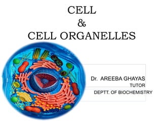
- 1. CELL & CELL ORGANELLES Dr. AREEBA GHAYAS TUTOR DEPTT. OF BIOCHEMISTRY
- 2. CHEMICAL MOLECULES OF LIFE Life is composed of lifeless chemicals molecules. Mainly six elements: Carbon, Hydrogen, Oxygen, Nitrogen, Phosphorous, And Sulfur. (90%) of the dry weight of the human body. A single cell of the bacterium E.coli contains about 6,000 different organic compounds. Human beings contains 1,00,000 different types of organic compounds.
- 3. COMPLEX BIOMOLECULES BIOMOLECULES BUILDING BLOCK MAJOR FUNCTIONS 1) Protein Amino acids Fundamental basis of structure and function cell 2) DNA Deoxyribonucleotides Repository of hereditary information 3) RNA Ribonucleotides Protein biosynthesis 4) Polysaccharide Glucose Storage form of energy to meet short term demands
- 4. Chemical Composition of Man Constituent Percent Weight(kg) Water 61.6 40 Protein 17.0 11 Lipid 13.8 9 Carbohydrates 1.5 1 Minerals 6.1 4
- 5. The Cell The cell is the structural and functional unit of living organisms; proposed by Schleiden & Schwann All animal tissues including human are also organized from collections of cells. Thus cell is the fundamental unit of life. Modern cell theory can be divided into the following fundamental statements: 1. All living things are made of cells. 2. Cells carry out the functions needed to support life. 3. Cells come only from other living cells.
- 6. TYPES OF CELLS The cells of the living kingdom may be divided into two categories. 1) Prokaryotes: pro-before ,karyon- nucleus Ex: bacteria 2) Eukaryotes: Eu-true , karyon- nucleus Ex: animals and plants
- 7. COMPARISON BETWEEN PROKARYOTIC & EUKARYOTIC CELLS Characteristic Prokaryotic Cell Eukaryotic Cell 1) Size Small Large 2) Cell Membrane Cell Is Enveloped By A Rigid Cell Wall Flexible Plasma Membrane 3) Sub Cellular Absent Present 4) Nucleus No defined nucleus. DNA present but not separated from rest of the cell; Histone absent. Present 5) Energy Metabolism Absent Present 6) Cell Division By binary division Mitosis 7) Cytoplasm Undifferentiated Contains various membrane bound organelles ; such as Mitochondria, Golgi bodies , Lysosomes & Peroxisomes
- 8. Cell has three major components: 1. Nucleus 2. Cytoplasm with its organelles –– Mitochondria –– Endoplasmic reticulum –– Golgi apparatus –– Lysosomes –– Peroxisomes 3. Plasma membrane (cell membrane) THE CELL ORGANELLES
- 9. 1) NUCLEUS 1) Nucleus was first discovered by ROBERT BROWN 2) Nucleus is the control centre of the cell; it contains the DNA organized into chromosomes which carry genetic information. 3) The nucleus is surrounded by a double membrane called nuclear envelope. 4) The outer membrane is fused with the endoplasmic reticulum at multiple sites. 5) Nuclear pores (multiprotein complexes) occur at points where the outer and inner membranes are connected.
- 11. DNA replication and RNA transcription of DNA occur in the nucleus. The nucleolus is non-membranous and contains RNA polymerase, RNAase, ATPase and other enzymes but no DNA polymerase. Nucleolus is the site of— - Synthesis of ribosomal RNA (r-RNA). -Assembling of ribosome subunits FUNCTION OF NUCLEUS
- 12. 2) MITOCHONDRIA 1) They are spherical, oval or rod-like bodies. 2) Erythrocytes are an exception which derive their ATP from glycolysis due to lack of mitochondria. 3) Mitochondria are the powerhouse of the cell, where energy released from oxidation of food stuffs is trapped as chemical energy in the form of ATP.
- 14. Structure Of Mitochondria The mitochondrion is bounded by two concentric membranes that have different properties and biological functions. Mitochondrial Membranes (a) Outer mitochondrial membrane: Consists mostly of phospholipids , cholesterol & protein (Porin). (b) Inner mitochondrial membrane: Are very rich in proteins. The inner membrane projects inwards into folds that are called cristae .
- 15. (c) Intermembrane space: is the space between the outer and inner membranes. The outer membrane is freely permeable to small molecules, the intermembrane space has about the same ionic composition as the cytosol. (d) Mitochondrial matrix: The region enclosed by the inner membrane is known as the mitochondrial matrix.
- 17. Functions Of Mitochondria The inter-membrane space contains several enzymes involved in nucleotide metabolism. The gel-like matrix (mitosol) consists of enzymes required for the metabolic pathways (Glycolysis) of oxidation of pyruvate (pyruvate dehydrogenase). The mitochondrial matrix is the site of most of the reactions of the citric acid cycle and fatty acid oxidation.
- 18. Components of electron transport system and oxidative phosphorylation that are responsible for the synthesis of ATP are embedded in inner membrane. Mitochondria contain their own DNA, (mtDNA), which in human encodes 13 respiratory chain proteins, small and large rRNAs and enough tRNAs to translate all codons. Mitochondria have a key role in aging; cytochrome c,an initiator of apoptosis.
- 19. MITOCHINDRIA-CLINICAL ASPECTS Mitochondrial DNA can be damaged by free radicals. Age related degenerative disorders –Parkinson’s disease, cardiomyopathy. Mutational defects leading to Mitochondrial myopathies- Leber’s hereditary optic neuropathy (LHON) Mitochondrial encephalomyopathy, lactic acidosis and stroke-like episodes (MELAS) Myoclonus epilepsy with ragged red fibers (MERRF)
- 20. 3) ENDOPLASMIC RETICULUM Eukaryotic cells are characterized by several membrane complexes that are interconnected by separate organelles. These organelles are involved in protein synthesis, transport, modification, storage and secretion. It is a network of interconnecting membranes enclosing channels or cisternae, that are continuous from outer nuclear envelope to outer plasma membrane.
- 21. TYPES OF ER There are two kinds of endoplasmic reticulum (ER): (i) Rough surfaced ER, They are coated with ribosomes. Near the nucleus, this type of ER merges with the outer membrane of the nuclear envelope. (ii) Smooth surfaced ER: They do not have attached ribosomes.
- 22. Functions of ER (a) Function of rough ER: Rough ER synthesises membrane lipids, and secretory proteins. These proteins are inserted through the ER membrane into the lumen of the cisternae where they are modified and transported through the cell. (b) Function of smooth ER: Smooth endoplasmic reticulum is involved: (i) In lipid synthesis (ii) Modification and transport of proteins synthesised in the rough ER.
- 23. ER STORAGE DISEASES ERSD are defects in the secretory protein encoded by mutated gene- Lipoprotein lipase deficiency Abetalipoproteinemia Disorders of lipid metabolism Growth hormone receptors deficiency Hereditary hypothyroidism
- 24. 4) GOLGI APPARATUS Camillo Golgi described the structure in 1898. The Golgi organelle is a network of flattened smooth membranes and vesicles. In Golgi apparatus proteins are processed, modified and prepared for export from the cell. It works in association with endoplasmic reticulum, where proteins for certain destinations are synthesized.
- 25. FUNCTION 1. Golgi bodies are secretory cell organelles. 2. The modified proteins are sorted, packaged and transported to destination inside or outside the cell. Golgi apparatus plays the role of post office mail sorting room, the mail in this case being newly synthesized proteins. 3. Post- translational modification of proteins occurs in golgi lumen. 4. Golgi bodies & ER give rise to primary lysosomes & peroxisomes. 5. Acrosome of sperm cell is derived from golgi bodies.
- 26. Golgi bodies-clinical aspects Wilson's disease Menkes disease Duchenne muscular dystrophy
- 27. 5) LYSOSOMES Lysosomes contain packet of enzymes. Lysosomes are digestive tract of the cell, they are involved in digestion of proteins, lipids, carbohydrates and nucleic acids. The lysosomal enzymes have an optimum pH around 5. These enzymes are a. Polysaccharide hydrolysing enzymes (alpha-glucosidase, alpha-fucosidase, beta-galactosidase, beta- glucuronidase, hyaluronidase, lysozyme) b. Protein hydrolysing enzymes (cathepsins, collagenase, elastase, peptidases) c. Nucleic acid hydrolysing enzymes (ribonuclease, deoxyribonuclease)
- 29. Lysosomal enzymes destroy the foreign substances and pathogens like bacteria ,virus that are engulfed by the cells of immune system. Destroys aging cell organelles that are no longer required by the host cell. They are capable of self-destruction of the cell (autophagy). Lysosomes are also called ‘suicide bags’ and play an important role in apoptosis After death, they play role in postmortem autolysis. During development, play an important role in the formation of specialized tissues such as fingers and toes. For example, lysosomes digest the webbed tissues that join fingers and toes in the embryo. FUNCTION
- 32. 6) PEROXISOMES 1) Peroxisomes ,also known as microbodies, are single membrane cellular cell. 2) These vesicles are concerned with the metabolism of hydrogen peroxide (H2O2),hence the name peroxisome. 3) They contain such enzymes that forms, uses and destroy the H2O2. 4) The H2O2 produced in the cell are highly toxic & is decomposed into H2O and oxygen by the enzyme Catalase present in them.
- 33. FUNCTION OF PEROXISOME Contains Catalase which causes breakdown of H2O2. They are present in hepatic cells takes part in breakdown (oxidation) of- Long chain fatty acid D-amino acid to ethanol Uric acid to allantoin They also help in synthesis of glycolipids, plasmalogens, isoprenoids & bile.
- 34. 7) CYTOSOL AND CYTOSKELETON 1) The cellular matrix is collectively referred to as cytosol. 2) The cytoplasmic filaments are of three types: 1) microtubules 2) micro filaments 3) intermediate filaments. The cytoskeleton play a role in movement of cell organelles from one position to other within the cell.
- 37. The plasma membrane is an envelop surrounding the cell. It separates and protect the cell from the external environment. Plasma membrane also provide a connecting system between the cell and its environment . BIOLOGICAL MEMBRANES
- 38. Chemical Composition The membranes are composed of lipids, proteins, and carbohydrates. The actual composition differs from tissue to tissue. Large globular protein molecules are interspreaded in this lipid bilayer.
- 40. Structure Of Membranes A lipid bilayer model originally proposed for membrane structure in 1935 by Danielle and Davson has been modified.
- 41. FLUID MOSAIC MODEL The membrane is sometimes referred to as a fluid mosaic. The membrane mosaic is fluid because most of the interactions among its components are non-covalent, leaving individual lipid and protein molecules free to move laterally in the plane of the membrane
- 43. The lipid of the membrane provides a barrier that obstructs the movements of water and water-soluble substances from one cell compartment to another. The integral protein molecules in the membrane do penetrate all the way through the membrane, organized into actual pores, for passage of specific substances through the membrane.
- 44. The approximate composition of cell membrane is: Protein: 55% Phospholipids: 25% Cholesterol: 13% Other lipids: 4% Carbohydrate: 3%
- 45. Lipid of the membrane The basic lipid bilayer is composed of phospholipid molecules. One end of each phospholipid molecule (head group) is soluble in water that is it is hydrophilic, the phosphate end. The other end (tail group) is soluble only in fats; that is, it is hydrophobic, the free fatty acid end.
- 47. A and B: Structure of phospholipid. (A) A common glycerophospholipid; (B) Diagrammatic representation of phospholipid. A B
- 48. The principle phospholipids in the membrane are: Glycerophospholipids: • phosphatidylcholine, • phosphatidylethanolamine, and • phosphatidylserine Sphinogophospholipid: sphingomyelin.
- 49. The lipid composition varies among different cell types, with phosphatidylcholine being the major plasma membrane phospholipid in most cell types. Plasma membrane for example, is enriched in cholesterol and contains no detectable cardiolipin (diphosphatidylglycerol lipid) Mitochondrial membrane is very low in cholesterol and sphingolipids but that contain cardiolipin.
- 50. CHOLESTEROL The cholesterol molecules in the membrane are also lipid in nature, interspreaded between the phospholipids & maintains membrane fluidity The presence of cholesterol and the cis unsaturated fatty acids in the membrane prevent the hydrophobic chains from packing too closely together. Loss of membrane fluidity affect integral proteins, such as ion channels and receptors for neurotransmitters involved conducting the nerve impulse.
- 51. Most of the membrane proteins are glycoproteins. Two types of membrane proteins: 1. Integral membrane proteins- protrudes all the way through the membrane. They are very firmly associated with the lipid bilayer. 2. Peripheral membrane proteins: attached either to membrane surface or to integral proteins.; do not penetrate through the membrane. MEMBRANE PROTEINS
- 52. They are associated with the membrane through electrostatic interactions and hydrogen bonding with the hydrophilic domains of integral proteins and with polar head groups of membrane lipids
- 53. Functions of Membrane Proteins Peripheral proteins function as enzymes or as controllers of transport of substances through the cell membrane. Integral membrane proteins – 1. Function as channels (pores) through which water molecules and water soluble substances, especially ions, can diffuse between extracellular and intracellular fluids. 2. Act as carrier proteins transporting substances that could not penetrate the lipid bilayer. 3. As receptors for hormones and neurotransmitter. 4. Role in cell signalling.
- 54. Membrane carbohydrates Membrane carbohydrates occur in combination with proteins or lipids in the form of glycoproteins or glycolipids. Some of the proteins and lipids on the external surface of the membrane contain short chains of carbohydrate (oligosaccharides) that extent into the aqueous medium.
- 56. proteoglycans are loosely attached to the outer surface of the cell. the entire outside surface of the cell has often a loose carbohydrate coat glycoproteins and glycolipids called the glycocalyx. Carbohydrate constitutes 2% to 10% of the weight of cell membrane.
- 57. Functions of Membrane carbohydrates Many of the carbohydrates have a negative electrical charge, which gives most cells an overall negative surface charge that repels other negative objects and restricts the uptake of hydrophobic compounds. The glycocalyx of some cell attaches to the glycocalyx of other cells, thus attaching cells to one another. act as hormone receptor such as insulin involved in immune reactions.
- 58. Marker Enzymes For Different Organelles The purity of isolated subcellular fraction is assessed by the analysis of marker enzymes. Marker enzymes are the enzymes that are located exclusively in a particular fraction, and thus become characteristic of that fraction.
- 60. Thank you
- 61. MEMBRANE TRANSPORT Dr. AREEBA GHAYAS TUTOR DEPTT. OF BIOCHEMISTRY
- 62. MEMBRANE TRANSPORT Cell membranes form closed compartments around the cytoplasm to define cell boundaries. The cell membrane has selective permeability. Selective membrane permeability is conferred by specific transporters/carriers and channels.
- 65. PASSIVE TRANSPORT Transport is from higher concentration to lower concentration.( Downhill transport) The substance pass through the membrane from both sides. Transport does not require energy in the form of ATP.
- 66. TYPES OF PASSIVETRANSPORT A. Simple Diffusion B. Facilitated Diffusion 1) Channel mediated 2) Carrier mediated C. Osmosis
- 68. A. Simple Diffusion It is a passive transport. From High concentration to low concentration. (downhill transport). Kinetic energy used for the movement of molecules. Small& unchargedmolecules likeO2, CO2, salts , urea, ammonia & amino acids. No channel proteins or carrier proteins required (movement through the bilayer). Diffusion stops when equilibrium is achieved
- 70. 1) Solute moves along the concentration gradient from higher to lower concentration (downhill transport). 2) No energy is required. 3) Occurs through the mediation of carrier or channel proteins.(hence,2 types) 4) Sometimes regulated by hormones. E.g. GLUT4 regulated by insulin in muscle and adipose tissue. 5) Follows Saturation kinetics contrast from simple diffusion. ex: glucose, galactose, leucine, phenylalanine B. FACILITATED DIFFUSION:
- 71. A ping pong model is put forth to explain the occurrence of facilitated diffusion. Mechanism Of Facilitated Diffusion:
- 72. According to this mechanism, :- carrier / channel transport protein exists in two conformation, in the ping conformation it is exposed to the side with high solute concentration. This allow the binding of solute to specific sites on the carrier protein. The protein changes in pong conformation , exposed to the side with low solute concentration where the solute molecule is released.
- 73. C. OSMOSIS Osmosis is the diffusion of water across the semi permeable membrane. The direction of osmosis depends on the concentration of solutes inside and outside the cell.
- 76. OSMOTIC PRESSURE
- 77. ACTIVE TRANSPORT Active transport occurs against a concentration gradient from low to high concentration (uphill transport). Energy in the form of ATP is required. Carrier mediated process like facilitated diffusion. Follows Saturation kinetics contrast from simple diffusion. The most important primary Active transport systems are ion pumps.
- 78. Types Of Active Transport
- 84. Ouabain Digoxin Clinical Aspect of Na-K ATP Pump
- 90. VESICULAR TRANSPORT Occurs by fusion of formed vesicles or formation of new vesicles. Macromolecules (such as sugars, amino acids, hormones, neurotransmitters, cellular secretions, etc) cannot be transported by diffusion or active transport process. Clathrin, coating proteins, dynamin & docking proteins are the vesicular transport proteins used for formation & transport of vesicles.
- 91. Types Of Vesicular Transport 1) Endocytosis: formation of vesicle from the cell membrane (intake) 2) Exocytosis: fusion of vesicle from the cell membrane (release)
- 93. 1) Endocytosis: Intake of macromolecules by the cells. Approximately 2% of the exterior surface of plasma membrane possesses characteristic Coated-pits. The pits can be internalized to form coated vesicles which contain an unusual protein called Clathrin. E.g., The uptake of LDL molecules by the cells. Occurs by constitutive pathway
- 94. Uptake Of LDL Molecules
- 95. Endocytosis occurs when the plasma membrane is pulled inwards and will form a “pocket” around a particular substance. The substance will become enclosed in the vesicle which is then pinched off and begins moving through the cytoplasm. Cells can bring in solids and liquids using this process.
- 98. 2. Exocytosis: Release of macromolecules from the cells to outside. 1. In golgi bodies, Macromolecules are packaged into a vesicle. 2. The vesicles in cytoplasm are transported to the plasma membrane. 3. Vesicles fuses with plasma membrane 4. Contents of the vesicle are released into the external environment of the cell. Role of exocytosis is to secrete substances being produced or excrete waste products. The secretion of hormone occur by Exocytosis. e.g. Insulin
- 100. Diseases due to loss of membrane transport systems: Hartnups disease Cystinuria Rickets
Editor's Notes
- Electrochemical gradient energy is used