
Recomendados
Más contenido relacionado
Más de cartlidge
Más de cartlidge (20)
Assessment statements h2
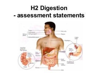
- 1. H2 Digestion - assessment statements
- 2. H.2.1 State that digestive juices are secreted into the alimentary canal by glands, including salivary glands, gastric glands in the stomach wall, the pancreas and the wall of the small intestine. H.2.2 Explain the structural features of exocrine gland cells. (Include the secretory cells grouped into acini and ducts, and the ultrastructure of secretory cells as seen in electron micrographs) H.2.3 Compare the composition of saliva, gastric juice and pancreatic juice. H.2.4 Outline the control of digestive juice secretion by nerves and hormones, using the example of secretion of gastric juice. (Limit this to the initial release of gastric juice under nerve stimulation after sight or smell of food, and sustained release under the influence of gastrin secreted when food is in the stomach)
- 3. H.2.5 Outline the role of membrane-bound enzymes on the surface of epithelial cells in the small intestine in digestion. (Some digestive enzymes (for example, maltase) are immobilized in the exposed plasma membranes of epithelial cells in intestinal villi) H.2.6 Outline the reasons for cellulose not being digested in the alimentary canal. H.2.7 Explain why pepsin and trypsin are initially synthesized as inactive precursors and how they are subsequently activated. H.2.8 Discuss the roles of gastric acid and Helicobacter pylori in the development of stomach ulcers and stomach cancers.
- 4. H.2.9 Explain the problem of lipid digestion in a hydrophilic medium and the role of bile in overcoming this. (Lipid molecules tend to coalesce and are only accessible to lipase at the lipid–water interface. Bile molecules have a hydrophilic end and a hydrophobic end, and thus prevent lipid droplets coalescing. The maximum surface is exposed to lipases. The need for lipase to be water-soluble and to have an active site to which a hydrophobic substrate binds should be mentioned)