Midterm Extra Office Hours and Techniques
•
0 recomendaciones•395 vistas

The document provides information about midterm exams and extra office hours for a class. It announces that the first midterm exam will be on Thursday, February 15 at 6pm in room 155 Dwinelle, and that review sessions will be held during the regular class time that day. It also notes that the professor will not have office hours on February 13.

Recomendados
Recomendados
Más contenido relacionado
La actualidad más candente
La actualidad más candente (20)
Similar a Midterm Extra Office Hours and Techniques
Similar a Midterm Extra Office Hours and Techniques (20)
Más de dream10f
Más de dream10f (20)
Último
Último (20)
Midterm Extra Office Hours and Techniques
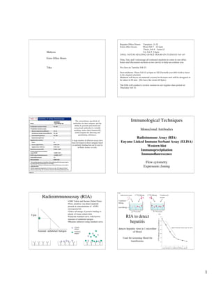
- 1. 1 Midterm Extra Office Hours Take Regular Office Hours: Tuesdays 11-12 Extra office hours: Wed, Feb 7 12-1pm Thurs, Feb 8 11am-12 Fri, Feb 9 2-4pm I WILL NOT BE HOLDING OFFICE HOURS ON TUESDAY Feb 13!! Dina, Tim, and I encourage all confused students to come to our office hours and discussion sections so we can try to help un-confuse you. No class on Tuesday Feb 13. First midterm: Thurs Feb 15 at 6pm in 155 Dwinelle (not 2050 VLSB as listed in the original schedule). Midterm will focus on material covered in lectures and will be designed to be taken in 90 min. (We have the room till 8pm.) The GSIs will conduct a review session in our regular class period on Thursday Feb 15. The extraordinary specificity of antibodies for their antigens, and the ability to generate polyclonal and monoclonal antibodies to virtually anything, makes them fantastically useful reagents for detecting and quantitating substances. A large number of different assays have been developed to detect antigens based on antibody binding that can be used in in fluids, tissues, or cells. Immunological Techniques Monoclonal Antibodies Radioimmune Assay (RIA) Enyzme Linked Immune Sorbant Assay (ELISA) Western blot Immunoprecipitation Immunofluorescence Flow cytometry Expression cloning Radioimmunoassay (RIA) Cpm Amount unlabeled Antigen •1960 Yalow and Berson (Nobel Prize) •Very sensitive: can detect material present at concentrations of <0.001 micrograms/ml. •Takes advantage of protein binding to plastic of tissue culture dish. •Generate standard curve with known amounts of unlabeled antigen •Measure unknown using standard curve. Labeled antigen Unlabeled antigen RIA to detect hepatitis detects hepatitis virus in 1 microliter of blood. Used for screening blood for transfusions.
- 2. 2 Enzyme-linked immune sorbant assay (ELISA) Colorometric assay Capture antigen using plate-bound antibody Add second specific antibody-enzyme conjugate Detection by secondary antibodies conjugated to enzymes (alkaline phosphatase, horse radish peroxidase, β-galactosidase). Break- down of substrate by enzyme produces a visible color. Variations of the ELISA method ELISA based pregnancy test
- 3. 3 Real life example of use of ELISA assay (from your reading assignment: “A Toll-like receptor recognizes bacterial DNA” Hemmi 2000 Nature v408 p 740 Figure 3b) Isolate macrophage from wild type and TLR9 mutant mice. Stimulate with CpG (mammalian DNA is methylated at CpG residues, and so differs from unmethylated bacterial DNA) Peptidoglygan (PGN) and Lipopolysaccaride (LPS) trigger innate immune responses through other TLRs and serve as positive controls. Measure cytokine production (TNF-α) in culture supernatants by ELISA Concentration of the cytokine TNF-α as measured by ELISA • The ELISPOT assay can be used to determine the number of cells within a sample that are secreting a particular cytokine. Western Blot Gel electrophoresis separates proteins by size, so Western blot not only provides quantitation, but also provides information about the molecular weight of antigen. SDS-polyacrylamide gel electrophoresis to separate a complex mixture of proteins based on their molecular weight. Transfer proteins from gel to a membrane sheet. After transferring proteins from gel to a membrane sheet, use labeled antibodies to your protein of interest to detect the relevant band. Labeled antibodies bind to band containing your protein of interest. Detect labeled antibody using colorimentic assay.
- 4. 4 Immunoprecipitation + Protein A or Protein G Anti- antibody Protein A and Protein G: bacterial cell wall proteins that binds to Ig. Immunoprecipitation: variations of the method. Magnetic beads coupled to antibodies can be used to isolate proteins from solution, or cells from a suspension. And check out “A Toll-like receptor recognizes bacterial DNA” Hemmi 2000 Nature v408 p 740 Figure 3 f and g For real-life examples of Immunoprecipitations and Western blots to analyze TLR signaling intermediates in wild type and TLR9- macrophage in response to different TLR agonists. Immunoaffinity Chromatography Immunofluorescence direct indirect Fluorochrome-conjugated specific antibody Unconjugated specific antibody Fluorochrome-conjugated secondary-antibody (anti-Ig antibody)
- 5. 5 Immunofluorescence can provide spatial information about cells or molecules that react with antibodies. Anti-IgM stain of B cells Immunological Techniques Monoclonal Antibodies Radioimmune Assay (RIA) Enyzme Linked Immune Sorbant Assay (ELISA) Western blot Immunoprecipitation Flow cytometry Expression cloning Mixture of cells labeled with fluorescent antibodies Flow cytometry can be used to determine the number of cells within a sample that react with a particular antibody (or antibodies) Fluorescence intensity (red) Cell number Fluorescence intensity Cell number Unstained cells (negative control) Stained cells 30% positive < 1 % positive Fluorescence intensity (green) Cell number Stained cells 40% positive Flow cytometric analysis of cells stained with 2 different labeled antibodies Fluorescence intensity (red) Fluorescenceintensity(green) 2 parameter dot plot: 1 parameter histograms: 20% “double Positive” 20% Green only 10% red only 50% “double negative” Analysis of cells stained with labeled antibodies Dot plots Contour plots histograms Amt. IgD Number of cells Number of cells
- 6. 6 The Power of Flow Cytometry Quantitative: Accurately determine relative fluorescent levels (proteins levels) on individual cells. Accurately determine the number of fluorescent cells within a population. Sensitive: Analysis can be performed with <104 cells. Flexible: Fluorescent labeled antibodies specific for many cell surface proteins are readily available. Can simultaneously stain for >4 markers. Real life example of use of flow cytometry (from your reading assignment: “A Toll-like receptor recognizes bacterial DNA” Hemmi 2000 Nature v408 p 740 Figure 3d) Fluorescence intensity Wild type TLR9-/- CD40 CD80 CD86 MHC-II Isolate macrophage from wild type and TLR9 mutant mice. Stimulate with CpG (or LPS as positive control). Examine expression of activation markers by flow cytometry. Note: CD40, CD90, CD86, and MHC-II are all important for T cell activation by macrophage (innate immunity stimulating adaptive immunity). Numberofcells The generation of monoclonal antibodies specific for cell surface proteins, coupled with flow cytometry, provides a powerful tool for identifying different lymphocyte populations. Flow cytometry can be used to identify different kinds of leukemia. (“CD” nomenclature) Fluorescence activated Cell- sorting (FACS) using the flow cytometer Ultrasonic nozzle vibrator Cell suspension Sheath fluid Drop-charging signal Cell collectorCell collector Flask for undeflected droplets Cell- sorting using the flow cytometer laser
- 7. 7 Magnetic beads coupled to antibodies can also be used to purify cells (an alternative to FACS, that is especially useful for processing larger numbers of cells.) Flow cytometry is used here to monitor the composition of the cell sample before and after separation. Expression cloning genes in E. coli Cell expressing Protein of interest Purified mRNA cDNA cDNA library in Bacterial expression vector Specific antibody Bacterial Colonies, Each Expressing One cDNA Bacteria expressing cDNA of interest Expression Cloning •Make antiserum against gene-product of interest •Make cDNA expression library from cell line expressing gene •Transfect library into cell line which does not normally express gene-product •Select cells expressing product by antibody-binding •Recover cDNA expression vector •Repeat. Recover. Sequence. Victory! A a result of V(D)J recombination every mature B cell expresses a unique antibody. Encounter with an antigen leads to clonal expansion of B cells with a particular specificity. Where we have been and where we are going Innate Immunity Antibodies and antigens I (emphasis on antibody structure) Antibodies and antigens II (emphasis on antigen-antibody binding interactions) Techniques based on antibodies V(D)J recombination B cell development and function V(D)J Recombination Discovery of Ig gene rearrangements Structure of antibody genes (RSS) Role of RAG proteins and DNA repair machinery
- 8. 8 Variability of Ig Sequences HV1 HV2 HV3 The puzzle of antibody diversity • Limitless array of Ig sequences (too large to be encoded in genome) • Variation limited to V domain. • Identical V segment could be associated with two different C regions (myeloma protein with γ and µ chains) • Germ-line vs somatic variation models • Dreyer and Bennett (1965) the 2 gene model; a violation of the “one gene, one polypeptide” rule • 1976: the emerging tools of molecular biology open the way for the breakthrough. . . The breakthrough paper Review of Southern blot method Restriction endonucleases cleave at specific sequences in DNA and can be used to generate a physical map of DNA. (e.g. EcoRI cleaves at the sequence: 5’-GAATTC-3’) Surprising Southern blot liver skin spleen myeloma1 myeloma2 myeloma3 Surprising Southern blot liver skin spleen myeloma1 myeloma2 myeloma3 probe Vκ1 Vκ 2 Cκ Jκ1Jκ2Jκ3Jκ4 Cκ
- 9. 9 Detecting Ig gene rearrangement using Southern blot V(D)J Recombination Discovery of Ig gene rearrangements Structure of antibody genes (RSS) Evidence for role of RAG proteins and DNA repair machinery VK gene segments JK gene segments CK exons Light chains encoded by 2 gene loci: kappa and lambda Each light chain encoded by 3 kinds of gene segments: V (variable), J (joining), C (constant) A V and J segment are brought together by somatic DNA rearrangement process: “V(D)J recombination” Multigene organization of Ig genes: light chain genes VH gene segments First 96 aa’s of Ig HC DH gene segments 3-6 aa’s HC JH gene segments 10-12 aa’s HC CH exons Multigene organization of Ig genes: heavy chain genes Heavy chains encoded by a single gene locus. Each heavy chain encoded by 4 kinds of gene segments: V(variable), D (diversity), J (joining), C (constant) V, D, and J segments are brought together by somatic DNA rearrangement process: “V(D)J recombination” Multigene organization of Ig genes V(D)J Rearrangement: light chain Gene rearrangement Transcription VK gene segments JK gene segments CK exons RNA splicing mRNA V and J gene segments brought together in DNA before transcription. (RNA slicing removes introns.)
- 10. 10 V(D)J Rearrangement: heavy chain VH gene segments First 96 aa’s of Ig HC DH gene segments 3-6 aa’s HC JH gene segments 10-12 aa’s HC CH exons Gene rearrangement Transcription RNA splicing mRNA V, D, and J gene segments brought together in DNA before transcription. (RNA slicing removes introns.) V(D)J Rearrangement VH gene segments First 96 aa’s of Ig HC DH gene segments 3-6 aa’s HC JH gene segments 10-12 aa’s HC CH exons Gene rearrangement Variable domain exon Constant domain exons Combinatorial Diversity in humans Junctional diversity (flexible joining of segments, P and N region additions at junctions) also contributes substantially to the total diversity of antibodies. Gene rearrangement juxtaposes promoter and enhancers Promoters: relatively short nucleotide sequences within ~200 bp of transcriptional start site that initiate transcription in a certain direction. Enhancers: nucleotide sequences located up-stream or down-stream of a gene that activated the promoter in an orientation independent manner.
- 11. 11 Ig promoters are actively transcribed when they are brought close to enhancers due to gene rearrangement. Allelic exclusion of Ig genes: ensures that most B cell will express a single antibody specificity (allele: two or more alternative forms of a gene.) Rearranging gene segments are flanked by a conserved “rearrangement signal sequence” Gene-segment 7-mer 9-mer12 or 23nt heptamer spacer nonamer RSS Also known as “one turn RSS” and “two turn RSS”. Rearranging gene segments are flanked by a conserved “rearrangement signal sequence” The 12/23 Rule Only gene segments flanked by RSSs with dissimilar spacers can undergo V(D)J recombination with one another. Ensures that V segments don’t join with other Vs, that VH don’t join with JH, etc. V(D)J Recombination: Reactants & Products
- 12. 12 Flexibility in joining of gene segments contributes to junctional diversity. Note most rearrangements are non- productive! (Only 1/3 rearrangements preserves the correct reading frame of the J segment.) P nucleotides are generated by resolution of hairpin structures. N nucleotide are added by an enzyme called terminal deoxynucleotide transferase (TdT). P and N nucleotide addition also contribute to junctional diversity.