General and Systemic Pathology Concepts-A Global Overview
•
125 recomendaciones•9,442 vistas

General and Systemic Pathology Concepts-A Global Overview

Recomendados
Más contenido relacionado
La actualidad más candente
La actualidad más candente (20)
Destacado
Destacado (20)
Similar a General and Systemic Pathology Concepts-A Global Overview
Similar a General and Systemic Pathology Concepts-A Global Overview (20)
Más de Imhotep Virtual Medical School
Más de Imhotep Virtual Medical School (20)
Último
Último (20)
General and Systemic Pathology Concepts-A Global Overview
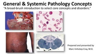
- 1. General & Systemic Pathology Concepts Prepared and presented by Marc Imhotep Cray, M.D. “A broad-brush introduction to select core concepts and disorders.”
- 2. Marc Imhotep Cray, M.D. Topical Outline 2 Introduction to Pathology Cell & Tissue Injury and Inflammation Neoplasia Cardiovascular System Respiratory System Gastrointestinal System Renal System Nervous System Musculoskeletal System Endocrine System
- 4. 4 General pathology is the study of mechanisms of disease, with emphasis on etiology and pathogenesis. Systematic pathology is the study of diseases as they occur within particular organ systems-it involves: Etiology Pathogenesis Epidemiology, macro- and microscopic appearance Specific diagnostic features Natural history and Sequelae Clinical pathology is often referred to as laboratory medicine and includes a number of diagnostic disciplines.
- 5. Marc Imhotep Cray, M.D. 5 Pathology provides the basis for understanding: The mechanisms of disease The classification of diseases The diagnosis of diseases The basis of treatment Monitoring the progress of disease Determining prognosis Understanding complications
- 6. Marc Imhotep Cray, M.D. Systematized Nomenclature of Medicine 6 SNOMED-standard classification of disease-considers following aspects: Topography Morphology Etiology Function Disease Procedure Occupation
- 7. 7 Techniques of Pathology Gross pathology – macroscopic investigation and observation of disease Light microscopy – thin section of wax or plastic permeated tissues, snap- frozen tissues Histochemistry – microscopy of treated tissue sections (to distinguish cell components) Immunohistochemistry and immunofluorescence – tagged antibodies (monoclonal better) Electron microscopy Biochemical techniques – e.g. fluid and electrolyte balance, serum enzymes Cell cultures – also allowing cytogenetic analysis Medical microbiology – direct microscopy, culturing and identification Molecular pathology – in situ hybridization (specific genes/mRNA), polymerase chain reaction (PCR)
- 8. Cell & Tissue Injury and Inflammation 8
- 9. Marc Imhotep Cray, M.D. Basic Concepts Cellular and tissue growth is a normal component of normal physiology Complex intra- and intercellular signaling mechanisms control rate and extent of growth Many disease processes are characterized by alterations in rate and control of cellular and tissue turnover Defects in these normal control mechanisms may lead to disease states such as neoplasia 9
- 10. Marc Imhotep Cray, M.D. Basic Concepts (2) 10 There are several ways in which constituents of body can alter in size in association with a normal physiological mechanism or as part of a disease process Cells and tissues may increase in size via o Hyperplasia= usually results from increased physiologic demands or hormonal stimulation or o Hypertrophy=in response to increased physiologic or pathophysiologic demands A decrease in size occurs via atrophy= causes (1) disuse (2) denervation(3) ischemia (4) nutrient starvation (5) interruption of endocrine signals (6) & persistent cell injury
- 11. Marc Imhotep Cray, M.D. Basic Concepts (3) 11 Metaplasia= is process whereby differentiated (i.e. mature) cells change from o Examples: Chronic irritation of bronchial mucosa by cigarette smoke leads to conversion of ciliated columnar epithelium to stratified squamous epithelium • Vitamin A is necessary to maintain epithelia Related: Ethiopian National Vitamin A Deficiency Survey Report, 2008. o Barrett’s esophagus Specialized intestinal metaplasia=replacement of nonkeratinized stratified squamous epithelium w intestinal epithelium (nonciliated columnar w goblet cells in distal esophagus • Due to chronic reflux esophagitis (GERD) • Associated w risk of esophageal adenocarcinoma
- 12. Marc Imhotep Cray, M.D. Basic Concepts (4) Cells and Tissues Insults 12 Cells and tissues may be damaged by a range of insults: physical (trauma and extremes of heat) chemical (e.g. acid) neoplastic (e.g. cancers infiltrating adjacent tissue) infective (e.g. bacterial pneumonia) immune (e.g. autoimmune diseases rheumatoid arthritis) iatrogenic (e.g. drugs causing gastric ulceration)
- 13. Marc Imhotep Cray, M.D. Inflammation (1) 13 Evolution of Inflammation Engulfment/entrapment Neutralization of irritant Elimination of injurious agent Definition= A local response to infection or injury Inflammation is a complex reaction of a tissue and its microcirculation to a pathogenic insult characterized by generation of inflammatory mediators and movement of fluid & leukocytes from blood into extravascular tissues It is a major component of response to cellular and tissue injury
- 14. Marc Imhotep Cray, M.D. Inflammation (2) 14 Inflammation Characterized by o increased blood flow (redness and warmth: rubor and calor) o swelling (tumor) and o pain (dolor) within affected area o systemic effects including malaise and pyrexia
- 15. Marc Imhotep Cray, M.D. The inflammatory response (3) 15 Is fundamentally a protective/defensive response Persists until inciting stimulus is removed & mediators are dissipated or inhibited Can be potentially harmful: Anaphylactic shock (peanut allergy) Systemic inflammatory response syndrome (SIRS) Is closely intertwined with repair Therapeutic strategies target critical control points in inflammatory pathways
- 16. Marc Imhotep Cray, M.D. Inflammation: “the players” (5) 16
- 17. Marc Imhotep Cray, M.D. Acute Inflammation: major components (4) 17 Vascular changes: Vasodilation and increased blood flow Increased vascular permeability Cellular events: Leucocyte transmigration Phagocytosis Chemical mediators (acute & chronic)
- 18. Marc Imhotep Cray, M.D. Acute inflammation 18 Acute inflammation occurs during early phase of a reaction to cellular/tissue damage It is characterized histologically by presence of acute inflammatory cells (neutrophils) within affected tissue Acute inflammation may resolve if underlying stimulus is removed, or it may progress to chronic inflammation
- 19. Marc Imhotep Cray, M.D. Acute inflammation (2) 19 Acute inflammation occurs through release of inflammatory mediators from damaged tissues and other cells This leads to a combination of increased vascular permeability and chemotaxis: attraction of inflammatory cells to area secondary to release of chemicals from site of inflammation
- 20. Marc Imhotep Cray, M.D. Cardinal Signs of Inflammation (6) 20 Redness (rubor) Swelling (tumor) Heat (calor) Pain (dolor) Loss of function (functio laesa) (fifth cardinal sign added by Virchow)
- 21. Marc Imhotep Cray, M.D. Cardinal Signs 21 Patient with a Methicillin-resistant Staphylococcus aureus wound infection, and classic signs of inflammation Rubin R and Strayer DS Eds. Rubin’s Pathology: Clinicopathologic Foundations of Medicine, 6th Ed. Baltimore: Lippincott Williams & Wilkins, 2012.
- 22. Marc Imhotep Cray, M.D. Cardinal Signs 22 X-ray of previous patient showing non-union of fracture Holes are from orthopedic screws Rubin R and Strayer DS Eds. Rubin’s Pathology: Clinicopathologic Foundations of Medicine, 6th Ed. Baltimore: Lippincott Williams & Wilkins, 2012.
- 23. Marc Imhotep Cray, M.D. Cardinal Signs 23 Bone scan of same patient, showing uptake in area of active inflammation Rubin R and Strayer DS Eds. Rubin’s Pathology: Clinicopathologic Foundations of Medicine, 6th Ed. Baltimore: Lippincott Williams & Wilkins, 2012.
- 24. Marc Imhotep Cray, M.D. Blood Cells and Platelets 24
- 25. Marc Imhotep Cray, M.D. Production of blood cells by bone marrow 25Widmaier, EP. Vander’s Human Physiology : The Mechanisms of Body Function. 13th Ed. McGraw-Hill, 2014.
- 26. Marc Imhotep Cray, M.D. Light micrograph of a human blood smear 26Widmaier, EP. Vander’s Human Physiology : The Mechanisms of Body Function. 13th Ed. McGraw-Hill, 2014.
- 27. Marc Imhotep Cray, M.D. Cells of Inflammation 27 Leukocytes (WBCs) are major cellular participants in inflammation and include Neutrophils T and B lymphocytes Monocytes-macrophages Eosinophils Mast cells and basophils Each cell type has specific functions but they overlap and change as inflammation progresses Inflammatory cells and resident tissue cells interact with each other in a continuous response during inflammation
- 28. Marc Imhotep Cray, M.D. Cells of inflammation: morphology & function (1) 28 Neutrophil Rubin R and Strayer DS Eds. Rubin’s Pathology: Clinicopathologic Foundations of Medicine, 6th Ed. Baltimore: LLW, 2012.
- 29. Marc Imhotep Cray, M.D. Effector functions of neutrophils 29Rubin R and Strayer DS Eds. Rubin’s Pathology: Clinicopathologic Foundations of Medicine, 6th Ed. Baltimore: LLW, 2012.
- 30. Marc Imhotep Cray, M.D. Cells of inflammation: morphology & function (2) 30 Endothelial cell Rubin R and Strayer DS Eds. Rubin’s Pathology: Clinicopathologic Foundations of Medicine, 6th Ed. Baltimore: LLW, 2012.
- 31. Marc Imhotep Cray, M.D. Cells of inflammation: morphology & function (3) 31 Monocyte/macrophage Rubin R and Strayer DS Eds. Rubin’s Pathology: Clinicopathologic Foundations of Medicine, 6th Ed. Baltimore: LLW, 2012.
- 32. Marc Imhotep Cray, M.D. More cells of inflammation: morphology and function (4) 32 Rubin R and Strayer DS Eds. Rubin’s Pathology: Clinicopathologic Foundations of Medicine, 6th Ed. Baltimore: LLW, 2012.
- 33. Marc Imhotep Cray, M.D. More cells of inflammation (5) 33Rubin R and Strayer DS Eds. Rubin’s Pathology: Clinicopathologic Foundations of Medicine, 6th Ed. Baltimore: LLW, 2012.
- 34. Marc Imhotep Cray, M.D. More cells of inflammation: morphology and function (6) 34 Rubin R and Strayer DS Eds. Rubin’s Pathology: Clinicopathologic Foundations of Medicine, 6th Ed. Baltimore: LLW, 2012.
- 35. Marc Imhotep Cray, M.D. Acute inflammation (7) 35 Densely packed (PMNs) with multilobed nuclei (arrows) Rubin R and Strayer DS Eds. Rubin’s Pathology: Clinicopathologic Foundations of Medicine, 6th Ed. Baltimore: LLW, 2012.
- 36. Marc Imhotep Cray, M.D. Acute Inflammation (8) 36 1. Vasodilation/ increased blood flow 2. Deposition of fibrin and other plasma proteins (exudate) 3. Transmigration and accumulation of neutrophils
- 37. Marc Imhotep Cray, M.D. Acute Inflammation (9) 37 Vasodilation Slowing of circulation Stasis and margination
- 38. Marc Imhotep Cray, M.D. Stasis and Margination 38 PMNs at margin of a vessel in acutely inflamed tissue Rubin R and Strayer DS Eds. Rubin’s Pathology: Clinicopathologic Foundations of Medicine, 6th Ed. Baltimore: LLW, 2012.
- 39. Marc Imhotep Cray, M.D. Chronic inflammation 39 Chronic inflammation may occur de novo or develop as a sequel to acute inflammation especially if source of cellular/tissue damage persists It is characterized histologically by presence of chronic inflammatory cells: lymphocytes, plasma cells and macrophages
- 40. Marc Imhotep Cray, M.D. Chronic inflammation (2) 40 Granulomatous inflammation is a special form of chronic inflammation characterized histologically by presence of granulomas localized collections of macrophages Multinucleate giant cells may also be present Causes of granulomatous inflammation include tuberculosis fungal infections tissue reactions to foreign material and specific diseases such as sarcoidosis and Crohn’s disease
- 41. Marc Imhotep Cray, M.D. Chronic inflammation (3) 41 Lymphocytes (double- headed arrow), plasma cells (arrows) and a few macrophages (arrowheads) are present Rubin R and Strayer DS Eds. Rubin’s Pathology: Clinicopathologic Foundations of Medicine, 6th Ed. Baltimore: Lippincott Williams & Wilkins, 2012.
- 42. Marc Imhotep Cray, M.D. Consequences of inflammation: definitions 42 Several definitions help in understanding of consequences of inflammation: ■ Edema is accumulation of fluid in extravascular space and interstitial tissues ■ An effusion is excess fluid in body cavities (e.g., peritoneum or pleura) ■ A transudate is edema fluid with a low protein content (specific gravity <1.015) ■ An exudate is edema fluid with a high protein conc. (specific gravity >1.015), frequently contains inflammatory cells Exudates are seen early in acute inflammation and are produced by mild injuries, such as sunburn or traumatic blisters
- 43. Marc Imhotep Cray, M.D. Consequences of inflammation: definitions (2) 43 ■ A serous exudate, or effusion, is characterized by absence of a prominent cellular response and has a yellow, straw-like color ■ Serosanguineous refers to a serous exudate, or effusion, that contains red blood cells and has a reddish tinge
- 44. Marc Imhotep Cray, M.D. Consequences of inflam: definitions (3) 44 ■ A fibrinous exudate has large amounts of fibrin due to activation of coagulation system o When a fibrinous exudate occurs on a serosal surface, such as pleura or pericardium, it is termed “fibrinous pleuritis” or “fibrinous pericarditis” ■ A purulent exudate or effusion contains prominent cellular components o Purulent exudates and effusions are often associated with pathologic conditions, such as pyogenic bacterial infections, in which polymorphonuclear neutrophils (PMNs) predominate ■ In suppurative inflammation, a purulent exudate is with significant liquefactive necrosis it is equivalent of pus
- 45. 45 Vascular Leakage Rubin R and Strayer DS Eds. Rubin’s Pathology: Clinicopathologic Foundations of Medicine, 6th Ed. Baltimore: Lippincott Williams & Wilkins, 2012.
- 46. 46 Margination, rolling, activation and adhesion Transmigration (diapedesis) Migration toward site of injury along a chemokine gradient Leukocyte Extravasation and Phagocytosis
- 47. Marc Imhotep Cray, M.D. Leukocyte Extravasation & Phagocytosis: Animation 47
- 48. 48 Local inflammatory events occurring in response to a wound Widmaier, EP. Vander’s Human Physiology : The Mechanisms of Body Function. 13th Ed. McGraw-Hill, 2014.
- 49. 49 Chemical Mediators of Inflammation Tissue injury stimulates production of inflammatory mediators in plasma & release into circulation Additional factors are generated by tissue cells & inflammatory cells Vasoactive and chemotactic mediators promote edema and recruit inflammatory cells to site of injury Rubin R and Strayer DS Eds. Rubin’s Pathology: Clinicopathologic Foundations of Medicine, 6th Ed. Baltimore: Lippincott Williams & Wilkins, 2012
- 50. Marc Imhotep Cray, M.D. Chemical Mediators of Inflammation (2) 50 Chemicals that are released from damaged tissues and inflammatory cells orchestrates inflammatory process e.g. histamine, prostaglandins, leukotrienes & TNF-α Protein cascades originating within plasma are also important in regulating response to tissue injury e.g. coagulation, fibrinolytic, complement and kinin cascades
- 51. Marc Imhotep Cray, M.D. Inflammation Resolution 51 Resolution of inflammation is associated with organization of inflammatory reaction: granulation tissue formation and myofibroblast proliferation followed by A variable degree of collagen deposition (fibrous scarring) o Collagen deposition more pronounced if inflammatory process has been prolonged
- 52. Marc Imhotep Cray, M.D. Tissue Injury and Healing 52 Tissue injury is usually followed by hemostasis= inflammatory response tissue restructuring w a variable degree of scarring Factors impairing healing include: old age poor nutritional state excessive tissue damage poor apposition of wound edges (or bony fragments after a fracture) presence of foreign material poor blood supply infection
- 53. 53 1.Tissue injury results in immediate and prolonged vascular changes. Chemical mediators and damaged tissue cells stimulate vasodilation and vascular injury leading to 2. leakage of fluid into tissues (edema) 3. Platelets are activated to initiate clot formation and hemostasis and increase vascular permeability via histamine release 4. Vascular endothelial cells contribute to clot formation, anchor circulating neutrophils via upregulated adhesion molecules and retract to allow increased vascular permeability to plasma and inflammatory cells at same time 5. microbes (red rods) initiate activation of the complement cascade, which, along with soluble mediators from macrophages, 6. recruits neutrophils to site of tissue injury. 7. Phagocytosis (See next sequence of slides.): Neutrophils and macrophages eliminate microbes and remove damaged tissue so that repair can begin Rubin R and Strayer DS Eds. Rubin’s Pathology: Clinicopathologic Foundations of Medicine, 6th Ed. Baltimore: Lippincott Williams & Wilkins, 2012. Summary of inflam. response to injury
- 54. 54 Chemistry of Phagocytosis Activated neutrophils and macrophages kill phagocytosed microbes (and damaged tissue) by action of microbicidal molecules in phagolysosomes Three classes of microbicidal molecules are most important 1. Reactive oxygen species (ROS)=highly reactive oxidizing agents that destroy microbes (& other cells) Called respiratory burst b/c it occurs during oxygen consumption (cellular respiration) 2. Nitric oxide 3. Proteolytic enzymes
- 55. 55 Chemistry of Phagocytosis (2) Reactive oxygen species (ROS) Oxygen (O2) has a major role as the terminal electron acceptor in mitochondria It is reduced from O2 to H2O and resultant energy is harnessed as an electrochemical potential across mitochondrial inner membrane Conversion of O2 to H2O entails transfer of four electrons three partially reduced species, representing transfers of varying numbers of electrons, are intermediate between O2 and H2O These are O2 − = superoxide (one electron); H2O2= hydrogen peroxide (two electrons); OH•= hydroxyl radical (three electrons)
- 56. 56 Phagocytosis and intracellular destruction of a microbe Widmaier, EP. Vander’s Human Physiology : The Mechanisms of Body Function. 13th Ed. McGraw-Hill, 2014.
- 57. Marc Imhotep Cray, M.D. 57 Phagocytosis & intracellular destruction of a microbe (2) Abbas AK, Lichtman AH, Pillai S. Cellular And Molecular Immunology. Saunders-Elsevier, 2015.
- 58. 58 A scanning electron microscope image of a single neutrophil (yellow), engulfing anthrax bacteria (orange) http://upload.wikimedia.org/wikipedia/com mons/f/f2/Neutrophil_with_anthrax_copy.jpg Widmaier, EP. Vander’s Human Physiology : The Mechanisms of Body Function. 13th Ed. McGraw- Hill, 2014. Scanning electron microscope (SEM) images of a single neutrophil and macrophage (LR) engulfing bacterium. Phagocytosis illustrated
- 59. Marc Imhotep Cray, M.D. Phagocyte respiratory burst (oxidative burst) 59 Primary free radical–generating system is phagocyte oxidase system Involves activation of phagocyte NADPH oxidase complex (e.g., in neutrophils, monocytes) which utilizes O2 as a substrate Plays an important role in immune response rapid release of reactive oxygen species (ROS) NADPH plays a role in both creation and neutralization of ROS Myeloperoxidase (produces hypochlorite) is a blue-green heme-containing pigment that gives sputum its color
- 60. 60 Phagocyte oxidase system (Redox RXN) Phagocyte oxidase is a multisubunit enzyme that is assembled in activated phagocytes mainly in phagolysosomal membrane activated by many stimuli, including IFN-γ and signals from TLRs Function of phagocyte oxidase is to reduce molecular oxygen into ROS* such as superoxide radicals (O2−) with reduced form of nicotinamide adenine dinucleotide phosphate (NADPH) acting as a cofactor Superoxide is enzymatically dismutated into hydrogen peroxide which is used by enzyme myeloperoxidase to convert normally unreactive halide ions into reactive hypohalous acids (hypochlorite) that are toxic for bacteria *Other ROS include H2O2= hydrogen peroxide & OH•= hydroxyl radical
- 61. Marc Imhotep Cray, M.D. Phagocyte respiratory burst (2) 61 Le T and Bhushan V. Microbiology. In: First Aid for the USMLE Step 1 2016. McGraw-Hill, 2016.
- 62. 62 Oxidative stress “a key trigger for cell & tissue injury and adaptive responses” For human life, oxygen is both a blessing and a curse Without it, life is impossible, but some of its derivatives are partially reduced oxygen species that can react with, and damage, virtually any molecule they reach i.e., ROS (free radicals) Reactive Oxygen Species N.B. ROSs causes of cell and tissue injury in many settings (Illust.) Copstead LC, Banksia JL. Pathophysiology, 5th Ed. St. Louis, Missouri: Saunders-Elsevier, 2013. Of note: Increased free radicals in heart can occur post MI reperfusion. Such toxic oxygen radicals are released from neutrophils when blood flow is restored following ischemia= Reperfusion injury
- 63. 63 Phagocyte respiratory burst (3) Phagocytic cell disorder Deficiency of one of components of phagocyte oxidase results in CGD (chronic granulomatous disease) = an X-linked inherited deficiency Phagocytes can utilize H2O2 generated by invading organisms & convert it to ROS Catalase-negative bacteria are effectively killed b/c microbes produce small amounts of peroxide leading to microbial death however CGD patients are at risk for infection by catalase ⊕ species (e.g., S aureus, Aspergillus [fungus]) capable of neutralizing their own H2O2 leaving phagocytes without ROS for fighting infections Related notes: Pyocyanin of P. aeruginosa functions to generate ROS to kill competing microbes Lactoferrin is a protein found in secretory fluids and neutrophils that inhibits microbial growth via iron chelation
- 64. Marc Imhotep Cray, M.D. Immune System: Protection from harmful microorganisms 64 Complex systems exist to protect body from microorganisms Some of these systems are innate and have a broad-based action (non-specific) while others are acquired as result of an adaptive immune response act more specifically Functions of immune system are carried out by immunoreactive cells circulating within blood and present within tissues (See inflammation section above) as well as by circulating antibodies
- 65. Marc Imhotep Cray, M.D. Innate and Adaptive Immunity 65 Defense against microbes is mediated by early reactions of innate immunity and later responses of adaptive immunity Innate immunity (also called natural or native immunity) provides early line of defense against microbes consists of cellular and biochemical defense mechanisms in place even before infection and respond rapidly to infections React to products of microbes and injured cells they respond in same way to repeated exposures
- 66. Marc Imhotep Cray, M.D. Mechanisms of innate immunity 66 Target structures common to groups of related microbes & do not distinguish fine differences betw microbes (non-specific) Principal components of innate immunity are 1) physical and chemical barriers such as epithelia and antimicrobial chemicals produced at epithelial surfaces 2) phagocytic cells (neutrophils, macrophages), dendritic cells, and natural killer (NK) cells and other innate lymphoid cells 3) blood proteins, including complement system and other mediators of inflammation
- 67. Marc Imhotep Cray, M.D. Innate and Adaptive Immunity cont. 67 Adaptive immunity (also called specific or acquired immunity) stimulated by exposure to infectious agents and increase in magnitude and defensive capabilities with each successive exposure to a particular microbe b/c this form of immunity develops as a response to infection and adapts to infection called adaptive immunity defining characteristics of adaptive immunity are ability to distinguish different substances, called specificity, and ability to respond more vigorously to repeated exposures to same microbe, known as memory (anamnestic response) unique components of adaptive immunity are cells called lymphocytes and their secreted products such as antibodies
- 68. Marc Imhotep Cray, M.D. Innate and adaptive immunity illustrated. 68Abbas AK, Lichtman AH, Pillai S. Cellular And Molecular Immunology. Saunders-Elsevier, 2015.
- 69. 69 Types of Adaptive Immune Responses There are two types of adaptive immune responses, called humoral immunity and cell-mediated immunity mediated by different components of the immune system and function to eliminate different types of microbes Humoral immunity is mediated by molecules in blood and mucosal secretions, called antibodies produced by cells called B lymphocytes (also called B cells) o Antibodies recognize microbial antigens, neutralize infectivity of microbes, and target microbes for elimination by various effector mechanisms Humoral immunity is the principal defense mechanism against extracellular microbes and their toxins b/c secreted antibodies can bind to these microbes and toxins and assist in their elimination (e.g. bacterial infections) o Antibodies themselves are specialized and may activate different mechanisms to combat microbes (effector mechanisms)
- 70. 70 Types of Adaptive Immune Responses cont. Cell-mediated immunity (also called cellular immunity) is mediated by T lymphocytes (also called T cells) Intracellular microbes, such as viruses and some bacteria, survive and proliferate inside phagocytes and other host cells, where they are inaccessible to circulating antibodies Defense against such infections is a function of cell-mediated immunity which promotes destruction of microbes residing in phagocytes or killing of infected cells to eliminate reservoirs of infection Some T lymphocytes also contribute to eradication of extracellular microbes by recruiting leukocytes that destroy these pathogens and by helping B cells make effective antibodies
- 71. Marc Imhotep Cray, M.D. Types of adaptive immunity illust. 71Abbas AK, Lichtman AH, Pillai S. Cellular And Molecular Immunology. Saunders-Elsevier, 2015.
- 72. Marc Imhotep Cray, M.D. Active immunity and Passive immunity 72 Active immunity= Protective immunity against a microbe is usually induced by host’s response to microbe The form of immunity that is induced by exposure to a foreign antigen is called active immunity b/c immunized individual plays an active role in responding to antigen Individuals and lymphocytes that have not encountered a particular antigen are said to be naïve implying they are immunologically inexperienced; contrastly Individuals who have responded to a microbial antigen and are protected from subsequent exposures to that microbe are said to be immune N.B. Only active immune responses generate immunologic memory.
- 73. Marc Imhotep Cray, M.D. Active immunity and Passive immunity cont. 73 Passive immunity= Immunity conferred on an individual by transferring serum or lymphocytes from a specifically immunized individual, a process known as adoptive transfer Recipient of such a transfer becomes immune to particular antigen without ever having been exposed to or having responded to that antigen thus, called passive immunity o Passive immunization = useful method for conferring resistance rapidly, without having to wait for an active immune response to develop A physiologically important example of passive immunity transfer of maternal antibodies through placenta to fetus enables newborns to combat infections before they develop ability to produce antibodies themselves
- 74. Marc Imhotep Cray, M.D. Active and passive immunity illustrated 74Abbas AK, Lichtman AH, Pillai S. Cellular And Molecular Immunology. Saunders-Elsevier, 2015.
- 75. Marc Imhotep Cray, M.D. Autoimmune diseases 75 Autoimmune diseases occur when immune system attacks ‘self’ cells and tissues this is referred to as a breakdown of “immune tolerance” This leads to inflammation and tissue damage, which may be o highly localized (e.g. type 1 diabetes mellitus) or o generalized (e.g. systemic lupus erythematosus)
- 76. Marc Imhotep Cray, M.D. Immune System Defects 76 Defects may occur within immune system May be: congenital (e.g. severe combined immunodeficiency) or acquired (e.g. reaction to chemotherapy, infection with human immunodeficiency virus (HIV)) May affect: a specific component of immune system or have more widespread effects within several components Defects usually lead to increased susceptibility to a range of infections
- 77. Marc Imhotep Cray, M.D. Mechanisms of Cell Death: Apoptosis vs Necrosis 77 There are two major mechanisms by which cells can die Apoptosis (programmed cell death) is an energy-requiring process leading to death of individual cells, which does not incite an inflammatory reaction o Apoptosis may be physiological or pathological in nature Necrosis does not require energy, usually affects groups of cells and typically incites an inflammatory reaction usually acute in nature
- 78. Marc Imhotep Cray, M.D. Cells and Tissue Degenerative Processes 78 Various degenerative processes can occur within cells and tissues as a result of disease states, for example: Calcification may occur if serum calcium conc. is chronically elevated (‘metastatic’ calcification) or within an abnormal tissue (e.g. a tumor or focus of chronic inflammation ‘dystrophic’ calcification Amyloid is an insoluble protein with a β-pleated sheet structure that is deposited either locally or in a widespread manner in various chronic disease states such as chronic inflammatory conditions (e.g. tuberculosis) or low-grade neoplasms of B-lymphocyte lineage (e.g. lymphoplasmacytic lymphoma) Other forms of degenerative change include glycogen accumulation, hyaline change and myxomatous change
- 79. 79 Cells and Tissue Pigment Accumulation Hemosiderin is an iron-containing pigment that may be deposited in tissues following red cell destruction and hemoglobin breakdown (e.g. after a hemorrhage) or w/in organs such as liver in genetic hemochromatosis hemosiderin granules impart yellow to brown color of healing bruise Lipofuscin (or lipochrome) is a wear-and-tear pigment that is deposited in organs such as heart and liver Melanin is produced by melanocytes in skin and is commonly found in tumors showing melanocytic differentiation (e.g. malignant melanoma) Bilirubin is a bile pigment that accumulates in jaundice, either in conjugated or unconjugated form (yellow sclera & skin= icterus) Anthracosis is a black color comes from carbon pigments in dust inhaled over years, engulfed by macrophages, and sent via lymphatics to nodes It looks bad but does not compromise lung function Smokers will have more anthracosis an accumulation exogenous
- 80. Marc Imhotep Cray, M.D. Shock 80 Shock is a clinical condition characterized by a fast pulse rate (usually > 100 beats/min) and a low blood pressure (systolic blood pressure usually < 100 mmHg) Common types of shock are hypovolemic (low blood volume, e.g. in hemorrhage), cardiogenic (heart pump failure, e.g. in myocardial infarction) septic (severe infection) Less common types are anaphylactic (type I hypersensitivity reaction, e.g. penicillin allergy) neurogenic (loss of sympathetic vasomotor tone, e.g. in a spinal cord injury)
- 81. Marc Imhotep Cray, M.D. Body protective mechanisms 81 Body possesses many mechanisms that aim to protect against potentially injurious agents These mechanisms may be o Behavioral o Anatomical or o Immunological
- 82. Marc Imhotep Cray, M.D. Congenital diseases vs Inherited diseases 82 Congenital diseases are those that are present at birth Inherited diseases are those passed on from parents via transfer of a genetic defect (e.g. familial adenomatous polyposis) Congenital diseases may be inherited from parents but may also occur though chromosomal abnormalities that originate during gametogenesis or fertilization (e.g. Down’s syndrome) or ‘insults’ sustained by fetus before birth (e.g. congenital infections)
- 83. 83 Neoplasia
- 84. Marc Imhotep Cray, M.D. Neoplasia 84 Neoplasia means “new growth” and indicates presence of cells or tissues showing evidence of abnormally controlled or disordered growth Neoplasms comprise cells that show differentiation along one or more pathways of development Benign vs Malignant Benign neoplasms expand locally but do not invade adjacent tissues or spread to distant sites, while Malignant neoplasms (cancers) invade adjacent tissues and spread to distant sites
- 85. 85 Neoplasia (2) Preneoplastic and neoplastic cellular changes Neoplasia Uncontrolled, clonal proliferation of cells Can be benign or malignant Dysplasia Disordered, non-neoplastic cell growth Used only with epithelial cells Mild dysplasia is usually reversible Severe dysplasia usually progresses to carcinoma in situ Differentiation degree to which a malignant tumor resembles its tissue of origin Well-differentiated tumors closely resemble their tissue of origin poorly differentiated look almost nothing like their tissue of origin Anaplasia Complete lack of differentiation of cells in a malignant neoplasm
- 86. Marc Imhotep Cray, M.D. Neoplasia (3) 86 Genetic and environmental factors influence development of neoplasia Most germline (i.e. inherited and present in all cells) genetic influences on neoplasm development are polygenic in nature, while A minority of neoplasms occur in association with a clearly defined inherited defect in a single gene (monogenic) Neoplasms vary in their relative incidence between populations and different geographical areas as a result of differences in gene pools and environmental contributors to disease development
- 87. Marc Imhotep Cray, M.D. Neoplasia (4) 87 Neoplasm development is characterized by accumulation of genetic defects within neoplastic cells In some neoplasms, this sequence is well characterized In others specific genetic mutations are found sufficiently commonly that their detection may be used to confirm the diagnosis of tissue type or to help to determine likely biological behavior of neoplasm (i.e. how aggressively the neoplasm is likely to grow)
- 88. Marc Imhotep Cray, M.D. Neoplasia (5) 88 Benign tumors may compress adjacent tissue but do not invade it Malignant tumors grow locally, infiltrate adjacent tissue and metastasize via lymphatic channels and blood vessels to distant sites Benign tumors can cause death by compressing vital structures (e.g. within brainstem) but otherwise generally possess a much better prognosis than malignant tumors
- 89. Marc Imhotep Cray, M.D. Neoplasia (6) 89 Malignant tumors commonly cause extensive local tissue damage but tumor metastasis to distant sites is often key process that causes death in advanced malignancy Benign and malignant tumors may also produce chemicals such as hormones and, therefore, be associated with clinical symptoms of hormone excess Called a “paraneoplastic syndrome”
- 90. 90 Neoplasia (7) Clinical and pathological features of neoplasms can indicate whether they are benign or malignant in nature Histopathological examination of malignant neoplasms is important to determine how aggressively neoplasm is likely to grow and metastasize Features such as tumor type grade (histological assessment of aggressiveness) size and presence of lymph node metastases are most commonly assessed features used to predict biological behavior of malignant neoplasms (See Grading & Staging, slides # 74 & 75.)
- 91. Marc Imhotep Cray, M.D. Neoplasia (8) 91 Most cancers (>90%) arise from "epithelial" tissues, such as inside lining of colon, breast, lung or prostate These are referred to as carcinomas and usually affect older people Contrastly, sarcomas are tumors that arise from "mesenchymal" tissues such as bone, muscle, connective tissue, cartilage and fat
- 92. Marc Imhotep Cray, M.D. Neoplasia (9) Lung cancer 92 Lung cancer is an aggressive neoplasm for which cigarette smoking is major risk factor Almost all lung cancers are carcinomas Neoplasm can invade local structures including mediastinum and chest wall and commonly metastasizes to distant sites Many patients present when disease is at an advanced local stage or with widespread metastases and when surgical removal is not possible
- 93. 93 Klatt EC. Robbins and Cotran Atlas of Pathology, 3rd Ed. Philadelphia: Saunders, 2015.
- 94. 94 Klatt EC. Robbins and Cotran Atlas of Pathology, 3rd Ed. Philadelphia: Saunders, 2015.
- 95. 95 Klatt EC. Robbins and Cotran Atlas of Pathology, 3rd Ed. Philadelphia: Saunders, 2015.
- 96. 96 Klatt EC. Robbins and Cotran Atlas of Pathology, 3rd Ed. Philadelphia: Saunders, 2015.
- 97. 97 Klatt EC. Robbins and Cotran Atlas of Pathology, 3rd Ed. Philadelphia: Saunders, 2015.
- 98. Bronchogenic carcinoma, gross The large carcinoma ( ) in the upper lobe is arising in a lung with centriacinar emphysema, suggesting cigarette smoking as the risk factor There are patchy infiltrates in lower lobe representing pneumonia, likely from central airway obstruction by this large mass There is inferior congestion, likely exacerbated by heart failure Klatt EC. Robbins and Cotran Atlas of Pathology, 3rd Ed. Philadelphia: Saunders, 2015.
- 99. 99
- 100. 100Klatt EC. Robbins and Cotran Atlas of Pathology, 3rd Ed. Philadelphia: Saunders, 2015.
- 101. 101 Klatt EC. Robbins and Cotran Atlas of Pathology, 3rd Ed. Philadelphia: Saunders, 2015.
- 102. Marc Imhotep Cray, M.D. Neoplasia (10) Breast cancer 102 Breast cancer is second most common malignancy in women (only exceeded by lung cancer in populations where cigarette smoking is common) Almost all breast cancers are carcinomas Most often present as breast masses and invade local structures including skin and breast wall as well as metastasizing to local lymph nodes and distant sites While breast cancer is an important cause of mortality among middle aged and older women modern advances in therapy have significantly improved outcome
- 103. 103 Klatt EC. Robbins and Cotran Atlas of Pathology, 3rd Ed. Philadelphia: Saunders, 2015.
- 104. 104Klatt EC. Robbins and Cotran Atlas of Pathology, 3rd Ed. Philadelphia: Saunders, 2015.
- 105. 105 Klatt EC. Robbins and Cotran Atlas of Pathology, 3rd Ed. Philadelphia: Saunders, 2015.
- 106. 106
- 107. 107Klatt EC. Robbins and Cotran Atlas of Pathology, 3rd Ed. Philadelphia: Saunders, 2015.
- 108. 108 Klatt EC. Robbins and Cotran Atlas of Pathology, 3rd Ed. Philadelphia: Saunders, 2015.
- 109. 109 Klatt EC. Robbins and Cotran Atlas of Pathology, 3rd Ed. Philadelphia: Saunders, 2015.
- 110. Marc Imhotep Cray, M.D. Neoplasia (11) Colorectal cancer 110 Colorectal cancer is one of three most common cancers in Western populations it is likely that environmental factors, including Western diet with low roughage, contribute to this Almost all colorectal cancers are carcinomas These neoplasms grow locally and pts. may present w rectal bleeding, a change in bowel habit or w acute abdominal symptoms caused by bowel obstruction or perforation Metastasis to local lymph nodes and distant sites (most commonly liver) may occur Surgical removal when disease is localized to bowel wall is often associated with a favorable outcome
- 111. 111 Klatt EC. Robbins and Cotran Atlas of Pathology, 3rd Ed. Philadelphia: Saunders, 2015.
- 112. 112 Klatt EC. Robbins and Cotran Atlas of Pathology, 3rd Ed. Philadelphia: Saunders, 2015.
- 113. 113 Klatt EC. Robbins and Cotran Atlas of Pathology, 3rd Ed. Philadelphia: Saunders, 2015.
- 114. 114Klatt EC. Robbins and Cotran Atlas of Pathology, 3rd Ed. Philadelphia: Saunders, 2015.
- 115. Marc Imhotep Cray, M.D. Neoplasia (12) Prostatic cancer 115 Prostatic cancer is increasing in incidence among middle-aged and elderly men although this may partly reflect increased detection of disease in its early stages in screening programs Almost all prostatic cancers are carcinomas May invade local pelvic structures and metastasize to distant sites, especially bone While advanced prostatic cancer is commonly fatal, localized disease (most commonly identified by screening) may be curable with prostatectomy Progression of advanced disease may be slowed with hormonal therapy
- 116. 116 Klatt EC. Robbins and Cotran Atlas of Pathology, 3rd Ed. Philadelphia: Saunders, 2015.
- 117. 117 Klatt EC. Robbins and Cotran Atlas of Pathology, 3rd Ed. Philadelphia: Saunders, 2015.
- 118. 118 Klatt EC. Robbins and Cotran Atlas of Pathology, 3rd Ed. Philadelphia: Saunders, 2015.
- 119. 119 Klatt EC. Robbins and Cotran Atlas of Pathology, 3rd Ed. Philadelphia: Saunders, 2015.
- 120. 120 Klatt EC. Robbins and Cotran Atlas of Pathology, 3rd Ed. Philadelphia: Saunders, 2015.
- 121. 121 Klatt EC. Robbins and Cotran Atlas of Pathology, 3rd Ed. Philadelphia: Saunders, 2015.
- 122. 122 Klatt EC. Robbins and Cotran Atlas of Pathology, 3rd Ed. Philadelphia: Saunders, 2015.
- 123. 123
- 124. 124 Klatt EC. Robbins and Cotran Atlas of Pathology, 3rd Ed. Philadelphia: Saunders, 2015.
- 125. 125 Klatt EC. Robbins and Cotran Atlas of Pathology, 3rd Ed. Philadelphia: Saunders, 2015.
- 126. 126 Klatt EC. Robbins and Cotran Atlas of Pathology, 3rd Ed. Philadelphia: Saunders, 2015.
- 127. 127 Klatt EC. Robbins and Cotran Atlas of Pathology, 3rd Ed. Philadelphia: Saunders, 2015.
- 128. 128 Klatt EC. Robbins and Cotran Atlas of Pathology, 3rd Ed. Philadelphia: Saunders, 2015.
- 129. Marc Imhotep Cray, M.D. Neoplasia (13) 129 Certain neoplasms occur primarily in childhood e.g. neuroblastoma and nephroblastoma Elderly individuals develop wear-and-tear diseases osteoarthritis atherosclerosis-associated conditions e.g. ischemic heart disease [IHD]) and Elderly individuals are at increased risk of many neoplasms
- 130. 130 Neoplasia (14) Neoplasm development is commonly associated with genetic abnormalities within neoplastic tissue however, proportion of neoplasms that occur as a result of a single inherited germline genetic abnormality (i.e. a mutation present within all of cells making up an individual) is relatively low Examples include inherited predispositions to breast cancer and colorectal cancer o Although relatively uncommon, these inherited syndromes are important since affected individuals may develop cancer at a young age and sometimes develop multiple cancers o Identification of affected families may allow cancer prevention programs and/or detection of cancers at an early stage
- 131. Marc Imhotep Cray, M.D. Neoplasia (15) Tumor grade vs stage 131 Grade Degree of cellular differentiation and mitotic activity on histology Range from low grade (well differentiated) to high grade (poorly differentiated, undifferentiated or anaplastic) Stage Degree of localization/spread based on site and size of 1° lesion, spread to regional lymph nodes, presence of metastases Based on clinical (c) or pathology (p) findings Example: cT3N1M0 Stage almost always has more prognostic value than grade
- 132. Marc Imhotep Cray, M.D. TNM staging system 132 TNM staging system (Stage = Spread): T = Tumor size N = Node involvement M = Metastases Each TNM factor has independent prognostic value M factor often most important
- 133. Marc Imhotep Cray, M.D. Disease screening 133 Disease screening means attempting to detect disease processes at an early (asymptomatic) stage when prompt treatment should result in an improved prognosis Diseases are required to fit various criteria in order to be suitable for screening US screening programs are currently in place for neoplastic diseases such as breast & cervical cancer & for non-neoplastic diseases such as neonatal hypothyroidism and phenylketonuria (PKU)
- 134. Marc Imhotep Cray, M.D. Disease and Extremes of Age 134 Body is particularly susceptible to certain conditions at extremes of age For example Premature babies possess immature body systems and are prone to infections and specific difficulties associated with organs that are not fully developed (e.g. respiratory failure, gut failure) Elderly individuals are at increased risk of many neoplasms, atherosclerosis-associated conditions, osteoarthritis etc.
- 136. Marc Imhotep Cray, M.D. Atherosclerosis 136 Atherosclerosis is a very common disease process occurring within arteries, especially large elastic arteries and their major branches Earliest lesions comprise ‘fatty streaks’ within arterial intima Established atherosclerotic plaques comprise a “cap” of fibrous tissue beneath which are pools of fat, foamy macrophages and smooth muscle cells Dystrophic calcification is common in older lesions Plaque surface may ulcerate (plaque rupture) leading to a thrombus that coats plaque acute vascular occlusion See: Atherosclerosis and Thrombosis Illustrated Notes Online version - Offline
- 137. 137 Arteriosclerosis Arteriosclerosis is a general term for several disorders that cause thickening and loss of elasticity in the arterial wall Atherosclerosis, the most common form, is also most serious b/c it causes coronary artery disease and cerebrovascular disease Atherosclerosis is patchy intimal plaques (atheromas) in medium-sized and large arteries plaques contain lipids, inflammatory cells, smooth muscle cells, and connective tissue Coronary artery with atherosclerotic narrowing, microscopic Normal coronary artery, microscopic From:WebpathCardiovascularPathologyimageplates
- 138. Marc Imhotep Cray, M.D. Ischemic heart disease (IHD) 138 IHD is leading cause of death among adults within Western populations It occurs secondary to narrowing of one or more of coronary arteries most commonly as a result of atherosclerotic changes Ischemic heart disease commonly results in angina and may lead to myocardial infarction and/or cardiac failure Sudden death may occur with or without evidence of MI
- 139. Marc Imhotep Cray, M.D. Diagnostic Classifications & Terminology 139 Anatomic Diagnosis= Atherosclerosis (ASHD) Etiologic Diagnosis= Coronary Heart Disease (CHD, IHD, CAD) Physiologic Diagnosis= e.g., Angina Pectoris Functional Diagnosis= Stable vs Unstable Angina vs MI [STEMI vs NSTEMI]=ACS
- 140. Marc Imhotep Cray, M.D. Coronary heart disease (CHD or IHD) Defined (Etiologic Dx) 140 Coronary heart disease proper circulation of blood and oxygen are not provided to heart and surrounding tissue due to a narrowing of small blood vessels, which normally supply heart with blood and oxygen
- 141. 141 Causes (Anatomic Dx) Typical cause of coronary heart disease is atherosclerosis takes place with plaque and fatty build up on artery walls narrowing vessels
- 143. 143 Pathobiology of Atherosclerosis (pathogenesis) When excess cholesterol deposits on cells and on the inside walls of blood vessels it forms an atherosclerotic plaque First step of atherosclerosis is injury to endothelium results in atherosclerotic lesion formation When plaque ruptures blood clots form lead to decreased blood flow resulting in cardiovascular events (ACS/MI) Coronary artery, severe atherosclerosis, gross Coronary artery, mild atherosclerosis, gross From:WebpathCardiovascularPathologyimageplates
- 144. 144 Pathobiology of Atherosclerosis (2) Symptoms develop when growth or rupture of plaque reduces or obstructs blood flow Diagnosis is clinical and confirmed by angiography, or other imaging tests Treatment includes risk factor management and dietary modification, physical activity, antiplatelet drugs, and antiatherogenic drugs Heart and LAD coronary artery with recent thrombus, gross Anterior surface of heart demonstrates an opened left anterior descending coronary artery Within lumen of coronary can be seen a dark red recent coronary thrombosis The dull red color to myocardium as seen below glistening epicardium to lower right of thrombus is consistent with underlying myocardial infarction From: Webpath Cardiovascular Pathology image plates
- 145. Marc Imhotep Cray, M.D. Risk Factors for Atherosclerosis 145 Risk factors atherosclerosis include: Dyslipidemia (hypercholesterolemia/LDL-C) diabetes mellitus cigarette smoking family history sedentary lifestyle obesity Hypertension Positive Family Hx CVD & premature death Lipoprotein(a) [abbreviated Lp(a)] o Apparently, only men, but not women, are affected by this risk
- 146. Marc Imhotep Cray, M.D. Treatment 146 Coronary heart disease Tx methods may include: (depends on presenting Physiologic Dx) 1. Angioplasty with stenting 2. Coronary artery bypass surgery (CABG) 3. Medication 4. Minimally invasive heart surgery 5. Proper diet and exercise 6. Quitting smoking 7. Treatment of other comorbidities, HTN, DM, Obesity
- 147. 147 Klatt EC. Robbins and Cotran Atlas of Pathology, 3rd Ed. Philadelphia: Saunders, 2015.
- 148. Marc Imhotep Cray, M.D. Cerebrovascular disease 148 Apart from ischemic heart disease, atherosclerosis also commonly affects carotid and intracranial arteries leading to cerebrovascular disease (e.g. strokes [CVA], vascular dementia) while aortic and iliac artery atherosclerosis leads to aortic aneurysm formation and peripheral vascular disease (e.g. intermittent claudication and foot gangrene)
- 149. 149 Klatt EC. Robbins and Cotran Atlas of Pathology, 3rd Ed. Philadelphia: Saunders, 2015.
- 150. Marc Imhotep Cray, M.D. Thrombosis 150 Thrombosis occurs after activation of clotting cascade and is a vital physiological mechanism for limiting blood loss when hemorrhage occurs Thrombosis occurring as part of a disease process lead to local vascular occlusion (e.g. coronary artery thrombosis) or to distant vascular occlusion (thromboembolism, e.g. pulmonary thromboembolism secondary to deep vein thrombosis)
- 151. 151 Klatt EC. Robbins and Cotran Atlas of Pathology, 3rd Ed. Philadelphia: Saunders, 2015.
- 152. Marc Imhotep Cray, M.D. Embolism 152 An embolism occurs when an embolus migrates from one part of body and causes a blockage of a distant blood vessel embolus can be made up of materials other than a thrombus, for example o Air o Amniotic fluid o Fat or o Tumor tissue
- 153. 153 Klatt EC. Robbins and Cotran Atlas of Pathology, 3rd Ed. Philadelphia: Saunders, 2015.
- 154. Marc Imhotep Cray, M.D. Valvular Heart Disease 154 The mitral and aortic valves are valves most commonly affected by degenerative disease in adults Stenosis or incompetence of these valves may lead to cardiac failure and (apart from mitral stenosis) left ventricular cardiac hypertrophy aortic stenosis is a not uncommon cause of sudden death Rheumatic fever is an important cause of mitral valve stenosis in older patients Damaged cardiac valves are prone to secondary bacterial infection (endocarditis) which itself can lead to further valvular damage
- 155. 155 Klatt EC. Robbins and Cotran Atlas of Pathology, 3rd Ed. Philadelphia: Saunders, 2015.
- 156. 156 Klatt EC. Robbins and Cotran Atlas of Pathology, 3rd Ed. Philadelphia: Saunders, 2015.
- 157. 157 Viral Myocarditis and Cardiomyopathy Unusual conditions of myocardium such as viral myocarditis and cardiomyopathy (e.g. hypertrophic cardiomyopathy) are important causes of sudden death in young adults Obstructive hypertrophic cardiomyopathy (subset) asymmetric septal hypertrophy and systolic anterior motion of mitral valve, outflow obstruction, dyspnea, possible syncope In hypertrophic cardiomyopathy diastolic dysfunction ensues Cardiomyopathies may result from a genetic defect or secondary to cardiac muscle damage, following, for example viral myocarditis or chronic excess alcohol consumption (dilated cardiomyopathy) o In dilated cardiomyopathy systolic dysfunction ensues
- 158. 158
- 159. 159
- 160. 160
- 161. 161
- 162. 162
- 163. 163
- 164. Marc Imhotep Cray, M.D. Congenital heart disease 164 There are many forms of congenital heart disease resulting in anatomical abnormalities of heart (e.g. ventricular septal defect, valvular atresia) and associated structures (e.g. patent ductus arteriosus) Congenital heart defects leading to introduction of systemic venous blood directly into systemic arterial circulation commonly cause cyanosis
- 165. 165 Klatt EC. Robbins and Cotran Atlas of Pathology, 3rd Ed. Philadelphia: Saunders, 2015.
- 166. 166 Klatt EC. Robbins and Cotran Atlas of Pathology, 3rd Ed. Philadelphia: Saunders, 2015.
- 167. Marc Imhotep Cray, M.D. Cardiac failure 167 Cardiac failure occurs when heart is unable to eject blood sufficiently effectively during systole Common causes of heart failure include ischemic heart disease cardiac valvular disease hypertensive heart disease chronic lung disease Less common causes include pericardial constriction and dilated cardiomyopathy LV cardiac failure results in pulmonary vascular congestion and edema (PE) RV cardiac failure produces a raised jugular venous pressure, hepatic venous congestion & peripheral edema N.B. Under conditions of poor tissue perfusion, there will be more anaerobic glycolysis and more acidosis in cells throughout the body. The blood lactate rises in this condition.
- 168. 168Klatt EC. Robbins and Cotran Atlas of Pathology, 3rd Ed. Philadelphia: Saunders, 2015.
- 169. 169 Klatt EC. Robbins and Cotran Atlas of Pathology, 3rd Ed. Philadelphia: Saunders, 2015.
- 170. Marc Imhotep Cray, M.D. Hypertension 170 Hypertension is common, often asymptomatic and has many causes including Stress Obesity Renal artery stenosis and Hormonal defects such as Cushing’s syndrome and Conn’s syndrome Chronic hypertension is characterized by an imbalance in sodium and water homeostasis Untreated hypertension can lead to accelerated atherosclerosis and to end-organ damage, including hypertensive nephropathy, hypertensive heart disease and intracerebral hemorrhage
- 171. 171 Klatt EC. Robbins and Cotran Atlas of Pathology, 3rd Ed. Philadelphia: Saunders, 2015.
- 172. 172 Klatt EC. Robbins and Cotran Atlas of Pathology, 3rd Ed. Philadelphia: Saunders, 2015.
- 173. 173 Klatt EC. Robbins and Cotran Atlas of Pathology, 3rd Ed. Philadelphia: Saunders, 2015.
- 174. 174 Klatt EC. Robbins and Cotran Atlas of Pathology, 3rd Ed. Philadelphia: Saunders, 2015.
- 176. Marc Imhotep Cray, M.D. Pneumonia 176 Pneumonia means inflammation within lung and most commonly occurs as a result of an infection Many microorganisms may infect lung tissue, but among most common are viruses and bacteria: bacteria resulting in most common and severe forms of pneumonia
- 177. Marc Imhotep Cray, M.D. Pneumonia (2) 177 Pneumonia may be acquired within community or while in hospital and these circumstances are associated with different infective organisms Pneumonia may primarily involve one pulmonary lobe (lobar pneumonia) or be more widespread and centered on respiratory bronchioles (bronchopneumonia) o Bronchopneumonia is a common terminal event in pts. w other serious diseases
- 178. 178Klatt EC. Robbins and Cotran Atlas of Pathology, 3rd Ed. Philadelphia: Saunders, 2015.
- 179. 179 Klatt EC. Robbins and Cotran Atlas of Pathology, 3rd Ed. Philadelphia: Saunders, 2015.
- 180. 180Klatt EC. Robbins and Cotran Atlas of Pathology, 3rd Ed. Philadelphia: Saunders, 2015.
- 181. 181 Klatt EC. Robbins and Cotran Atlas of Pathology, 3rd Ed. Philadelphia: Saunders, 2015.
- 182. 182Klatt EC. Robbins and Cotran Atlas of Pathology, 3rd Ed. Philadelphia: Saunders, 2015.
- 183. Marc Imhotep Cray, M.D. Tuberculosis 183 Tuberculosis affects millions of individuals worldwide and most commonly occurs in developing countries There is a strong association between tuberculosis and HIV infection particularly in Africa Tuberculosis is caused by Mycobacterium tuberculosis bacterium and is classically associated w extensive tissue necrosis and granulomatous inflammation TB Infection may be localized (e.g. to lung) or widespread latter is commonly fatal Treatment usually requires prolonged therapy with multiple special antibiotics
- 184. Marc Imhotep Cray, M.D. Pulmonary tuberculosis: primary vs secondary 184 Ghon complex is typical of primary tuberculosis and consists of a subpleural granuloma, usually involving lower part of upper lobe or upper part of lower lobe, and ipsilaterally enlarged hilar lymph nodes, which also contain tuberculous granulomas Secondary tuberculosis (Sec) typically presents in form of apical lesions Damjanov I, Pathology Secrets 3rd ed. Mosby-Elsevier, 2009.
- 185. 185 Klatt EC. Robbins and Cotran Atlas of Pathology, 3rd Ed. Philadelphia: Saunders, 2015.
- 186. 186 Klatt EC. Robbins and Cotran Atlas of Pathology, 3rd Ed. Philadelphia: Saunders, 2015.
- 187. 187Klatt EC. Robbins and Cotran Atlas of Pathology, 3rd Ed. Philadelphia: Saunders, 2015.
- 188. 188 Klatt EC. Robbins and Cotran Atlas of Pathology, 3rd Ed. Philadelphia: Saunders, 2015.
- 189. 189 Klatt EC. Robbins and Cotran Atlas of Pathology, 3rd Ed. Philadelphia: Saunders, 2015.
- 190. 190 Klatt EC. Robbins and Cotran Atlas of Pathology, 3rd Ed. Philadelphia: Saunders, 2015.
- 191. 191 Klatt EC. Robbins and Cotran Atlas of Pathology, 3rd Ed. Philadelphia: Saunders, 2015.
- 192. Marc Imhotep Cray, M.D. Chronic obstructive pulmonary disease (COPD) 192 COPD is characterized by presence of emphysema (lung tissue destruction) and chronic bronchitis (excess bronchial mucus and airway wall thickening) in variable proportions There is a strong association with cigarette smoking Disease is chronic, results in an ‘obstructive’ pulmonary function defect & is often complicated by pulmonary infection Death eventually occurs through respiratory failure, sepsis or right ventricular cardiac failure
- 193. 193 Klatt EC. Robbins and Cotran Atlas of Pathology, 3rd Ed. Philadelphia: Saunders, 2015.
- 194. 194 Klatt EC. Robbins and Cotran Atlas of Pathology, 3rd Ed. Philadelphia: Saunders, 2015.
- 195. 195 Klatt EC. Robbins and Cotran Atlas of Pathology, 3rd Ed. Philadelphia: Saunders, 2015.
- 196. Marc Imhotep Cray, M.D. Asthma 196 Asthma is a reversible obstructive pulmonary airway defect associated with bronchial smooth muscle hypersensitivity and excess bronchial mucus production An acute asthma attack is characterized by bronchoconstriction and airway blockage by mucus plugs leads to wheezing and in very severe cases respiratory failure (status asthmaticus) Treatment with inhaled bronchodilators (e.g. β2- adrenoceptor agonists) and anti-inflammatory agents (e.g. inhaled steroids) is effective in majority of pts.
- 197. 197 Klatt EC. Robbins and Cotran Atlas of Pathology, 3rd Ed. Philadelphia: Saunders, 2015.
- 198. 198 Klatt EC. Robbins and Cotran Atlas of Pathology, 3rd Ed. Philadelphia: Saunders, 2015.
- 199. 199 Klatt EC. Robbins and Cotran Atlas of Pathology, 3rd Ed. Philadelphia: Saunders, 2015.
- 200. 200 Klatt EC. Robbins and Cotran Atlas of Pathology, 3rd Ed. Philadelphia: Saunders, 2015.
- 201. Marc Imhotep Cray, M.D. Restrictive Lung Disease (RLD) 201 Diseases that make lung tissue stiffer result in restrictive lung disease: lungs are unable to expand fully and total lung capacity (TLC) is reduced Conditions most commonly associated with a restrictive lung function defect include fibrosis (e.g. cryptogenic fibrosing alveolitis, asbestosis)
- 202. 202 Klatt EC. Robbins and Cotran Atlas of Pathology, 3rd Ed. Philadelphia: Saunders, 2015.
- 203. 203 Klatt EC. Robbins and Cotran Atlas of Pathology, 3rd Ed. Philadelphia: Saunders, 2015.
- 204. 204 Klatt EC. Robbins and Cotran Atlas of Pathology, 3rd Ed. Philadelphia: Saunders, 2015.
- 206. Marc Imhotep Cray, M.D. Barrett esophagus 206 Chronic GERD (gastroesophageal reflux disease) with esophageal mucosal injury can lead to metaplasia of normal esophageal squamous mucosa into gastric- type columnar mucosa, but with intestinal-type goblet cells= known as Barrett esophagus Ten percent of patients with chronic gastric reflux may develop Barrett esophagus Ulceration leads to bleeding and pain inflammation with stricture may ensue
- 207. 207 Klatt EC. Robbins and Cotran Atlas of Pathology, 3rd Ed. Philadelphia: Saunders, 2015.
- 208. 208 Klatt EC. Robbins and Cotran Atlas of Pathology, 3rd Ed. Philadelphia: Saunders, 2015.
- 209. 209 Klatt EC. Robbins and Cotran Atlas of Pathology, 3rd Ed. Philadelphia: Saunders, 2015.
- 210. 210 Klatt EC. Robbins and Cotran Atlas of Pathology, 3rd Ed. Philadelphia: Saunders, 2015.
- 211. 211 Klatt EC. Robbins and Cotran Atlas of Pathology, 3rd Ed. Philadelphia: Saunders, 2015.
- 212. Marc Imhotep Cray, M.D. Peptic ulcer disease (PUD) 212 PUD is common in Western populations and involves mucosal ulceration within stomach and duodenum Helicobacter pylori infection is by far the most common underlying cause Peptic ulcers cause abdominal pain while complications include GI hemorrhage and perforation of gastric or duodenal wall Perforation usually causes peritonitis but Perforation into pancreas may cause acute pancreatitis
- 213. Marc Imhotep Cray, M.D. Internal and external features of stomach 213 Drake RL, et al. Gray’s Atlas Of Anatomy, 2nd Ed. Churchill Livingstone, 2015.
- 214. 214Klatt EC. Robbins and Cotran Atlas of Pathology, 3rd Ed. Philadelphia: Saunders, 2015.
- 215. 215 Klatt EC. Robbins and Cotran Atlas of Pathology, 3rd Ed. Philadelphia: Saunders, 2015.
- 216. 216 Klatt EC. Robbins and Cotran Atlas of Pathology, 3rd Ed. Philadelphia: Saunders, 2015.
- 217. 217 Klatt EC. Robbins and Cotran Atlas of Pathology, 3rd Ed. Philadelphia: Saunders, 2015.
- 218. 218 Klatt EC. Robbins and Cotran Atlas of Pathology, 3rd Ed. Philadelphia: Saunders, 2015.
- 219. 219 Klatt EC. Robbins and Cotran Atlas of Pathology, 3rd Ed. Philadelphia: Saunders, 2015.
- 220. 220 Klatt EC. Robbins and Cotran Atlas of Pathology, 3rd Ed. Philadelphia: Saunders, 2015.
- 221. 221 Klatt EC. Robbins and Cotran Atlas of Pathology, 3rd Ed. Philadelphia: Saunders, 2015.
- 222. 222 Klatt EC. Robbins and Cotran Atlas of Pathology, 3rd Ed. Philadelphia: Saunders, 2015.
- 223. Marc Imhotep Cray, M.D. 223Moore KL, Dalley AF, Agur A. MOORE Clinically Oriented Anatomy, 7th ed. LLW, 2014. Abdominal contents in situ and in relation to alimentary system
- 224. Marc Imhotep Cray, M.D. Malabsorption 224 Malabsorption of nutrients from food may be caused by pancreatic exocrine insufficiency (e.g. chronic pancreatitis) or a specific or generalized defect w/i luminal GIT o Specific defects include pernicious anemia [damage to intrinsic factor (IF)] producing parietal cells w/i specialized gastric mucosa) o generalized defects include post-infectious diarrhea (damage to small intestinal microvillous brush border)
- 225. Marc Imhotep Cray, M.D. Gallstones 225 Gallstones are very common They occur when cholesterol or bile pigments crystallize within concentrated bile and usually form within gallbladder Complications include acute and chronic cholecystitis obstructive jaundice and acute pancreatitis
- 226. 226 Klatt EC. Robbins and Cotran Atlas of Pathology, 3rd Ed. Philadelphia: Saunders, 2015.
- 227. 227 Klatt EC. Robbins and Cotran Atlas of Pathology, 3rd Ed. Philadelphia: Saunders, 2015.
- 228. Marc Imhotep Cray, M.D. Acute & Chronic Pancreatitis 228 Acute pancreatitis is a potentially life-threatening condition that most commonly occurs secondary to alcohol abuse and/or gallstones Chronic pancreatitis is an insidious condition that most commonly develops secondary to chronic alcohol abuse Both conditions can lead to pancreatic exocrine (and sometimes endocrine) insufficiency
- 229. 229 Klatt EC. Robbins and Cotran Atlas of Pathology, 3rd Ed. Philadelphia: Saunders, 2015.
- 230. 230 Klatt EC. Robbins and Cotran Atlas of Pathology, 3rd Ed. Philadelphia: Saunders, 2015.
- 231. 231 Klatt EC. Robbins and Cotran Atlas of Pathology, 3rd Ed. Philadelphia: Saunders, 2015.
- 232. 232 Klatt EC. Robbins and Cotran Atlas of Pathology, 3rd Ed. Philadelphia: Saunders, 2015.
- 233. 233 Klatt EC. Robbins and Cotran Atlas of Pathology, 3rd Ed. Philadelphia: Saunders, 2015.
- 234. 234 Klatt EC. Robbins and Cotran Atlas of Pathology, 3rd Ed. Philadelphia: Saunders, 2015.
- 235. Marc Imhotep Cray, M.D. Diabetes Mellitus: Type 1 vs Type 2 235 T1DM occurs secondary to autoimmune destruction of pancreatic insulin producing beta cells in islet T1DM develops most commonly in children and young adults as a result of a combination of an inherited genetic predisposition to autoimmune disease plus a triggering factor that may be a viral infection
- 236. Marc Imhotep Cray, M.D. Diabetes Mellitus: Type 1 vs Type 2 cont. 236 T2DM occurs primarily though increasing resistance of peripheral tissues to insulin and it typically develops in middle-aged and elderly people where it is closely associated with obesity DM may also occur as a secondary phenomenon in conditions such as Cushing’s disease or as a side effect of treatments such as steroid therapy
- 237. Marc Imhotep Cray, M.D. Acute & Chronic Complications of DM 237 Acute complications of DM include hyperglycemia with ketoacidosis (type 1 diabetes) or hyperosmolar coma (type 2 diabetes) and hypoglycemia hypoglycemia occurs secondary to therapy (i.e. insulin replacement in type 1 or oral hypoglycemic agents in type 2) Chronic complications of DM include an increased susceptibility to infections, accelerated atherosclerosis and microvascular angiopathy leading to retinopathy and forming a component of diabetic nephropathy
- 238. Marc Imhotep Cray, M.D. Liver Fatty Change, Hepatitis & Cirrhosis 238 Fatty change is a common liver condition with many causes, including excess alcohol consumption, DM, obesity, drug reactions and various other forms of metabolic disturbance Cirrhosis is nodular transformation of liver characterized by hepatocyte regeneration together with bands of fibrous scar tissue causes for cirrhosis include chronic alcohol abuse, viral hepatitis and autoimmune conditions (e.g. autoimmune hepatitis, primary biliary cirrhosis)
- 239. 239 Klatt EC. Robbins and Cotran Atlas of Pathology, 3rd Ed. Philadelphia: Saunders, 2015.
- 240. 240 Klatt EC. Robbins and Cotran Atlas of Pathology, 3rd Ed. Philadelphia: Saunders, 2015.
- 241. 241 Klatt EC. Robbins and Cotran Atlas of Pathology, 3rd Ed. Philadelphia: Saunders, 2015.
- 242. 242 Klatt EC. Robbins and Cotran Atlas of Pathology, 3rd Ed. Philadelphia: Saunders, 2015.
- 243. 243 Rubin R and Strayer DS Eds. Rubin’s Pathology: Clinicopathologic Foundations of Medicine, 6th Ed. Baltimore: Lippincott Williams & Wilkins, 2012.
- 244. 244 Klatt EC. Robbins and Cotran Atlas of Pathology, 3rd Ed. Philadelphia: Saunders, 2015.
- 245. Cirrhosis and portal hypertension Cirrhosis diffuse bridging fibrosis and regenerative nodules disrupt normal architecture of liver increase risk for hepatocellular carcinoma (HCC) Etiologies include alcohol (60–70% of cases in US), nonalcoholic steatohepatitis, chronic viral hepatitis, autoimmune hepatitis, biliary disease, genetic / metabolic disorders Portal hypertension increase pressure in portal venous system Etiologies include cirrhosis (most common cause in Western countries), vascular obstruction (e.g., portal vein thrombosis, Budd- Chiari syndrome), schistosomiasis Le T and Bhushan V. Microbiology. In: First Aid for the USMLE Step 1 2016. McGraw-Hill, 2016.
- 246. 246 Klatt EC. Robbins and Cotran Atlas of Pathology, 3rd Ed. Philadelphia: Saunders, 2015.
- 247. 247 Klatt EC. Robbins and Cotran Atlas of Pathology, 3rd Ed. Philadelphia: Saunders, 2015.
- 248. 248
- 249. 249 Renal System
- 250. Marc Imhotep Cray, M.D. Urinary tract infections 250 UTIs are much more common in females than males and usually occur secondary to infection with fecal bacteria such as Escherichia coli Infections commonly involve bladder (causing cystitis) but may also involve kidneys (causing pyelonephritis) Predisposing factors include female gender, urinary calculi and urinary stasis UTIs are a common cause of septicemia, especially within the elderly
- 251. 251 Klatt EC. Robbins and Cotran Atlas of Pathology, 3rd Ed. Philadelphia: Saunders, 2015.
- 252. 252 Klatt EC. Robbins and Cotran Atlas of Pathology, 3rd Ed. Philadelphia: Saunders, 2015.
- 253. Marc Imhotep Cray, M.D. Glomerulonephritis 253 Glomerulonephritis means inflammation centered on glomeruli remainder of nephron may show secondary changes Glomerulonephritis may occur as an acute or chronic condition and causes nephritic syndrome (especially in children) nephrotic syndrome and renal failure (acute and chronic) There are multiple causes and several distinct histological subtypes, each with a different clinical outcome
- 254. 254 Klatt EC. Robbins and Cotran Atlas of Pathology, 3rd Ed. Philadelphia: Saunders, 2015.
- 255. 255 Klatt EC. Robbins and Cotran Atlas of Pathology, 3rd Ed. Philadelphia: Saunders, 2015.
- 256. 256 Klatt EC. Robbins and Cotran Atlas of Pathology, 3rd Ed. Philadelphia: Saunders, 2015.
- 257. 257 Klatt EC. Robbins and Cotran Atlas of Pathology, 3rd Ed. Philadelphia: Saunders, 2015.
- 258. 258 Klatt EC. Robbins and Cotran Atlas of Pathology, 3rd Ed. Philadelphia: Saunders, 2015.
- 259. 259 Klatt EC. Robbins and Cotran Atlas of Pathology, 3rd Ed. Philadelphia: Saunders, 2015.
- 260. 260 Klatt EC. Robbins and Cotran Atlas of Pathology, 3rd Ed. Philadelphia: Saunders, 2015.
- 261. 261 Nervous System
- 262. Marc Imhotep Cray, M.D. Increased intracranial pressure (ICP) 262 Raised ICP may occur secondary to intracranial hemorrhage (usually acute onset) or as a result of a space-occupying lesion such as a neoplasm (usually gradual onset) Early effects include cranial nerve compression (e.g. third nerve compression leading to pupillary dilatation) Later effects include herniation of brain tissue through an anatomical aperture (e.g. the foramen magnum), which when severe may lead to brainstem compression and death
- 263. 263 Klatt EC. Robbins and Cotran Atlas of Pathology, 3rd Ed. Philadelphia: Saunders, 2015.
- 264. 264 Klatt EC. Robbins and Cotran Atlas of Pathology, 3rd Ed. Philadelphia: Saunders, 2015.
- 265. 265 Klatt EC. Robbins and Cotran Atlas of Pathology, 3rd Ed. Philadelphia: Saunders, 2015.
- 266. 266 Klatt EC. Robbins and Cotran Atlas of Pathology, 3rd Ed. Philadelphia: Saunders, 2015.
- 267. 267 Klatt EC. Robbins and Cotran Atlas of Pathology, 3rd Ed. Philadelphia: Saunders, 2015.
- 268. 268 Klatt EC. Robbins and Cotran Atlas of Pathology, 3rd Ed. Philadelphia: Saunders, 2015.
- 269. 269 Klatt EC. Robbins and Cotran Atlas of Pathology, 3rd Ed. Philadelphia: Saunders, 2015.
- 270. 270 Le T and Bhushan V. Microbiology. In: First Aid for the USMLE Step 1 2016. McGraw-Hill, 2016.
- 271. 271 Klatt EC. Robbins and Cotran Atlas of Pathology, 3rd Ed. Philadelphia: Saunders, 2015.
- 272. 272 Klatt EC. Robbins and Cotran Atlas of Pathology, 3rd Ed. Philadelphia: Saunders, 2015.
- 273. Marc Imhotep Cray, M.D. Strokes (CVA) 273 CVA present clinically as sudden neurological defects and may be caused by intracranial hemorrhage (e.g. subarachnoid or intracranial hemorrhage) or cerebral infarction (usually secondary to thrombotic or embolic occlusion of a carotid or intracranial artery) Strokes may lead to death or permanent severe neurological defects but modern therapies can result in remarkable clinical recovery
- 274. Marc Imhotep Cray, M.D. Dementia 274 Dementia is a progressive global decline in intellectual capacity that occurs with increasing frequency with advancing age Two most commonly encountered forms are Alzheimer’s disease (AD) (sometimes familial) and Vascular (multi-infarct) dementia (VaD) Less common dementias are Huntington’s disease (an inherited condition) and Pick’s disease
- 275. 275 Klatt EC. Robbins and Cotran Atlas of Pathology, 3rd Ed. Philadelphia: Saunders, 2015.
- 276. 276 Klatt EC. Robbins and Cotran Atlas of Pathology, 3rd Ed. Philadelphia: Saunders, 2015.
- 277. 277 Klatt EC. Robbins and Cotran Atlas of Pathology, 3rd Ed. Philadelphia: Saunders, 2015.
- 278. Vascular (multi-infarct) dementia, gross Multiple vascular events, including embolic arterial occlusion, atherosclerosis with vascular narrowing and thrombosis, and hypertensive arteriolar sclerosis may lead to focal but additive loss of cerebral tissue Cumulative effect of multiple small areas of infarction ( ) may result in clinical findings equivalent to AD along with focal neurologic deficits or gait disturbances Vascular dementia marked by loss of higher mental function in a stepwise, not continuous, fashion Klatt EC. Robbins and Cotran Atlas of Pathology, 3rd Ed. Philadelphia: Saunders, 2015.
- 280. Marc Imhotep Cray, M.D. Osteoporosis & Osteomalacia 280 Osteoporosis is loss of bone matrix (density) and most commonly occurs in postmenopausal women hormone replacement therapy is an important prophylaxis against its development Osteomalacia is loss of bone mineralization and occurs b/c of poor dietary vitamin D intake or defects in vitamin D and calcium metabolism (e.g. chronic renal failure) Osteoporosis and osteomalacia predispose to fractures especially of hip, wrist and thoracolumbar spine
- 281. 281 DEXA (dual-energy x-ray absorptiometry) chart Bone mineral density (BMD) is best assessed with radiologic imaging, and dual-energy x-ray absorptiometry (DEXA) scans provide a standardized way of assessing risk for fracture from osteoporosis A graphical display of a DEXA scan for hip (femur) comparing BMD age and T-score (in standard deviations above or below comparable healthy young adult woman’s mean BMD) The asterisk representing a woman at age 48 is within expected range for age The circle marks BMD for a woman age 60 and is concerning for greater bone loss from osteopenia (−1 to −2.5) but not yet osteoporosis The X marks the BMD for a woman age 76 and is in range of osteoporosis (exceeding −2.5) with increased risk for fracture Klatt EC. Robbins and Cotran Atlas of Pathology, 3rd Ed. Philadelphia: Saunders, 2015.
- 282. 282 Klatt EC. Robbins and Cotran Atlas of Pathology, 3rd Ed. Philadelphia: Saunders, 2015.
- 283. 283 Klatt EC. Robbins and Cotran Atlas of Pathology, 3rd Ed. Philadelphia: Saunders, 2015.
- 284. Marc Imhotep Cray, M.D. Osteoarthritis 284 Osteoarthritis is a wear-and-tear condition most commonly affecting major weight-bearing joints and characterized by erosion of articular cartilage and osteophyte formation Predisposing factors include ‘excess’ physical activity (e.g. sports people) and prior damage to joint or associated bones both result in abnormal joint stresses
- 285. 285 Klatt EC. Robbins and Cotran Atlas of Pathology, 3rd Ed. Philadelphia: Saunders, 2015.
- 286. 286Klatt EC. Robbins and Cotran Atlas of Pathology, 3rd Ed. Philadelphia: Saunders, 2015.
- 287. Marc Imhotep Cray, M.D. Rheumatoid arthritis (RA) 287 Rheumatoid arthritis is a multisystem disorder comprising a symmetrical inflammatory polyarthritis together w extra-articular manifestations including pulmonary fibrosis and subcutaneous nodules
- 288. 288Klatt EC. Robbins and Cotran Atlas of Pathology, 3rd Ed. Philadelphia: Saunders, 2015.
- 289. 289 Klatt EC. Robbins and Cotran Atlas of Pathology, 3rd Ed. Philadelphia: Saunders, 2015.
- 290. 290 Klatt EC. Robbins and Cotran Atlas of Pathology, 3rd Ed. Philadelphia: Saunders, 2015.
- 291. 291 Endocrine System
- 292. Marc Imhotep Cray, M.D. Endocrine hormones pathologies 292 Endocrine hormones are key factors in regulation of metabolism, and correct regulation of their production is essential Excess endocrine hormone production results in conditions such as Cushing’s syndrome (excess glucocorticosteroids) Conn’s syndrome (excess mineralocorticoids) Graves’ disease (excess thyroid hormone) and Acromegaly (excess growth hormone) Insufficient endocrine hormone production results in conditions such as Addison’s disease (insufficient corticosteroids) and Hypothyroidism
- 293. 293 Practice Q&A
- 294. Marc Imhotep Cray, M.D. Question 1 294 A 45-year-old man has had a fever and dry cough for 3 days, and now has difficulty breathing and a cough productive of sputum. On physical examination his temperature is 38.5 C. Diffuse rales are auscultated over lower lung fields. A chest radiograph shows a right pleural effusion. A right thoracentesis is performed. The fluid obtained has a cloudy appearance with a cell count showing 15.500 leukocytes per microliter, 98% of which are neutrophils. Which of the following terms best describes his pleural process? A Serous inflammation B Purulent inflammation C Fibrinous inflammation D Chronic inflammation E Granulomatous inflammation
- 295. Marc Imhotep Cray, M.D. Answer 1 295 (A) Incorrect. A transudate in a serous effusion has few cells. (B) CORRECT. The neutrophils suggest an acute process; the fluid is characteristic for an exudate. Such a large amount of purulent exudate in the pleural space can be termed an empyema. (C) Incorrect. Fibrin can often accompany acute inflammatory processes, but a process with so many neutrophils is best characterized as a purulent exudate. (D) Incorrect. Chronic inflammation has a preponderance of mononuclear cells, not neutrophils. (E) Incorrect. A granulomatous response is characterized by mononuclear cells.
- 296. Marc Imhotep Cray, M.D. Question 2 296 A 56-year-old man has had increasing difficulty breathing for the past week. On physical examination he is afebrile. Auscultation of his chest reveals diminished breath sounds and dullness to percussion bilaterally. There is 2+ pitting edema present to the level of his thighs. A chest radiograph reveals bilateral pleural effusions. Which of the following laboratory test findings is he most likely to have? A Hypoalbuminemia B Glucosuria C Neutrophilia D Anemia E Hypernatremia
- 297. Marc Imhotep Cray, M.D. Answer 2 297 (A) CORRECT. The decrease in oncotic pressure from decreased serum albumin, the blood protein that accounts for most of the oncotic pressure, can be significant. This can be a cause for edema and fluid transudates. Too little circulating protein doesn't keep in or draw water into the vasculature (B) Incorrect. Glucosuria with diabetes mellitus can explain loss of free water with dehydration, not edema. (C) Incorrect. Neutrophilia suggests an acute inflammatory response, which can produce localized edema in the area of inflammation. (D) Incorrect. Anemia reduces oxygen carrying capacity; if severe, it could eventually lead to a high output congestive heart failure that would initially involve mainly the left heart, with consequent pulmonary congestion and edema. (E) Incorrect. An increased serum sodium suggests loss of free water and dehydration, not edema.
- 298. Marc Imhotep Cray, M.D. Question 3 298 43. A 48-year-old woman goes to her physician for a routine physical examination. A 4 cm diameter non-tender mass is palpated in her right breast. The mass appears fixed to the chest wall. Another 2 cm non-tender mass is palpable in the left axilla. A chest radiograph reveals multiple 0.5 to 2 cm nodules in both lungs. Which of the following classifications best indicates the stage of her disease? A T1 N1 M0 B T1 N0 M1 C T2 N1 M0 D T3 N0 M0 E T4 N1 M1
- 299. Marc Imhotep Cray, M.D. Answer 3 299 (A) Incorrect. This classification is for a small primary cancer with nodal metastases but no distant metastases. (B) Incorrect. This classification is for a small primary cancer with no lymph node metastases but with distant metastases. (C) Incorrect. This classification is for a larger primary cancer with nodal metastases but no distant metastases. (D) Incorrect. This classification is for a larger primary cancer with no metastases to either lymph nodes or to distant sites. (E) CORRECT. She has a large invasive (high T) primary tumor mass with axillary node (N > 0) and lung metastases (M1).
- 300. Marc Imhotep Cray, M.D. Question 4 300 Review of a series of surgical pathology reports indicates that a certain type of neoplasm is diagnosed as grade I on a scale of I to IV. Clinically, some of the patients with this neoplasm are found to have stage I disease. Which of the following is the best interpretation of a neoplasm with these designations? A Unlikely to be malignant B Arising from epithelium C May spread via lymphatics and bloodstream D Has an in situ component E Well-differentiated and localized
- 301. Marc Imhotep Cray, M.D. Answer 4 301 (A) Incorrect. Criteria for malignancy must be satisfied first, then grading and staging follow. (B) Incorrect. Grading and staging are most useful for epithelial malignancies, but are not reserved specifically for them. (C) Incorrect. It may indeed spread to lymph nodes, particularly if it is a carcinoma, or distant sites, but is less likely to do so if it has a low grade and it remains small and localized. (D) Incorrect. It may have an in situ component, but the behavior of most neoplasms is judged by the worst part of it, and stage I puts it beyond in situ. (E) CORRECT. A well-differentiated and localized neoplasm usually has both a low grade and low stage. In such cases surgery is more likely to be curative.
- 302. Marc Imhotep Cray, M.D. Question 5 302 A 55-year-old man has a 30-year history of poorly controlled diabetes mellitus. He has had extensive black discoloration of skin and soft tissue of his right foot, with areas of yellowish exudate, for the past 2 months. Staphylococcus aureus is cultured from this exudate. A below-the-knee amputation is performed. The amputation specimen received in the surgical pathology laboratory is most likely to demonstrate which of the following pathologic abnormalities? A Neoplasia B Gangrene C Coagulopathy D Hemosiderosis E Caseation
- 303. Marc Imhotep Cray, M.D. Answer 5 303 (A) Incorrect. A neoplasm is a mass lesion. (B) CORRECT. Gangrenous necrosis is a typical complication of diabetes mellitus with marked peripheral vascular disease. Gangrene is a form of coagulative necrosis that involves a body part, including several tissues. The infection adds an element of liquefactive necrosis, best described as 'wet gangrene. (C) Incorrect. Such a disorder, with either thrombosis or hemorrhage, would be more likely manifested throughout the body. Coagulopathy is not a feature of diabetes mellitus (D) Incorrect. Hemosiderin may form locally from remote hemorrhage. With iron overload, it collects in tissues of the mononuclear phagocyte system. (E) Incorrect. Caseation is a part of granulomatous inflammation. Caseating granulomas are soft, cheesy, and white.
- 304. Marc Imhotep Cray, M.D. Question 6 304 The lifestyle patterns of healthy persons from 20 to 30 years of age are studied. A subset of these persons have a lifestyle characterized by consumption of a lot of pizza and very little physical exercise. Which of the following tissue changes is most likely to develop in this subset of persons as a consequence of this lifestyle? A Fatty metamorphosis of liver B Pancreatic fat necrosis C Fatty degeneration of myocardium D Hypertrophy of adipocyte E Metaplasia of muscle to adipose tissue
- 305. Marc Imhotep Cray, M.D. Answer 6 305 (A) Incorrect. Fatty change in the liver is due to toxic and metabolic derangements, such as those that occur with malnutrition or alcoholism. (B) Incorrect. Pancreatic fat necrosis may occur from injury from inflammation or trauma. (C) Incorrect. Fatty change in the heart is a consequence of toxic or hypoxic events. (D) CORRECT. The fat cells (adipocytes) increase in size (hypertrophy) with obesity in adults, and this is the predominant effect of weight gain. (E) Incorrect. Muscle does not typically undergo metaplasia in response to weight gain. Adipocytes in fascial planes and around the muscle can increase in size. The muscle may atrophy in response to the sedentary lifestyle.
- 306. Marc Imhotep Cray, M.D. Question 7 306 A 44-year-old woman has had episodes of right upper quadrant pain during the past 2 weeks. Her stools have become pale in color over the past 3 days. Laboratory studies show a serum total bilirubin of 9.7 mg/dL. A cholangiogram shows that a gallstone has passed into the common bile duct, resulting in obstruction of the biliary tract. Which of the following cellular alterations is most likely to be visualized on her skin surfaces? A Hemosiderosis B Calcification C Lipofuscin deposition D Icterus E Steatosis
- 307. Marc Imhotep Cray, M.D. Answer 7 307 (A) Incorrect. Excessive iron can be accumulated through increased absorption, increased intake, or prolonged transfusion therapy. (B) Incorrect. Dystrophic calcification can occur in areas of tissue damage, as in granulomatous diseases. The liver is not a typical spot for metastatic calcification. (C) Incorrect. Steatosis occurs with direct injury to hepatocytes, not biliary tract obstruction (D) CORRECT. She probably has a 'jaundiced' appearance to her sclerae and skin due to the increased amount of bilirubin. The bile pigments impart a yellow color to the tissues. She has biliary tract obstruction from cholelithiasis and choledocholithiasis. (E) Incorrect. Fatty change is a process that occurs in the liver, and biliary tract obstruction does not typically cause it.
- 308. Marc Imhotep Cray, M.D. Question 8 308 A 45-year-old man has a traumatic injury to his forearm and incurs extensive blood loss. On physical examination in the emergency department his blood pressure is 70/30 mm Hg. Which of the following cellular changes is most likely to represent irreversible cellular injury as a result of this injury? A Epithelial dysplasia B Cytoplasmic fatty metamorphosis C Nuclear pyknosis D Atrophy E Anaerobic glycolysis F Autophagocytosis
- 309. Marc Imhotep Cray, M.D. Answer 8 309 (A) Incorrect. Although dysplasia can be a premalignant condition, it is still reversible. (B) Incorrect. Fatty change is potentially a reversible condition. (C) CORRECT. The hypotension leads to diminished tissue perfusion with ischemic injury. Nuclear chromatin clumping is reversible, but nuclear pyknosis is not. (D) Incorrect. 'Downsizing' of the cell in atrophy is reversible. (E) Incorrect. A lack of sufficient oxygen may lead to anaerobic metabolism, but this can be temporary until the hypoxia is relieved. (F) Incorrect. The cell 'downsizes' with autophagocytosis of cytoplasmic organelles, via its own lysosomes, but the cell does not die.
- 310. Marc Imhotep Cray, M.D. Question 9 310 A 73-year-old man suffers a "stroke." On physical examination he cannot move his right arm. A cerebral angiogram demonstrates occlusion of the left middle cerebral artery. An echocardiogram reveals a thrombus within a dilated left atrium. Which of the following is the most likely pathologic alteration from this event that has occurred in his brain? A Cerebral softening from liquefactive necrosis B Pale infarction with coagulative necrosis C Predominantly the loss of glial cells D Recovery of damaged neurons if the vascular supply is reestablished E Wet gangrene with secondary bacterial infection
- 311. Marc Imhotep Cray, M.D. Answer 9 311 (A) CORRECT. Liquefactive necrosis typifies brain infarction. The brain tissue contains abundant lipid. After the initial softening, tissue macrophages will increase and clear the debris, leaving a cystic space. Since neurons cannot regenerate, the size of the infarct determines the amount of functional loss. The brain has some capacity for rewiring, but this diminishes with age. (B) Incorrect. Infarction of most organs is accompanied by coagulative necrosis, but not the brain. (C) Incorrect. Neurons are far more sensitive to hypoxia than glial cells. (D) Incorrect. It is unlikely that the vascular supply can be reestablished in a matter of minutes. (E) Incorrect. Gangrenous necrosis is more typical of a body part, such as a toe or a foot
- 312. Marc Imhotep Cray, M.D. Question 10 312 A 30-year-old woman is claiming in a civil lawsuit that her husband has abused her for the past year. A workup by her physician reveals a 2 cm left breast mass. There is no lymphadenopathy. No skin lesions are seen, other than a bruise to her upper arm. An excisional biopsy of the breast mass is performed. On microscopic examination, the biopsy shows fat necrosis. This biopsy result is most consistent with which of the following etiologies? A Physiologic atrophy B Breast trauma C Lactation D Radiation injury E Hypoxic injury
- 313. Marc Imhotep Cray, M.D. Answer 10 313 (A) Incorrect. At age 30 she is premenopausal. (B) CORRECT. Fat necrosis is seen with trauma to the breast, and her lawyer will make good use of that documentation. The pattern of multiple injuries of differing ages at different sites suggests abuse. (C) Incorrect. Lactation leads to a physiologic hyperplasia of the breast with increase in lobules. (D) Incorrect. A variety of vascular and parenchymal changes can occur with radiation injury. (E) Incorrect. The breast is not a site for hypoxic injury.
- 314. 314 THE END See next slide for links to tools and resources for further study.
- 315. Marc Imhotep Cray, M.D. Tools & resources for further study : 315 eNotes: IVMS General Pathology Lecture Notes.pdf Images: IVMS-Gross Pathology, Histopathology, Microbiology and Radiography High Yield Image Plates.pdf Atlas: Klatt EC. Robbins and Cotran Atlas of Pathology 3rd Ed. Elsevier-Saunders, 2015. WebPath Website: http://www-medlib.med.utah.edu/WebPath/webpath.html Textbooks: Kumar V and Abbas AK. Robbins and Cotran Pathologic Basis of Disease 8th ed. Philadelphia: Saunders, 2014. Rubin R and Strayer DS Eds Baltimore: Lippincott Williams & Wilkins, 2012.