SlideShare a Scribd company logo
1 of 41
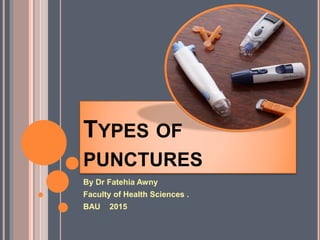
TYPES OF
PUNCTURES
By Dr Fatehia Awny
Faculty of Health Sciences .
BAU 2015
 Skin puncture Earlobe puncture.
Types of punctures:
Finger puncture heel stick
1. for patients in whom venous access is difficult,
2. when small quantities of blood are sufficient for
testing.
In newborns, heel sticks are the preferred
collection method for small volumes of
blood.
Why Perform a Skin Puncture?
CAPILLARY COLLECTIONS MAY BE
PREFERABLE OVER VENIPUNCTURE:
 Severely burned patients
 Obese patients
 Patients with thrombotic tendencies
 Elderly patients or others in whom superficial veins
are very fragile or inaccessible
 Patients performing self-testing
 Point-of-care testing
 Newborn testing
 Patients who have a paralyzing fear of needles
 Differences Between Skin Puncture Blood and
Venipuncture Blood!!!!!!!!!!!!!!!!!!!!!!!
FINGER PUNCTURE:
Preparation for finger stick
1. Place all collection materials
on top of disposable pad.
Open the lancet, alcohol
swabs, gauze, bandage, and
other items. Have all items
ready for blood collection.
FINGER PUNCTURE:
CHOOSE THE FINGER CAREFULLY
 Best locations for a finger
stick is the 3rd and 4th
fingers of the non-
dominant hand.
 Avoid the 2nd and 5th
fingers if possible.
 Perform the stick off to
side of the center of the
finger.
 NEVER use the tip or
center of the finger.
FINGER PUNCTURE:
Massage or Warm the site
• Avoid fingers that are cold,
cyanotic, swollen, scarred
or covered with a rash.
• Massage to warm the
finger and increase blood
flow by gently squeezing
from hand to fingertip 5-6
times.
FINGER PUNCTURE:
Clean and DRY the site
 Cleanse fingertip
with 70% isopropyl
alcohol
 Wipe dry with clean
gauze or allow to air
dry.
 Caution: Alcohol can
falsely elevate or lower
blood glucose results.
FINGER PUNCTURE:
Hold the finger in an upward
position and lance the
finger (across the
fingerprint) between the
side and the pad with the
proper size lancet
(adult/child). Press firmly
on the finger when making
the puncture. Doing so
will help you to obtain the
amount of blood you need.
FINGER PUNCTURE:
Finger Stick location
• Using a sterile
lancet, make a skin
puncture just off the
center of the finger
pad.
• Wipe away the first
drop of blood (which
tends to contain
excess tissue fluid).
5. Apply slight pressure
to start blood flow.
Blot the first drop of
blood on a gauze pad
and discard in
appropriate biohazard
container.
FINGER PUNCTURE:
Keep the finger in a downward
position and gently massage it
(but do not “milk”) to maintain
blood flow.
FINGER PUNCTURE:
SUMMARY
 If child is <12 months of age, the lancet
 must have a depth of 2 mm or less, and a
 fingerstick MAY NOT be performed – must
 do a heel stick instead
 The heel is used for dermal punctures on infants
less than 1 year of age because it
 contains more tissue than the finger, and has not
yet become callused from walking.
 No nerves ,bones or tendons near by
WHY HEEL STICK?
 Hatched areas (arrows)
indicate safe areas for
puncture site.
heel stick:
 Warm site with soft cloth
moistened with warm water
(up to 41
o
C) for 3 – 5
minutes.
HEEL STICK:
 Cleanse site with
alcohol prep.
 Wipe DRY with sterile
gauze pad.
HEEL STICK:
Puncture heel.
Wipe away first
blood drop with
sterile gauze pad.
Allow another
LARGE blood drop
to form.
HEEL STICK:
 Mainly used for bleeding time .
 Principle
 The bleeding time is the time it takes for a small
standardized wound, introduced into the capillary bed
of the finger or earlobe,to stop bleeding. It is
dependent upon:
1- the elasticity of the skin and capillary vessels,
2-the efficiency of the tissue fluids
3-the mechanical and chemical action of the
thrombocytes (blood platelets).
EARLOBE PUNCTURE:
 Make a small wound 3 mm deep in the lateral aspect of
a fingertip or the lower portion of the ear lobe, using a
suitable lancet. The wound should be sufficiently deep
to give a free flow of blood without squeezing.
 Start the stop watch immediately after the puncture is
made. Though the stop watch is started a few seconds
after puncturing the skin, very little error results in the
bleeding time.3.
 At intervals of one-half minute gently blot the blood from
the wound with a piece of filter paper, being careful not
to touch the skin.4.

EARLOBE PUNCTURE:
 The interval of time between the puncture and the
cessation of bleeding is the bleeding time. The
blood should fail to appear on the filter paper in 1-3
minutes. All abnormal findings should be
rechecked.
NORMAL : 1-3 minutes
EARLOBE PUNCTURE:
Specimen Types
 Blood
– Proper collection vial
– Collection comprised of two bottles:
Aerobic
Anaerobic
25
Blood culture bottles.
26
SPECIMEN TYPES
29
PHLEBOTOMY
 Phlebotomy procedure
 Important to all laboratory testing
 Sample’s quality determines accuracy of its final result
 Anticoagulant
 Mixed with blood to prevent coagulation
 Three anticoagulants are used in the hematology
laboratory
30
PHLEBOTOMY
 Anticoagulant
 EDTA – prevents coagulation by chelating Ca2+
 Use for tests: CBC, Hct, Plt, Retic, peripheral blood smear
examination, flow cytometry
 Sodium citrate – prevents coagulation by binding Ca2+
 Used for coagulation studies
 Lithium heparin – prevents coagulation by interacting
with antithrombin
 Used for osmotic fragility: not appropriate for routine
hematology testing
31
EQUIPMENT
 Sample collection tubes
 Evacuated sample collection tubes
 Sterile
 Color coded to indicate type of anticoagulant present or the
lack of anticoagulant
 Only small amounts of sample for analysis necessary
 Interior of a sample collection tube is a vacuum
 Capillary punctures
 Microcollection tubes
 Do not contain a vacuum
32
CAPILLARY PUNCTURE
 Collection tubes
 Should be properly labeled with patient’s name, unique
ID number, date, and time of collection
33
SPECIMEN COLLECTION
 Anticoagulant of choice
 Sodium citrate, 3.2%
 Ratio of anticoagulant : whole blood is 1:9
 > 55% hematocrit
 Smaller volume of plasma relative to citrate
 Excess free citrate binds calcium in the test procedure
 Falsely prolonged clotting times
 Adjust citrate concentration
 Citrate tubes are available that draw 4.5 mL, 2.7 mL, 1.8 mL
34
SPECIMEN COLLECTION
 Anticoagulant to blood ratio
 1:9 ratio critical for valid results
 If under-filled – ↑ citrate, bind calcium in test procedure
 Falsely prolonged test results
 If overfilled – insufficient calcium bound
 Clotting can occur in tube
 Falsely prolonged results
 Accurate labeling of tube
 Include pre-or postinfusion, time of draw
35
SPECIMEN COLLECTION
 Specimen draw time
 Check patient history of receiving blood products
 If testing done within the half-life of administered clotting factor
then test may measure transfused component as well as the
patient's component
 Fibrinolytic factors – diurnal variability
 Platelet studies – medication history
36
SPECIMEN PROCESSING
 Plasma for coagulation testing
 Citrated whole blood is centrifuged
 Platelet-poor plasma (PPP)
 Platelet-rich plasma (PRP)
37
SPECIMEN PROCESSING
 Platelet-poor plasma
 Citrated specimen centrifuged for 15 minutes at 2500xg
 < 10 x 109/L platelets
 Depending on coagulation instrumentation
 Leave plasma on top of packed cells or
 Remove plasma with plastic pipette – plastic tube with cap
 Leave small amount of plasma on top
38
SPECIMEN PROCESSING
 Use platelet-poor plasma because
 Platelets contain PF4
 Neutralizes heparin
 Platelets contain phospholipids
 Affect LA testing and factor assay testing
 Platelets contain proteases
 Alter results for von Willebrand testing
 Any clot in specimen
 Specimen unacceptable
 After removing PPP, twirl applicator stick in packed cells to
detect clots
39
SPECIMEN PROCESSING
 Platelet-poor plasma
 Separated plasma
 Stored at 18-24°C or 2-8°C for up to 4 hours
 If testing is delayed > 4 hours
 Stored at -20°C for up to 1 week
 Stored at -70°C for up to 6 months
40
SPECIMEN PROCESSING
 Platelet-poor plasma
 Frozen specimens
 Thawed rapidly at 37°C
 Excessive heating can destroy Factor V and VIII
 Never store in self-defrost freezers
 Special testing may require special collection and
storage procedures
41

More Related Content

What's hot

Blood collection and anticoagulants
Blood collection and anticoagulantsBlood collection and anticoagulants
Blood collection and anticoagulantsJanani Mathialagan
 
collection of blood sample and preanalytical errors
collection of blood sample and preanalytical errorscollection of blood sample and preanalytical errors
collection of blood sample and preanalytical errorsUtkarsh Sharma
 
Blood Banking.. Experiment # 2
Blood Banking.. Experiment # 2Blood Banking.. Experiment # 2
Blood Banking.. Experiment # 2raymund120590
 
Introduction to haematology laboratory
Introduction to haematology laboratoryIntroduction to haematology laboratory
Introduction to haematology laboratoryHussein Al-tameemi
 
3 blood collection by capillary method
3 blood collection by capillary method3 blood collection by capillary method
3 blood collection by capillary methodnawakhar neupane
 
Safety rules for histopathology lab
Safety rules for histopathology labSafety rules for histopathology lab
Safety rules for histopathology labDr. Waqas Nawaz
 
Merits and demerits of different fixatives
Merits and demerits of different fixativesMerits and demerits of different fixatives
Merits and demerits of different fixativesRoohi1234
 
Forward and reverse grouping by Negash Alamin
Forward and reverse grouping  by Negash AlaminForward and reverse grouping  by Negash Alamin
Forward and reverse grouping by Negash AlaminNegash Alamin
 
Point of care testing ( POCT)
Point of care testing ( POCT)Point of care testing ( POCT)
Point of care testing ( POCT)binaya tamang
 
Copy Of Lesson (3) 3 Laboratory Bleedingtime
Copy Of Lesson (3) 3 Laboratory BleedingtimeCopy Of Lesson (3) 3 Laboratory Bleedingtime
Copy Of Lesson (3) 3 Laboratory BleedingtimeMiami Dade
 

What's hot (20)

Blood collection and anticoagulants
Blood collection and anticoagulantsBlood collection and anticoagulants
Blood collection and anticoagulants
 
Blood collection
Blood collectionBlood collection
Blood collection
 
Stool Examination
Stool ExaminationStool Examination
Stool Examination
 
collection of blood sample and preanalytical errors
collection of blood sample and preanalytical errorscollection of blood sample and preanalytical errors
collection of blood sample and preanalytical errors
 
Blood Banking.. Experiment # 2
Blood Banking.. Experiment # 2Blood Banking.. Experiment # 2
Blood Banking.. Experiment # 2
 
Blood sample collection
Blood sample collectionBlood sample collection
Blood sample collection
 
Introduction to haematology laboratory
Introduction to haematology laboratoryIntroduction to haematology laboratory
Introduction to haematology laboratory
 
3 blood collection by capillary method
3 blood collection by capillary method3 blood collection by capillary method
3 blood collection by capillary method
 
Compatability testing
Compatability testingCompatability testing
Compatability testing
 
Skin puncture method
Skin puncture methodSkin puncture method
Skin puncture method
 
STOOL EXAMINATION
STOOL EXAMINATIONSTOOL EXAMINATION
STOOL EXAMINATION
 
Phlebotomy
PhlebotomyPhlebotomy
Phlebotomy
 
Urine preservative
Urine preservative Urine preservative
Urine preservative
 
Safety rules for histopathology lab
Safety rules for histopathology labSafety rules for histopathology lab
Safety rules for histopathology lab
 
Merits and demerits of different fixatives
Merits and demerits of different fixativesMerits and demerits of different fixatives
Merits and demerits of different fixatives
 
Crossmatching
CrossmatchingCrossmatching
Crossmatching
 
Forward and reverse grouping by Negash Alamin
Forward and reverse grouping  by Negash AlaminForward and reverse grouping  by Negash Alamin
Forward and reverse grouping by Negash Alamin
 
Point of care testing ( POCT)
Point of care testing ( POCT)Point of care testing ( POCT)
Point of care testing ( POCT)
 
Blood collection sunil
Blood collection sunil Blood collection sunil
Blood collection sunil
 
Copy Of Lesson (3) 3 Laboratory Bleedingtime
Copy Of Lesson (3) 3 Laboratory BleedingtimeCopy Of Lesson (3) 3 Laboratory Bleedingtime
Copy Of Lesson (3) 3 Laboratory Bleedingtime
 

Viewers also liked

Viewers also liked (20)

Preparation of peripheral blood flim
Preparation of peripheral blood flimPreparation of peripheral blood flim
Preparation of peripheral blood flim
 
Windu skin puncture (1)
Windu skin puncture (1)Windu skin puncture (1)
Windu skin puncture (1)
 
1intro
1intro1intro
1intro
 
Sample collection and processig
Sample collection and processigSample collection and processig
Sample collection and processig
 
HEMATOLOGY: Laboratory Tests
HEMATOLOGY: Laboratory TestsHEMATOLOGY: Laboratory Tests
HEMATOLOGY: Laboratory Tests
 
Skrining pada bayi baru lahir
Skrining pada bayi baru lahirSkrining pada bayi baru lahir
Skrining pada bayi baru lahir
 
Acute Myeloid Leukemia (PCNA)
Acute Myeloid Leukemia (PCNA)Acute Myeloid Leukemia (PCNA)
Acute Myeloid Leukemia (PCNA)
 
Preparation and staining of peripheral blood smear
Preparation and staining of peripheral blood smearPreparation and staining of peripheral blood smear
Preparation and staining of peripheral blood smear
 
Roche Diagnostics
Roche DiagnosticsRoche Diagnostics
Roche Diagnostics
 
Blood donation
Blood donationBlood donation
Blood donation
 
Phlebotomy
PhlebotomyPhlebotomy
Phlebotomy
 
Stain of hematology
Stain of hematologyStain of hematology
Stain of hematology
 
Phlebotomy
PhlebotomyPhlebotomy
Phlebotomy
 
Phlebotomy
PhlebotomyPhlebotomy
Phlebotomy
 
LUMBER PUNCTURE By Dr Bashir Ahmed Dar Associate Professor Medicine Chinkipor...
LUMBER PUNCTURE By Dr Bashir Ahmed Dar Associate Professor Medicine Chinkipor...LUMBER PUNCTURE By Dr Bashir Ahmed Dar Associate Professor Medicine Chinkipor...
LUMBER PUNCTURE By Dr Bashir Ahmed Dar Associate Professor Medicine Chinkipor...
 
Electrolyte Analyzer.Pptx [Autosaved]
Electrolyte Analyzer.Pptx [Autosaved]Electrolyte Analyzer.Pptx [Autosaved]
Electrolyte Analyzer.Pptx [Autosaved]
 
Venipuncture Equipment Unit 2
Venipuncture Equipment Unit 2Venipuncture Equipment Unit 2
Venipuncture Equipment Unit 2
 
Cytochemical staining checked
Cytochemical staining checkedCytochemical staining checked
Cytochemical staining checked
 
Trans septal puncture
Trans septal punctureTrans septal puncture
Trans septal puncture
 
Peripheral Blood Smear
Peripheral Blood SmearPeripheral Blood Smear
Peripheral Blood Smear
 

Similar to Types of Punctures Guide

Sample Collection & Processing.pptxDr Rajju Tiwari M.D. biochemistry
Sample Collection & Processing.pptxDr Rajju Tiwari M.D. biochemistrySample Collection & Processing.pptxDr Rajju Tiwari M.D. biochemistry
Sample Collection & Processing.pptxDr Rajju Tiwari M.D. biochemistryRanjana187247
 
Collection of Blood and Anticoagula.pptx
Collection of Blood and Anticoagula.pptxCollection of Blood and Anticoagula.pptx
Collection of Blood and Anticoagula.pptxnandithapradeep92
 
Subjective assignment 2
Subjective assignment 2Subjective assignment 2
Subjective assignment 2amjad hussain
 
BLOOD COLLECTION TECHNIQUES.pptx
BLOOD COLLECTION TECHNIQUES.pptxBLOOD COLLECTION TECHNIQUES.pptx
BLOOD COLLECTION TECHNIQUES.pptxseeja2
 
Venous blood coolection
Venous  blood coolectionVenous  blood coolection
Venous blood coolectionHamidAltameme1
 
Laboratory Blood/ Specimen collection
Laboratory Blood/ Specimen collectionLaboratory Blood/ Specimen collection
Laboratory Blood/ Specimen collectionpriyanka raviraj
 
sample collection & rejection .pptx is used in hospital
sample collection & rejection .pptx is used in hospitalsample collection & rejection .pptx is used in hospital
sample collection & rejection .pptx is used in hospitalsathishmanoharan13
 
Hema I Chapter 3_phlebotomy.ppt
Hema I Chapter 3_phlebotomy.pptHema I Chapter 3_phlebotomy.ppt
Hema I Chapter 3_phlebotomy.pptderibew genetu
 
clinical parasitology-hemoparasites.pptx
clinical parasitology-hemoparasites.pptxclinical parasitology-hemoparasites.pptx
clinical parasitology-hemoparasites.pptxHammadBhatti20
 
III B.Sc.Clinical Laboratory Technology PRACTICAL.pptx
III B.Sc.Clinical Laboratory Technology PRACTICAL.pptxIII B.Sc.Clinical Laboratory Technology PRACTICAL.pptx
III B.Sc.Clinical Laboratory Technology PRACTICAL.pptxNoor Muhammed
 
BLOOD SAMPLE COLLECTION PPT.pptx
BLOOD SAMPLE COLLECTION PPT.pptxBLOOD SAMPLE COLLECTION PPT.pptx
BLOOD SAMPLE COLLECTION PPT.pptxanjalatchi
 
Blood sample collection procedure ppt
Blood sample collection procedure pptBlood sample collection procedure ppt
Blood sample collection procedure pptanjalatchi
 
Methods of Blood Collection and Anticoagulants
Methods of Blood Collection and AnticoagulantsMethods of Blood Collection and Anticoagulants
Methods of Blood Collection and AnticoagulantsShreya D Prabhu
 
IUC_Haematology_Lecture 3_Blood Collection.ppt
IUC_Haematology_Lecture 3_Blood Collection.pptIUC_Haematology_Lecture 3_Blood Collection.ppt
IUC_Haematology_Lecture 3_Blood Collection.pptKingslyNdanga1
 

Similar to Types of Punctures Guide (20)

Sample Collection & Processing.pptxDr Rajju Tiwari M.D. biochemistry
Sample Collection & Processing.pptxDr Rajju Tiwari M.D. biochemistrySample Collection & Processing.pptxDr Rajju Tiwari M.D. biochemistry
Sample Collection & Processing.pptxDr Rajju Tiwari M.D. biochemistry
 
Collection of Blood and Anticoagula.pptx
Collection of Blood and Anticoagula.pptxCollection of Blood and Anticoagula.pptx
Collection of Blood and Anticoagula.pptx
 
Subjective assignment 2
Subjective assignment 2Subjective assignment 2
Subjective assignment 2
 
Eritro intro01
Eritro intro01Eritro intro01
Eritro intro01
 
BLOOD COLLECTION TECHNIQUES.pptx
BLOOD COLLECTION TECHNIQUES.pptxBLOOD COLLECTION TECHNIQUES.pptx
BLOOD COLLECTION TECHNIQUES.pptx
 
Venous blood coolection
Venous  blood coolectionVenous  blood coolection
Venous blood coolection
 
Laboratory Blood/ Specimen collection
Laboratory Blood/ Specimen collectionLaboratory Blood/ Specimen collection
Laboratory Blood/ Specimen collection
 
sample collection & rejection .pptx is used in hospital
sample collection & rejection .pptx is used in hospitalsample collection & rejection .pptx is used in hospital
sample collection & rejection .pptx is used in hospital
 
Hema I Chapter 3_phlebotomy.ppt
Hema I Chapter 3_phlebotomy.pptHema I Chapter 3_phlebotomy.ppt
Hema I Chapter 3_phlebotomy.ppt
 
Blood examination
Blood examinationBlood examination
Blood examination
 
Blood collection
Blood collectionBlood collection
Blood collection
 
blood collection.pptx
blood collection.pptxblood collection.pptx
blood collection.pptx
 
clinical parasitology-hemoparasites.pptx
clinical parasitology-hemoparasites.pptxclinical parasitology-hemoparasites.pptx
clinical parasitology-hemoparasites.pptx
 
Ms REPORT
Ms REPORTMs REPORT
Ms REPORT
 
Ms REPORT
Ms REPORTMs REPORT
Ms REPORT
 
III B.Sc.Clinical Laboratory Technology PRACTICAL.pptx
III B.Sc.Clinical Laboratory Technology PRACTICAL.pptxIII B.Sc.Clinical Laboratory Technology PRACTICAL.pptx
III B.Sc.Clinical Laboratory Technology PRACTICAL.pptx
 
BLOOD SAMPLE COLLECTION PPT.pptx
BLOOD SAMPLE COLLECTION PPT.pptxBLOOD SAMPLE COLLECTION PPT.pptx
BLOOD SAMPLE COLLECTION PPT.pptx
 
Blood sample collection procedure ppt
Blood sample collection procedure pptBlood sample collection procedure ppt
Blood sample collection procedure ppt
 
Methods of Blood Collection and Anticoagulants
Methods of Blood Collection and AnticoagulantsMethods of Blood Collection and Anticoagulants
Methods of Blood Collection and Anticoagulants
 
IUC_Haematology_Lecture 3_Blood Collection.ppt
IUC_Haematology_Lecture 3_Blood Collection.pptIUC_Haematology_Lecture 3_Blood Collection.ppt
IUC_Haematology_Lecture 3_Blood Collection.ppt
 

Types of Punctures Guide

  • 1. TYPES OF PUNCTURES By Dr Fatehia Awny Faculty of Health Sciences . BAU 2015
  • 2.  Skin puncture Earlobe puncture. Types of punctures: Finger puncture heel stick
  • 3. 1. for patients in whom venous access is difficult, 2. when small quantities of blood are sufficient for testing. In newborns, heel sticks are the preferred collection method for small volumes of blood. Why Perform a Skin Puncture?
  • 4. CAPILLARY COLLECTIONS MAY BE PREFERABLE OVER VENIPUNCTURE:  Severely burned patients  Obese patients  Patients with thrombotic tendencies  Elderly patients or others in whom superficial veins are very fragile or inaccessible  Patients performing self-testing  Point-of-care testing  Newborn testing  Patients who have a paralyzing fear of needles  Differences Between Skin Puncture Blood and Venipuncture Blood!!!!!!!!!!!!!!!!!!!!!!!
  • 5.
  • 6. FINGER PUNCTURE: Preparation for finger stick 1. Place all collection materials on top of disposable pad. Open the lancet, alcohol swabs, gauze, bandage, and other items. Have all items ready for blood collection.
  • 7. FINGER PUNCTURE: CHOOSE THE FINGER CAREFULLY  Best locations for a finger stick is the 3rd and 4th fingers of the non- dominant hand.  Avoid the 2nd and 5th fingers if possible.  Perform the stick off to side of the center of the finger.  NEVER use the tip or center of the finger.
  • 8. FINGER PUNCTURE: Massage or Warm the site • Avoid fingers that are cold, cyanotic, swollen, scarred or covered with a rash. • Massage to warm the finger and increase blood flow by gently squeezing from hand to fingertip 5-6 times.
  • 9. FINGER PUNCTURE: Clean and DRY the site  Cleanse fingertip with 70% isopropyl alcohol  Wipe dry with clean gauze or allow to air dry.  Caution: Alcohol can falsely elevate or lower blood glucose results.
  • 10. FINGER PUNCTURE: Hold the finger in an upward position and lance the finger (across the fingerprint) between the side and the pad with the proper size lancet (adult/child). Press firmly on the finger when making the puncture. Doing so will help you to obtain the amount of blood you need.
  • 11. FINGER PUNCTURE: Finger Stick location • Using a sterile lancet, make a skin puncture just off the center of the finger pad. • Wipe away the first drop of blood (which tends to contain excess tissue fluid).
  • 12. 5. Apply slight pressure to start blood flow. Blot the first drop of blood on a gauze pad and discard in appropriate biohazard container. FINGER PUNCTURE:
  • 13. Keep the finger in a downward position and gently massage it (but do not “milk”) to maintain blood flow. FINGER PUNCTURE:
  • 14.
  • 16.  If child is <12 months of age, the lancet  must have a depth of 2 mm or less, and a  fingerstick MAY NOT be performed – must  do a heel stick instead
  • 17.  The heel is used for dermal punctures on infants less than 1 year of age because it  contains more tissue than the finger, and has not yet become callused from walking.  No nerves ,bones or tendons near by WHY HEEL STICK?
  • 18.  Hatched areas (arrows) indicate safe areas for puncture site. heel stick:
  • 19.  Warm site with soft cloth moistened with warm water (up to 41 o C) for 3 – 5 minutes. HEEL STICK:
  • 20.  Cleanse site with alcohol prep.  Wipe DRY with sterile gauze pad. HEEL STICK:
  • 21. Puncture heel. Wipe away first blood drop with sterile gauze pad. Allow another LARGE blood drop to form. HEEL STICK:
  • 22.  Mainly used for bleeding time .  Principle  The bleeding time is the time it takes for a small standardized wound, introduced into the capillary bed of the finger or earlobe,to stop bleeding. It is dependent upon: 1- the elasticity of the skin and capillary vessels, 2-the efficiency of the tissue fluids 3-the mechanical and chemical action of the thrombocytes (blood platelets). EARLOBE PUNCTURE:
  • 23.  Make a small wound 3 mm deep in the lateral aspect of a fingertip or the lower portion of the ear lobe, using a suitable lancet. The wound should be sufficiently deep to give a free flow of blood without squeezing.  Start the stop watch immediately after the puncture is made. Though the stop watch is started a few seconds after puncturing the skin, very little error results in the bleeding time.3.  At intervals of one-half minute gently blot the blood from the wound with a piece of filter paper, being careful not to touch the skin.4.  EARLOBE PUNCTURE:
  • 24.  The interval of time between the puncture and the cessation of bleeding is the bleeding time. The blood should fail to appear on the filter paper in 1-3 minutes. All abnormal findings should be rechecked. NORMAL : 1-3 minutes EARLOBE PUNCTURE:
  • 25. Specimen Types  Blood – Proper collection vial – Collection comprised of two bottles: Aerobic Anaerobic 25
  • 27.
  • 28.
  • 30. PHLEBOTOMY  Phlebotomy procedure  Important to all laboratory testing  Sample’s quality determines accuracy of its final result  Anticoagulant  Mixed with blood to prevent coagulation  Three anticoagulants are used in the hematology laboratory 30
  • 31. PHLEBOTOMY  Anticoagulant  EDTA – prevents coagulation by chelating Ca2+  Use for tests: CBC, Hct, Plt, Retic, peripheral blood smear examination, flow cytometry  Sodium citrate – prevents coagulation by binding Ca2+  Used for coagulation studies  Lithium heparin – prevents coagulation by interacting with antithrombin  Used for osmotic fragility: not appropriate for routine hematology testing 31
  • 32. EQUIPMENT  Sample collection tubes  Evacuated sample collection tubes  Sterile  Color coded to indicate type of anticoagulant present or the lack of anticoagulant  Only small amounts of sample for analysis necessary  Interior of a sample collection tube is a vacuum  Capillary punctures  Microcollection tubes  Do not contain a vacuum 32
  • 33. CAPILLARY PUNCTURE  Collection tubes  Should be properly labeled with patient’s name, unique ID number, date, and time of collection 33
  • 34. SPECIMEN COLLECTION  Anticoagulant of choice  Sodium citrate, 3.2%  Ratio of anticoagulant : whole blood is 1:9  > 55% hematocrit  Smaller volume of plasma relative to citrate  Excess free citrate binds calcium in the test procedure  Falsely prolonged clotting times  Adjust citrate concentration  Citrate tubes are available that draw 4.5 mL, 2.7 mL, 1.8 mL 34
  • 35. SPECIMEN COLLECTION  Anticoagulant to blood ratio  1:9 ratio critical for valid results  If under-filled – ↑ citrate, bind calcium in test procedure  Falsely prolonged test results  If overfilled – insufficient calcium bound  Clotting can occur in tube  Falsely prolonged results  Accurate labeling of tube  Include pre-or postinfusion, time of draw 35
  • 36. SPECIMEN COLLECTION  Specimen draw time  Check patient history of receiving blood products  If testing done within the half-life of administered clotting factor then test may measure transfused component as well as the patient's component  Fibrinolytic factors – diurnal variability  Platelet studies – medication history 36
  • 37. SPECIMEN PROCESSING  Plasma for coagulation testing  Citrated whole blood is centrifuged  Platelet-poor plasma (PPP)  Platelet-rich plasma (PRP) 37
  • 38. SPECIMEN PROCESSING  Platelet-poor plasma  Citrated specimen centrifuged for 15 minutes at 2500xg  < 10 x 109/L platelets  Depending on coagulation instrumentation  Leave plasma on top of packed cells or  Remove plasma with plastic pipette – plastic tube with cap  Leave small amount of plasma on top 38
  • 39. SPECIMEN PROCESSING  Use platelet-poor plasma because  Platelets contain PF4  Neutralizes heparin  Platelets contain phospholipids  Affect LA testing and factor assay testing  Platelets contain proteases  Alter results for von Willebrand testing  Any clot in specimen  Specimen unacceptable  After removing PPP, twirl applicator stick in packed cells to detect clots 39
  • 40. SPECIMEN PROCESSING  Platelet-poor plasma  Separated plasma  Stored at 18-24°C or 2-8°C for up to 4 hours  If testing is delayed > 4 hours  Stored at -20°C for up to 1 week  Stored at -70°C for up to 6 months 40
  • 41. SPECIMEN PROCESSING  Platelet-poor plasma  Frozen specimens  Thawed rapidly at 37°C  Excessive heating can destroy Factor V and VIII  Never store in self-defrost freezers  Special testing may require special collection and storage procedures 41