

Recomendados
Más contenido relacionado
La actualidad más candente
La actualidad más candente (20)
Similar a 6
Similar a 6 (20)
6
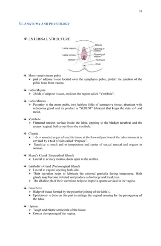
- 1. 31 VI. ANATOMY AND PHYSIOLOGY EXTERNAL STRUCTURE Mons veneris/mons pubis pad of adipose tissue located over the symphysis pubis, protect the junction of the pubic bone from trauma. Labia Majora 2folds of adipose tissues, encloses the region called “Vestibule”. Labia Minora Posterior to the mons pubis, two hairless folds of connective tissue, abundant with sebaceous gland and its product is “SEBUM” lubricant that keeps the skin soft and moist. Vestibule Flattened smooth surface inside the labia, opening to the bladder (urethra) and the uterus (vagina) both arises from the vestibule. Clitoris 1-2cm rounded organ of erectile tissue at the forward junction of the labia minora it is covered by a fold of skin called “Prepuce”. Sensitive to touch and to temperature and center of sexual arousal and orgasm in woman. Skene’s Gland (Paraurethral Gland) Lateral to urinary meatus, ducts open to the urethra. Bartholin’s Gland (Vulvovaginal Gland) Lateral to vaginal opening both side Their secretion helps to lubricate the external genitalia during intercourse. Both glands may become infected and produce a discharge and local pain. The alkaline ph of their secretions helps to improve sperm survival in the vagina. Fourchette Ridge of tissue formed by the posterior joining of the labia’s. Episiotomy is done on this part to enlarge the vaginal opening for the passageway of the fetus. Hymen Tough and elastic semicircle of the tissue. Covers the opening of the vagina.
- 2. 32 Perineum Diamond shape region between the anterior end of the labial fold and the anus posteriorly. Anus Distal end of the digestive tract, the outlet of the rectum. INTERNAL STRUCTURE Fallopian tube Arise from each upper corner of the uterine body , extend outward and backward until each opens at its distal end next to an ovary. 10cm long, conveys ovum from the ovaries to the uterus and to provide a place for fertilization of the ovum by sperm. 4PARTS: 1. Interstitial Most proximal division, 1 cm in length, 1mm in Diameter. Where ectopic pregnancy occur. 2. Isthmus Cut or scaled in tubular ligation or tubal sterilization. 3. Ampulla Longest portion 5cm long. Fertilization of ovum usually occurs in here. 4. Infundibulum Have fimbrae (small hairs) that helps to guide the ovum into fallopian tube. Vagina Allows passage of menstrual flow. Receives penis during intercourse. Lower portion of birth canal during delivery. Cervix Neck of the uterus, separates upper end of the vagina from the isthmus of the uterus.
- 3. 33 Uterus Pear shaped muscular organ that has three components the corpus (body), cervix (neck), fundus. Broad ligament/round ligament is the fibrous muscular cords that steady the uterus. 3 LAYERS Endometrium - Inner layer, where implantation of embryo occur. Myometrium - bulky middle of the uterus - plays important role in delivery to contract rhythmically - to force the body out. Perimetrium - outermost serious layer of the uterus. Ovary Grayish white, almond shaped female gonads. Produce mature and discharged ova. Maturation and maintenance of sex characteristics. PRODUCTION OF OVUM Ovary reveals tiny sack like structures called “Ovarian/Primary Follicle”. Each follicle consists of an immature egg “Oocyte”. Surrounded by one or more layers of very different cells “Follicle Cell”. As the egg developed, follicle begins to ripen/mature. Follicle enlarges and develops a fluid filled central region called “Antrum” also called “Vesicular/Graafian Follicle” and developing egg is ready to be ejected from the ovary an event called “Ovulation”. Ruptured follicle is transformed into a very small structure called “Corpus Luteum”, “Corpus Albican”. MENSTRUATION – Episodic uterine bleeding in response to cyclic hormonal changes. FOLLICLE STIMULATING HORMONE Stimulates ovary to produce Estrogen Follicle development of ovary Oocyte maturation
- 4. 34 Graafian follicle maturation Stimulates in increasing the amount of endometrial activity during luteal phase. LEUTINIZING HORMONE Triggers ovulation of egg Rupture follicle to produce Progesterone, Estrogen PROGESTERONE Hormone of mothers Necessary to maintain the endometrial lining of the uterus during pregnancy. Present in serum as early as 4th week of pregnancy as a result of continuation of Corpus Luteum. After placental synthesis begins at about 12th week, level of progesterone rises progressively during the remainder of pregnancy. Reduce the contractility of uterus during pregnancy, preventing premature labor. Reduce contractility is produced by a change in electrolytes (notably potassium and calcium), which decreases the contraction potential of the uterus. Progesterone withdrawal due to menstruation. Hormone for the stimulation of gall bladder. ESTROGEN Primarily ESTRIOL is produced as a second product of the syncytial cells of the placenta. Contributes to the woman mammary gland development in preparation for lactation and stimulates uterine growth to accommodate the developing fetus. Development of uterus, fallopian tube, vagina, and the development of the secondary sex characteristics. PROSTAGLANDIN Mediator of pain Secondary to the contraction of the lining of the uterus stimulating muscle to contract. INITIATION OF MENSTRUATION Hypothalamus is the main initiator of menstruation. Release of Gonadotrophic releasing hormone (leutinizing hormone releasing hormone) by the hypothalamus. Hypothalamus stimulates the menstrual cycle During childhood hypothalamus is very sensitive to high amount of estrogen produced by the adrenal gland that release of hormone is suppressed. During puberty hypothalamus is less sensitive to estrogen and feedback mechanism occur, GnRH is transmitted from hypothalamus to AnPG to stimulate the release of estrogen and progesterone, menstruation then occurs. PHASES OF MENSTRUATION MENSTRUAL PHASE Last for five days, decrease estrogen, 28-32 days(R), 22-44(IR) Stimulates the Hypothalamus Signals the Anterior Pituitary Gland to release Gonadotrophic hormone Stimulating the release of Follicle Stimulating Hormone and Leutinizing Hormone Follicle Stimulating Hormone stimulates ovary to develop ovarian follicle secreting high amount of estrogen
- 5. 35 Negative feedback mechanism goes to hypothalamus Suppressing the follicle stimulating hormone Leutinizing hormone acts to enhance Corpus Luteum Formation PROLIFERATIVE PHASE Increase in FSH, Estrogen, 6-14 days 1-8 fold of endomertium. Increase in follicular phase Primordial follicle stimulates ovarian follicle forming CLF Ovarian response phases Follicle maturation response to FSH stimulation CLF stimulates to develop from secondary/rupture follicle Increase in FSH,estrogen SECRETORY PHASE Increase in vascularity of endometrium 15-26 days After the ovum was released it is rupture in the form of CLF Endometrium lining thickens continuously Increase in capillary supply Skimming phase (no fertilization) Increase in progesterone, LH/CLF regress Follicle matured CNS decrease Estrogen and Progesterone ISCHEMIC PHASE 27-28 days Regression of corpus luteum formation ↓progesterone, ↓estrogen Constriction of artery of endometrium Increase in secretion of prostaglandins No fertailization(skimming phase) Endometrium shed of ruptured Menstruation begins EPISIOTOMY Surgical incision of the perineum that is made both to prevent tearing of the perineum and to release pressure of the fetal head. EPISIORAPHY Is the repair of the perineum through stitches an d with the use of chromic suture. TYPES : MER (Median episiotomy repair) heal more easily less blood loss less postpartal discomfort RMLER (Right medio lateral episiotomy repair) more on bleeding intense pain away from rectum, less complications DEGREES OF LACERATION 1st degree o small laceration, no stitches are required
- 6. 36 2nd degrees o laceration deep into the muscle, underneath 3rd degree o tear in the vaginal tissue o perineal skin/muscle that extend to the anal sphincter 4th degree o to the sphincter, tissue underneath o tear of the top of the vagina near the urethra/ paraurethral laceration. PLACENTA Latin “pancake” arises out of the continuing growth of trophoblast tissue. 15-20cm in diameter 2-3 cm in depth covering about half the surface area of the internal uterus at term. Role is it serves as a respiratory, excretory, and nutrition delivery system for the fetus. It produces several protein and steroid hormones that help maintain pregnancy and pave the way for the delivery of the baby. It also produces a hormone called hCG( human chorionic gonadotropin). 100 maternal uterine artery supply the mature placenta to provide enough blood for exchange. Woman lies on her left side to lift the uterus away the inferior vena cava. Placental circulation can be so sharply reduced to supine hypotension, if the women lies on her back and the weight of the uterus compresses the vena cava. Placenta weighs about 400-600g (1lb); containing 20-30 cotyledons. RELAXIN one of placental hormone causes mothers pelvic ligaments and the symphysis to relax and become more flexible which eases birth passage. CHORIONIC VILLI Once implantation is complete, Trophoblastic layer of cells of the Blastocyst begins to mature rapidly. 11th-12th day miniature villi that resemble proving fingers termed to be called as CHORIONIC VILLI. At term 200 villi will have formed. 2LAYER: SYNCYTIOTROPHOBLAST ▪ or the syncytial layer, the outer layer, ▪ produce placental hormone such as Hcg, Hpl, estrogen, progesterone. CYTOTROPHOBLAST ▪ Langhan’s layer, present as early as 12 days gestation. ▪ to protect the embryo and fetus from certain infectious organisms Such as the spirochete of syphilis. SIGNS OF PLACENTAL SEPARATION 1. Lengthening of the cord 2. Sudden gush of vaginal blood 3. Change in the size of the uterus (Calkin’s sign) 4. Rise of the fundus in the abdomen
- 7. 37 PLACENTAL SEPERATION Schultze presentation - shiny, seperates first at its center and last at its ends Dunkan presentation - placenta seperates first at its edge, bloody maternal signs BREAST mammary glands form from ectodermic tissue early in utero ACINAR cell where milk is deposited. LACTIFEROUS DUCTS it is the passageway of the milk. Nipple has 20-30 small opening through which milk is secreted. AMPULLA portion of duct located just posterior to the nipple serves as a reservoir for milk before breast feeding. AREOLA is the skin surrounding the nipples, a dark pigmentation. MONTGOMERY’S TUBERCLE– area appears rough on surface, sebaceous glands. LACTATING is the production of milk. TYPES OF MILK 1. NEONATAL MILK (WITCH MILK) 2. COLUSTRUM (contains antibody) 3. HIND MILK (after let down reflex) HUMAN PLACENTAL LACTOGEN Human Chorionic Somatomammotropin Hormone with both growth-promoting and lactogenic (milk-producing) properties. Produced by the placenta, beginning as early as the 6th week of pregnancy, ↑ to a peak level at term. It can be assayed in both maternal serum and urine. Promotes mammary gland (breast) growth in preparation for lactation in the mother. Important role is by regulating maternal glucose, protein, and fat levels so that adequate amounts of these nutrients are always available to the fetus.
- 8. 38 PROLACTIN With the delivery of the placenta,↓ progesterone, dramatically stimulating “PROLACTIN” an AnPG hormone. Acts on the Acinar cells of the mammary glands to stimulate the production of milk. When an infant sucks at the breast, nerve impulses travel from the nipple to the hypothalamus to stimulate the production of “PROLACTIN – RELEASING FACTOR.” This stimulates further active production of Prolactin. Othrer anterior thyroid stimulating hormone and growth hormone probably also play a role in growth of the mammary glands and their ability to secrete milk. CHLOASMA Extra pigment on the face that occurs from “MELANOCYTE- STIMULATING HORMONE”. Which might accompany pregnancy. AMNIOTIC MEMBRANE Smooth chorion. Outermost fetal membrane. Form the sac that contains the amniotic fluid. Forms beneath the chorion, to produce the fluid, support fluid. AMNIOTIC FLUID Shield fetus against pressure or a blow to the mother’s abdomen. Protects fetus from changes in temperature. Aids in muscular development, allows freedom of fetus to move. Helps baby’s lung grow and develops because baby breathes in fluid. Helps baby’s digestive system develop because baby swallow fluid. Protects umbilical cord from pressure, protecting the fetal oxygen supply. Fluid is slightly alkaline with a Ph of about 7.2. 800-1200 ml. Clear and transparent in color It contains electrolyte, minerals, vitamins, proteins, hormone and antibodies. Human growth hormone, human follicle stimulating hormone, human leutinizing hormone, human chorionic gonadotropin, thyroid hormone, insulin, glucagon, testosterone, progesterone, estradiol, cortisone. DECIDUA Corpus luteum continues to atrophy because of Hcg hormone created by the trophoblast cell. Causes uterine endometrium to continue to grow in thickness and vascularity instead of sloughing off this is termed as the DECIDUA or the discharged after giving birth. ▪ Decidua Basalis o Lies directly under the embryo trophoblast portion is lied where communication with the blood vessel and maternal occur. ▪ Decidua Capsularis o Stretches or encapsulates the surface of the trophoblast.. ▪ Decidua Vera
- 9. 39 o Remaining portion HUMAN CHORIONIC GONADOTROPIN Produced very early in pregnancy Produced by the developing embryo and then by the fetal part of the placenta. Stimulates ovary to continue producing estrogen and progesterone so that lining of the uterus is not sloughed off. Hormone that is secreted early in the first week of missed menstruation. Can be detected with the use of pregnancy kit or test.