Proton beam radiotherapy advantages over IMXT
•Descargar como PPT, PDF•
7 recomendaciones•1,822 vistas

The document discusses proton beam radiotherapy and its advantages over traditional x-ray radiotherapy. It describes the equipment used for proton beam therapy including cyclotrons, treatment rooms with rotating gantries, patient immobilization devices, and beam shaping tools. It also provides examples of treatment plans comparing proton and x-ray intensity modulated radiotherapy for tumors in brain and spine.

Recomendados
Más contenido relacionado
La actualidad más candente
La actualidad más candente (20)
Destacado
Destacado (20)
Similar a Proton beam radiotherapy advantages over IMXT
Similar a Proton beam radiotherapy advantages over IMXT (20)
Más de fondas vakalis
Más de fondas vakalis (20)
Último
Último (20)
Proton beam radiotherapy advantages over IMXT
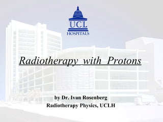
- 1. Radiotherapy with Protons by Dr. Ivan Rosenberg Radiotherapy Physics, UCLH
- 2. Why Proton Beam Radiotherapy ?
- 3. A Schematic Cyclotron Treatment Room 1 : isocentric gantry Treatment Room 2 horizontal beam Treatment Room 3: isocentric gantry Research beam; radiobiology, gantry design, engineering and fundamental physics Proton beam line
- 5. Patient Positioning for Charged Particles High precision in patient positioning is even more important for charged particle therapy, because of the sharp Bragg Peaks. Depending on the disease site, extensive immobilisation procedures need to be adopted
- 6. Head and Neck horizontal beam , LLMC
- 7. Extreme accuracy needed for eye melanoma treatments
- 8. Body mould immobilisation for pelvic sites
- 9. Patient Positioning for Charged Particles Patient in body mould in treatment position inside large proton beam gantry, LLMC. Auxiliary retractable X-ray tube inside machine head used to produce port films Deep false floor to allow gantry rotation
- 10. Compact gantry at the Paul Scherrer Institute, Switzerland
- 11. Practical Proton Radiotherapy Beams Range modulation of a proton beam
- 12. Practical Proton Radiotherapy Beams Range modulator (passive)
- 13. Practical Proton Radiotherapy Beams Collimators and beam shaping Irregular apertures are used to conform the beam to the shape of the tumour as projected along the direction of the incident beam they need to be thick enough to expend all the incident energy (thicker than the range)
- 14. Planning with Protons Treatments for Ocular Melanoma can be achieved most successfully with about 70 MeV protons. Because of its small volume, a single slightly modulated beam gives an ideal dose distribution
- 15. Practical Heavy Particle Radiotherapy Beams Beam Spreading by active scanning Tumor divided in iso-energy slices 2-D dose distribution on the actually scanned slice Bragg-peak Scanning magnets in x and y Z X Y proton beam Bragg peak On-line monitor system
- 16. Intensity Modulated Proton Treatment With active scanning, modulating the intensity and the energy depending on position, sophisticated plans can be achieved
- 17. 12 year old boy Delivered single field plan Some more Proton / IMXT comparisons Unpublished data 9 field IMRT plan 9 field IMRT – second try Factor 6 lower integral dose for protons
- 18. More IMPT vs. IMXT comparisons 64Gy to target volume, doses to brainstem and optic structures as low as possible IMXT ( 9 fields) IMPT ( 3 fields) Unpublished data z = 49 z = 49 z = 57 z = 57 z = 64 z = 64
- 19. Paraspinal tumour treated with IXRT vs. IPRT From Weber et al, Int. J. Radiation Oncology Biol. Phys., Vol. 58, No. 5, pp. 1596–1606, 2004
- 20. Paraspinal tumour treated with IXRT vs. IPRT From Weber et al, Int. J. Radiation Oncology Biol. Phys., Vol. 58, No. 5, pp. 1596–1606, 2004
- 22. The case for protons at UCLH UCLH practice is highly specialised and serves nearby National Hospital for Neurology and Neurosurgery, Great Ormond Street Children`s Hospital, ENT and Dental Hospital (Gray`s Inn Rd) and North London Cancer Network. Strong academic presence. Optimum Situation : University College Hospital near Euston Road/Station (excellent public transport access - train stations for Mid and North England/Scotland very close) . Surrounding medical support & expertise is most impressive: specialist medicine, imaging, surgery, medical oncology, research oncology. Several Large Radiation Oncology Centres nearby with large number of radiation oncology and medical physics experts. Use this expertise in patient selection, treatment planning and follow-up.