2. e1954 The Journal of Clinical Endocrinology Metabolism, 2021, Vol. 106, No. 5
IPS-to-peripheral (IPS:P) ACTH gradient, suggesting an ectopic source. The prolactin-
adjusted IPS:P ACTH ratio can improve differentiation between CD and ectopic ACTH
syndrome when there is a lack of proper IPS venous efflux. In patients who have unilateral
successful IPS cannulation, a contralateral source cannot be excluded.The value of the
intersinus ACTH ratio to predict tumor lateralization may be improved using a prolactin-
adjusted ACTH ratio, but this requires further evaluation.
Conclusion: A stepwise approach in performing and interpreting IPSS will provide
clinicians with the best information from this important but delicate procedure.
Key Words: IPSS, performance, interpretation, case-based review
Cushingsyndrome(CS)canbedividedintoACTH-dependent
and ACTH-independent hypercortisolism. Inferior petrosal
sinus sampling (IPSS) is the “gold standard” to determine
the source of hypercortisolism in ACTH-dependent CS.
IPSS is indicated following inconclusive imaging but posi-
tive, biochemical testing confirming ACTH-dependent
hypercortisolemia (1, 2). The sensitivity and specificity of
IPSS in diagnosing Cushing disease (CD) can range from
88% to 100% and 67% to 100%, respectively (3). IPSS may
not be required in patients who have a sellar mass 6 mm
when dynamic testing is suggestive of CD. A recent study
by Yogi-Morren et al. that included 104 patients with CD
and 26 patients with ectopic ACTH syndrome (EAS) showed
that pituitary tumors 6 mm have a 96% specificity for CD
(vs. EAS) (4, 5). Many clinicians have limited experience per-
forming and interpreting IPSS results. We provide readers a
summary of the current literature and offer strategies to help
optimize use of this important diagnostic test.
History of IPSS
IPSS was first performed at the Walter Reed Army Medical
Center in 1977, with selective and bilateral (but not sim-
ultaneous) catheterization via a transjugular approach (6).
This evolved to a multistep process that included unilat-
eral sampling (for localization) followed by bilateral and
simultaneous catheterization (for localization and lateral-
ization) (7). In 1985, Oldfield and colleagues demonstrated
a high rate of remission following hemihypophysectomy
to the side showing highest ACTH concentrations on IPSS
despite lack of radiological evidence of microadenoma, al-
though the number of patients was small (8). The utility of
IPSS has since been further improved by direct stimulation
of the corticotroph adenoma using corticotropin-releasing
hormone (CRH) or vasopressin (9, 10). In 1991, Oldfield
and his National Institutes of Health (NIH) colleagues pub-
lished on 281 patients with CS, a report that supported the
use of CRH to distinguish CD from EAS (11). Prolactin
measurement was suggested to measure pituitary venous
effluent and confirm successful cannulation in 2004 (12).
IPSS Procedure
Inferior petrosal sinus sampling is most often performed
under local anesthesia. The femoral veins are cannulated,
and microcatheters are placed in the bilateral petrosal
sinuses under fluoroscopic guidance. Venous angiography
is used to confirm correct catheter placement, as demon-
strated by retrograde flow of contrast into the contralateral
cavernous sinus (3). Simultaneous venous blood samples
are obtained from the petrosal sinuses and a peripheral vein.
The blood samples are drawn at baseline and then at 2, 5,
10, and 15 minutes following CRH (1 μg/kg, maximum
100 μg) or vasopressin (10 μg) administration. Some insti-
tutions may use slightly different timepoints for collection.
IPS catheter placement is reassessed at various intervals
during the procedure to ensure sustained cannulation. Our
neuroradiologist checks the position of the IPS catheters
at 5 minutes and once sampling is completed. If the cath-
eter locations appear unstable, imaging is obtained prior to
each blood draw. The patient is observed for several hours
postprocedure. There are rare reports of serious complica-
tions, the most notable being stroke and subarachnoid hem-
orrhage (13-16). These were believed related to aberrant
anatomy, leading to transient hypotension and/or vascular
injury during the procedure (15). IPSS should be aborted for
any significant intraoperative hypotension, hypertension, or
change in neurological status. The most common adverse
events are groin hematoma (4%) and transient headache
(occurs more often if the sinus is small) (17). IPSS is a tech-
nically challenging procedure and should be performed by
an experienced interventional or neuroradiologist.Although
prospective, randomized data are missing, we and others ex-
pect better results from centers that have a dedicated radi-
ologist who focuses on successful IPSS. A stepwise approach
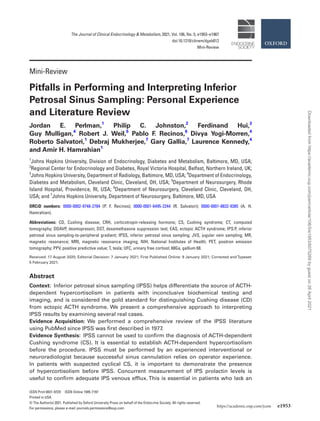
to IPSS interpretation is given in Fig. 1.
Some centers use DDAVP (desmopressin) for IPSS
because of lower cost and past unavailability of CRH.
DDAVP stimulates ACTH production via the V2R re-
ceptor subtype on corticotroph adenomas and elicits a
response similar to CRH (18-20). There is a theoretical
risk of false-positive results (positive IPS-to-peripheral
Downloaded
from
https://academic.oup.com/jcem/article/106/5/e1953/6075269
by
guest
on
26
April
2021
3. The Journal of Clinical Endocrinology Metabolism, 2021, Vol. 106, No. 5 e1955
[IPS:P] ACTH gradient) using DDAVP in a patient with
an ectopic, ACTH-producing tumor if the V2R receptor
is present (21). However, the clinical experience seems
similar using both CRH and DDAVP. Deipolyi et al.
showed comparable diagnostic results using identical
IPS:P ACTH gradients after DDAVP and CRH stimu-
lation (10). An earlier study by Tsagarakis et al. dem-
onstrated good IPSS sensitivity and specificity in 54
patients following administration of combination CRH
and DDAVP (22).
It is our preference to perform IPSS before starting any
antihypercortisolemic therapy, but patients may be re-
ferred to our center after already having been prescribed
these medications. If we determine that a patient on
antihypercortisolemic therapy needs IPSS, our practice is
to stop the medication and schedule the procedure once we
confirm hypercortisolemia on biochemical testing.
Anatomical Variations
There are anatomical variations in pituitary drainage that
can affect the success and interpretation of IPSS. The an-
terior pituitary drains from the cavernous into the inferior
petrosal sinuses. The inferior petrosal sinuses course pos-
terior and caudal and enter the jugular veins at the skull
base (23) (Fig. 2). It is not uncommon to have unequal
drainage of the cavernous sinuses. Some literature suggests
this anatomical variant may be present in 40% of individ-
uals (24, 25). There are other aberrations that make ana-
lysis much more difficult. The most notable variants are
as follows: (1) inferior petrosal anastomosis to vertebral
venous plexus; (2) no connection between the inferior pe-
trosal sinus and the jugular vein; and (3) a hypoplastic in-
ferior petrosal sinus (26). Previous studies have suggested
that microcatheters can be used to obtain accurate venous
sampling if abnormal anatomy is known or suspected (27).
It is also possible to obtain a direct sample from the cav-
ernous sinuses, but this approach has not proved superior
to IPSS (28-32). The literature indicates that the prevalence
of IPS abnormalities exceeds that of false-negative IPSS re-
sults, meaning that adequate catheterization is most often
feasible.
Case Examples
Case Presentation 1
A 50-year-old woman with progressive weight gain, mal-
aise, and arthralgias presented for evaluation. Her med-
ical history was notable for uncontrolled type 2 diabetes
mellitus, steatohepatitis, hypertension, hyperlipidemia, and
obstructive sleep apnea. Physical examination showed ab-
dominal obesity, facial plethora, and diffuse acanthosis
nigricans. Biochemical testing revealed 24-hour urinary free
cortisol (UFC) levels of 94.2 and 152.2 µg (0-50), ACTH
levels of 50 and 69 pg/mL (normal range, 10-60 pg/mL),
and a cortisol level of 13.8 µg/dL after 1-mg dexametha-
sone suppression test (DST). Sellar magnetic resonance
(MR) imaging demonstrated a 3 × 4-mm focus of early en-
hancement on the left side of the gland. IPSS was performed
Confirm endogenous ACTH-
dependent Cushing syndrome
IPS:P ACTH ratio 2 (pre-
CRH) or 3 (post-CRH)
YES NO
CENTRAL/PITUITARY
SOURCE
Consider using prolactin-
adjusted intersinus ACTH ratio
for adenoma lateralization
IPS:P prolactin ratio 1.3 (1.8)
NO
YES
ECTOPIC
CUSHING
SYNDROME
Adenoma 6mm on pituitary MRI
Calculate prolactin-
adjusted IPS:P
ACTH ratio
0.8-1.3
INDETERMINATE
0.8
LIKELY ECTOPIC
CUSHING SYNDROME
1.3-1.8
LIKELY
CENTRAL/PITUITARY
SOURCE
Figure 1. Our approach to differentiating the etiologies of endogenous, ACTH-dependent Cushing syndrome.
Downloaded
from
https://academic.oup.com/jcem/article/106/5/e1953/6075269
by
guest
on
26
April
2021
4. e1956 The Journal of Clinical Endocrinology Metabolism, 2021, Vol. 106, No. 5
using DDAVP; the neuroradiologist reported successful bi-
lateral IPS catheter placement (Table 1).
Can IPSS differentiate between ACTH-dependent and
pseudo-Cushing syndrome?
No, IPSS cannot differentiate between CD and pseudo-
Cushing states. A diagnosis of ACTH-dependent CS must
be established before a patient is referred for IPSS.
It is critical to establish a diagnosis of endogenous
hypercortisolism before pursuing IPSS. This is an invasive
procedure and the presence of a positive IPS:P ACTH ratio
may precipitate unnecessary surgical exploration. Pseudo-
Cushing states have a number of clinical associations
including: pregnancy, morbid obesity, severe psychological
stress (including major depressive disorder), uncontrolled
diabetes mellitus, chronic alcoholism, physical illness or in-
jury, and severe sleep apnea (33). It is thought that higher
brain centers stimulate CRH secretion in these conditions,
leading to activation of the entire hypothalamic–pitu-
itary–adrenal axis (34). The negative feedback inhibition
of cortisol on CRH and ACTH may restrain the resultant
hypercortisolemia. It is uncommon to see UFC elevations
4 times the upper limit of normal in patients with pseudo-
Cushing states (1). These same conditions can blunt the
circadian rhythm and cause abnormal late-night salivary
cortisol concentrations (35-38). Repeat biochemical testing
following resolution or treatment of the underlying dis-
ease process should be considered. Additional confirmatory
testing such as 2-day low-dose DST or a combined dexa-
methasone suppression/CRH stimulation test may also be
used to exclude pseudo-Cushing states. The endocrinology
team felt the hypercortisolism in this patient was not sec-
ondary to a pseudo-Cushing state and proceeded with an
IPSS study.
Researchers at the NIH have demonstrated that
there is significant overlap of ACTH concentrations and
IPS:P ACTH ratios between CD, normal volunteers, and
Table 1. IPSS results for case presentation 1
Time (min) ACTH
Peripheral (P) Petrosal sinus
Right Left ACTH ratio
R/P L/P
Baseline 52 811 469 15.6 9.0
+3 2 3015 271 1507.5 135.5
+5 59 4558 741 77.3 12.6
+10 90 2359 414 26.2 4.6
+15 90 1696 596 18.8 6.6
Abbreviations: L/P, left IPS/peripheral; R/P, right IPS/peripheral.
Figure 2. In this schematic, the infundibulum and pituitary gland are marked in red.The pituitary gland sits in the sella.The relevant venous struc-
tures are designated in blue.
Downloaded
from
https://academic.oup.com/jcem/article/106/5/e1953/6075269
by
guest
on
26
April
2021
5. The Journal of Clinical Endocrinology Metabolism, 2021, Vol. 106, No. 5 e1957
pseudo-Cushing states (39, 40). Yanovski et al. [37] com-
pared IPSS results from patients with CD (n
=
40) to
normal subjects (n
=
7) and those with pseudo-Cushing
states (n = 8) at baseline and 2 minutes post-CRH stimu-
lation (the institutional review board did not allow add-
itional sampling). There was crossover in IPS:P ACTH
ratios between the various conditions. This study confirms
that IPSS should not be used to distinguish pseudo-Cushing
states from CD. Pseudo-Cushing states must be eliminated
as potential causes of ACTH-dependent hypercortisolism
before referring a patient for IPSS (40).
Is the IPSS result in this patient consistent with Cushing
disease?
Yes, the patient has a central to peripheral ACTH ratio 3
following CRH stimulation. This result confirms CD. She
should be referred to an experienced neurosurgeon for sur-
gical exploration of the pituitary gland.
Cushing disease is confirmed when the basal IPS:P
ACTH ratio is 2 and/or the CRH-stimulated ratio is 3
(8, 41-43). The peripheral ACTH level at 3 minutes post-
CRH administration was low (2 pg/mL) and consistent
with laboratory error considering her other ACTH values.
ACTH must be collected in an EDTA tube and transported
swiftly to the laboratory on ice. The sample should then
be centrifuged to separate plasma and frozen (if not pro-
cessed). Delayed or improper handling of the samples
during transportation may result in falsely low levels (44).
In theory, if a pituitary tumor lacked any CRH receptors,
one may not see a rise in ACTH following CRH stimu-
lation. In such an event, the IPS:P ACTH ratio may still
be elevated before CRH administration. We have not ob-
served such a clinical scenario in the past (positive IPS:P
ACTH ratio before but not after CRH stimulation). This
might be because even a low density of CRH receptors
on corticotroph cells is enough to generate an increased
gradient after CRH stimulation in the highly concentrated
IPS environment.”
The patient had successful transsphenoidal resection of
a left-sided pituitary mass. Her cortisol nadired following
the procedure at 1.7 µg/dL and she required postoperative
glucocorticoids. An ACTH-positive corticotroph adenoma
was confirmed by surgical pathology.
Is it necessary to confirm hypercortisolism at the time
of IPSS?
Yes, IPSS may not be reliable if there is lack of
hypercortisolemia at the time of the procedure (39, 45-47).
The absence of sustained hypercortisolism can cause
misleading IPSS results. This can happen in a patient with
cyclical CD if cortisol is tested during a trough period. The
IPSS may reveal an absent IPS:P ACTH gradient because of
lack of ACTH production by the corticotroph adenoma and
suppression of normal corticotrophs secondary to recent
hypercortisolism.ThisIPSSresultmaymimicanectopicsource.
By contrast, incomplete suppression of normal corticotroph
function caused by intermittent ACTH production from a
cyclical ectopic tumor might produce a false-positive IPS:P
ACTH ratio (48, 49). For these reasons, we measure serum
cortisol the morning of scheduled IPSS and proceed only if the
value is 10 μg/dL (50). There are some centers that use mid-
night salivary cortisol to confirm hypercortisolism the night
before IPSS (45, 46). We know of several investigators who
require 6 weeks of consistent hypercortisolemia via 24-hour
UFC before performing the procedure.This approach requires
close monitoring of patients and clinical significance, cost, and
practicability require more detailed study.
Table2givesanexampleofIPSSresultsobtainedinasecond
patient with cyclical Cushing tested during a trough period
(49). The patient was confirmed to have cyclic CD by serial
measurements of 24-hour UFC, demonstrating 3 elevated and
2 normal/low values (Table 3).IPSS was performed using CRH.
Table 2. IPSS results obtained during a trough period in our patient with cyclical Cushing
Time (min) ACTH (pg/mL) Cortisol (μg/dL)
Peripheral (P) Petrosal sinus Peripheral (P)
Right Left ACTH ratio
R/P L/P
-10 8 73 9 9.1 1.1 6.2
-5 8 64 8 8 1
+2 7 145 7 21 1
+5 7 386 7 55 1
+10 7 471 8 67 1.1
+15 9 2 10 0.2 1.1
Abbreviations: L/P, left IPS/peripheral; R/P, right IPS/peripheral.
Downloaded
from
https://academic.oup.com/jcem/article/106/5/e1953/6075269
by
guest
on
26
April
2021
6. e1958 The Journal of Clinical Endocrinology Metabolism, 2021, Vol. 106, No. 5
There was unsuccessful left-sided IPS cannulation (Table 2).
Despite the significant IPS:P gradient on the right side, the au-
thors felt the results were uninterpretable because of a lack of
hypercortisolism during the procedure. This is evidenced by
the peripheral cortisol and ACTH levels of 6.2 μg/dL and 8 pg/
mL at the time of sampling, respectively (49). Swearingen et al.
reported 3 cases of false-positive IPSS findings (IPS:P ACTH
gradient suggestive of CD) in ectopic tumors. These patients
had low peripheral ACTH levels obtained during IPSS, making
the IPS:P ratios difficult to interpret (51).After 2 IPSS attempts,
during which he was not hypercortisolemic, our patient was
referred for pituitary exploration.The transsphenoidal surgery
(performed through the nose) yielded an ectopic corticotroph
adenoma suspended from the mucosa of the sphenoid sinus
(49). The IPSS results are difficult to interpret even retrospect-
ively because we cannot know the venous drainage pattern of
this ectopic adenoma.In these rare situations,IPSS may be con-
sistent with either CD or an ectopic ACTH syndrome.
Case Presentation 2
A 51-year-old woman with progressive weight gain, hair
loss, emotional lability, and new-onset type 2 diabetes mel-
litus was referred for possible hypercortisolism. Physical
examination revealed abdominal obesity, widespread
ecchymoses, excess supraclavicular fat, mild muscle wasting,
and proximal muscle weakness. Her biochemical results
were as follows: 24-hour UFC, 2000 µg (0-50); morning
cortisol 43.8 µg/dL; and ACTH 170 pg/mL following 8 mg
DST. Sellar MR imaging (MRI) showed a 5-mm right-sided
adenoma. She had an unremarkable chest computed tom-
ography (CT) scan. IPSS was performed using CRH; the
neuroradiologist reported successful bilateral IPS catheter
placement (Table 4).
Is the lack of an IPS:P ACTH gradient consistent with a
diagnosis of ectopic CS in this patient?
Absent a significant IPS:P ACTH ratio, the IPS prolactin
level should be used as a surrogate marker of appro-
priate catheterization/normal IPS venous efflux to avoid a
false-negative result.
IPS prolactin can help confirm correct catheter place-
ment during venous sampling. IPS catheterization can be
verified using an IPS:P prolactin ratio ≥ 1.3 to 1.8 before
and after CRH/DDAVP administration (12, 52). The pres-
ence of an IPS:P prolactin ratio 1.3 and the absence of sig-
nificant elevation should raise suspicion for either improper
cannulation or abnormal IPS venous efflux (53). Prolactin is
the most abundant anterior pituitary hormone and spatial
Table 4. IPSS results for case presentation 2
ACTH (pg/dL) Prolactin (ng/mL) Cortisol (μg/dL)
Peripheral (P) Petrosal sinus Peripheral (P) Petrosal sinus Peripheral (P)
RT LT ACTH ratio RT LT PRL ratio
Time (min) R/P L/P R/P L/P
-10 102 125 124 1.2 1.2 4.8 5.7 8.4 1.2 1.7 64
-5 100 222 111 2.2 1.1 5.6 8.3 6.3 1.5 1.1
+2 102 130 129 1.3 1.2 5.0 6.2 7.1 1.2 1.4
+5 143 220 191 1.5 1.3 5.3 6.3 6.7 1.1 1.2
+10 176 217 235 1.2 1.3 5.2 5.2 6.6 1.0 1.2
+15 197 224 232 1.1 1.2 5.0 5.5 8.5 1.1 1.1
Abbreviations: LT, left; L/P, left IPS/peripheral; RT, right; R/P, right IPS/peripheral.
Table 3. Biochemical testing for our patient diagnosed with cyclical ectopic corticotroph adenoma (49)
Measurements (units) Time
Presentation Day 2 Week 5 Week 17 Week 18 Week 19 Week 22
Urinary free cortisol (mcg/d)ª 119.6 (RAI) 110.2 (RAI) 35.9 (RAI) 343.0 (RAI) 789.1 (HPLC) 7.3 (HPLC) 692.4 (RAI)
Urinary creatinine (g/d) 1.7 1.6 1.6 1.9 1.9 1.8 1.8
Urinary volume (mL) 1329 1318 1382 1900 1250 2200 1300
RAI normal range: 20-100 mcg/d. HPLC normal range: 45 mcg/d.
Abbreviation: RAI, radioactive iodine.
a
Urinary free cortisol measurements were obtained by either RAI or HPLC.
Downloaded
from
https://academic.oup.com/jcem/article/106/5/e1953/6075269
by
guest
on
26
April
2021
7. The Journal of Clinical Endocrinology Metabolism, 2021, Vol. 106, No. 5 e1959
separation of normal lactotrophs from corticotropes within
the gland make it a good reference hormone (54). Previous
studies have advocated use of GH, B-endorphin, and TSH
for localization of ACTH-secreting microadenomas, but
interpreting these data remain challenging (55). These hor-
mones may also be suppressed in hypercortisolism (most
often ectopic ACTH syndrome) (56-58).
There are several possible reasons that radiological con-
firmation of IPS cannulation and IPS:P prolactin ratios
might be discordant. This incongruity is more often attrib-
utable to procedural abnormalities rather than sensitivity
of the IPS:P prolactin ratio (52). The most common proced-
ural problems include: (1) intermittent displacement of the
catheter tip (from the IPS); (2) aspiration of samples from
an aberrant collateral vessel (despite proper cannula place-
ment); and (3) the sample does not actually contain venous
blood from the pituitary gland (occurs when too small a
catheter is used to interrupt laminar flow). As with other
technically demanding procedures, the success of IPSS
often depends upon the experience of the interventional or
neuroradiologist (59).
The absence of an IPS:P ACTH gradient here (Table
4) suggests an ectopic source of hypercortisolism. The
only exception is the right IPS:P ACTH ratio at -5
minutes (2.2), a borderline positive value for CD. The
neuroradiologist confirmed catheter placement using
fluoroscopy. We routinely check IPS prolactin levels at
our institution to ensure proper IPS venous efflux before
concluding that the ACTH source is ectopic. Some cen-
ters limit procedural costs by storing serum during IPSS
and checking prolactin concentrations only if ACTH
gradients are absent.
What is the role of the prolactin-adjusted IPS:P ACTH
ratio when there is lack of IPS catheterization or
appropriate IPS venous efflux?
The prolactin-adjusted IPS:P ACTH ratio can improve
differentiation between CD and ectopic ACTH syndrome
in the absence of proper IPS venous efflux.
Findling and colleagues used prolactin as an index of
pituitary venous efflux in 3 cases of surgically proven CD
in whom IPSS failed to demonstrate an appropriate IPS:P
ACTH gradient (and an ectopic source could not be found).
The authors showed that a prolactin-adjusted IPS:P ACTH
ratio (dominant post-CRH IPS:P ACTH/ipsilateral pre-CRH
IPS:P prolactin) 0.8 would have identified these 3 patients
as having CD (12). The ratio was 0.6 in 5 EAS patients.
A 2013 retrospective study from the NIH examined prolactin-
adjusted IPS:P ACTH ratios in CRH-stimulated IPSS samples
from 29 patients with ACTH-dependent CS (60). They diag-
nosed CD using a prolactin-adjusted IPS:P ACTH ratio ≥ 1.3.
All ratios ≤ 0.8 corresponded to ectopic ACTH syndrome.
Prolactin-adjusted IPS:P ACTH ratios ranging from 0.8 to
1.3 did not discriminate between CD and ectopic ACTH syn-
drome.The authors concluded that a prolactin-adjusted IPS:P
ACTH ratio of: (1) ≤ 0.8 suggests EAS; (2) ≥ 1.3 indicates
CD; and (3) 0.8 to 1.3 needs further investigation (60). These
findings are similar to those of Grant et al (61).
The use of the prolactin-adjusted ACTH ratio has po-
tential limitations. The pre-CRH prolactin value does not
account for erroneous IPS sampling that may occur after
CRH injection because of repositioning of the catheter tip.
It is also possible that the prolactin-adjusted IPS:P ACTH
ratio in ectopic ACTH syndrome may mimic CD if there is
unsuccessful IPS cannulation (52). In case 2, using the base-
line sample at -10 minutes, the prolactin-adjusted IPS:P
ratio is 0.77 and below the threshold of 0.8, which could
indicate an ectopic source. If we use the second pre-CRH
prolactin value (drawn at the -5 minute timepoint), then the
ratio further decreases to 0.63 (very close to the 0.6 cutoff
suggested by Findling et al.) (52). Larger studies are needed
to confirm the role of the prolactin-adjusted IPS:P ACTH
ratio in ACTH-dependent CS and failed IPS cannulation.
Are there additional clues that may suggest lack of proper
IPS venous efflux during IPSS?
Yes, the absolute ACTH levels (pre- and post-CRH) in the
inferior petrosal sinuses and ACTH response to CRH in the
periphery may identify CD when there is lack of a signifi-
cant central ACTH gradient.
Wind et al. showed that baseline ACTH values 200 pg/
mL and peak post-CRH ACTH values 400 pg/mL may
indicate lack of proper IPS venous efflux (62). The index pa-
tient only achieved an ACTH 200 pg/mL (in the right IPS)
at -5 minutes, corresponding to a positive IPS:P ACTH ratio
( 2) at the same timepoint. Additional data suggest that
peripheral ACTH response to CRH may help identify false-
negative results (the presence of CD but absence of a central
IPS:P ACTH gradient) (46, 63). Swearingen et al. studied 9
patients with negative IPSS results and no known ectopic
source and evaluated peripheral ACTH response to CRH.
Eight of these patients had a significant rise in peripheral
ACTH ( 35%) following CRH administration. All 9 pa-
tients were later proven to have CD. These findings support
using CRH-stimulated peripheral ACTH levels to improve
the diagnostic accuracy of IPSS (51). In a recent study from
Germany, an increase of ≥ 43% in peripheral ACTH after
CRH administration had an 83% sensitivity and 94% speci-
ficity for CD (64). Previous studies that compared standard
CRH-stimulation testing (measuring cortisol) to IPSS yielded
mixed results (65, 66). In our case, the patient had a sig-
nificant increase in peripheral ACTH following CRH ad-
ministration, signalling a probable false-negative IPSS result
(Table 4).
Downloaded
from
https://academic.oup.com/jcem/article/106/5/e1953/6075269
by
guest
on
26
April
2021
8. e1960 The Journal of Clinical Endocrinology Metabolism, 2021, Vol. 106, No. 5
Our patient elected to pursue transsphenoidal resection
of the right-sided pituitary lesion. She was aware that it
might have been an incidental finding. Her cortisol and
ACTH levels decreased to normal (11.4 μg/dL and 19 pg/
mL) during the immediate postoperative period but she
did not become hypocortisolemic. Pathology confirmed the
presence of a corticotroph adenoma. The patient experi-
enced mild symptoms of (relative) adrenal insufficiency and
was discharged on physiologic hydrocortisone. She decided
to see her local endocrinologist for further postoperative
care and surveillance.
Case Presentation 3
A 30-year-old woman with progressive weight gain, fa-
tigue, muscle weakness, and skin changes suggestive of
hypercortisolism presented for endocrine evaluation. Her
biochemical testing was as follows: 24-hour UFCs of 824
and 3515 µg (0-50); random plasma ACTHs of 80 and 102
pg/mL, and cortisol of 44.8 µg/dL after 1 mg DST admin-
istration. Sellar MR failed to reveal a pituitary adenoma.
This patient’s whole-body imaging did not show any source
of possible ectopic ACTH syndrome. She underwent IPSS
using CRH (Table 5), during which the neuroradiologist
reported successful bilateral IPS catheter placement.
Is this IPSS result consistent with ectopic ACTH
syndrome?
No; the presence of a pituitary adenoma on the contralat-
eral side cannot be excluded when there is only unilateral
successful IPS catheterization.
The IPS:P prolactin ratios in this patient indicate lack of
appropriate venous efflux or failed cannulation on the right
side (all 1.3). Her IPSS results demonstrate, however, sig-
nificant peripheral ACTH response to CRH, suggestive
of central disease. She had transsphenoidal exploration,
which revealed a right-sided adenoma. The tumor stained
positive for ACTH on surgical pathology. The patient be-
came hypocortisolemic during the immediate postoperative
period confirming CD (2). If there are no intersinus anas-
tomoses, there may not be a positive contralateral IPS:P
ACTH gradient in CD (when there is no mixing of blood,the
ACTH values represent suppressed normal corticotrophs).
Our neuroradiologist has encountered this only once in his
15 years of experience. In the absence of a positive IPS:P
ACTH ratio, a unilateral successful IPS cannulation cannot
rule out the presence of a corticotroph adenoma on the
contralateral side.
Case Presentation 4
A 56-year-old woman with progressive weight gain, easy
bruising, and fatigue was evaluated for possible CS. Physical
examination revealed abdominal obesity, wide and viol-
aceous striae, excess dorsocervical and supraclavicular fat,
facial plethora, and thin extremities. Her medical history
was notable for type 2 diabetes mellitus, hypertension, and
hyperlipidemia. Her biochemical results showed midnight
salivary cortisols of 203 and 341 ng/dL (normal, 90);
24-hour UFC, 27.8 µg (0-50); morning cortisol, 27.2 µg/
dL; and morning ACTH, 126 pg/mL. Sellar MR demon-
strated bilateral adenomas (right = 4 mm, left = 2 mm). CT
scan of the chest and adrenals was negative. The patient
was referred for IPSS. The procedure was performed using
CRH and the neuroradiologist reported successful bilateral
IPS catheter placement (Table 6).
What is the value of the intersinus ACTH ratio in tumor
lateralization?
An intersinus ACTH gradient 1.4 has limited value in
predicting tumor lateralization (8).
Table 5. IPSS results for case presentation 3
Time (min) ACTH (pg/mL) Prolactin (ng/mL) Cortisol (μg/dL)
Peripheral (P) Petrosal sinus Peripheral (P) Petrosal sinus Peripheral (P)
RT LT ACTH ratio RT LT PRL ratio
R/P L/P R/P L/P
-10 73 79 89 1.1 1.2 14 13.4 24.1 1.0 1.8 29.1
-5 76 77 85 1.0 1.1 13.1 12.5 28.6 1.0 2.2
+2 83 108 117 1.3 1.4 13.3 12.5 35.2 1.1 2.8
+5 210 196 267 0.93 1.3 13.6 11.8 38.8 1.2 3.3
+10 246 326 315 1.3 1.3 13 13.3 34.6 0.98 2.6
+15 288 383 370 1.3 1.3 13.7 12.7 34.9 1.1 2.7
Abbreviations: LT, left; L/P, left IPS/peripheral; RT, right; R/P, right IPS/peripheral.
Downloaded
from
https://academic.oup.com/jcem/article/106/5/e1953/6075269
by
guest
on
26
April
2021
9. The Journal of Clinical Endocrinology Metabolism, 2021, Vol. 106, No. 5 e1961
There are limited data on the use of IPSS to lateralize pi-
tuitary adenomas. In 1985, Oldfield and colleagues proposed
an intersinus ACTH gradient of 1.4 (before CRH adminis-
tration) for ipsilateral lateralization of pituitary tumors (8).
This ratio has proven less reliable as more data have emerged,
including a large series of 501 patients published by NIH in
2013 (62).The literature reports correct tumor lateralization
using this ratio in 50% to 70% of cases (62, 67-69). There is
no evidence to suggest that lateralization improves following
CRH administration. In later work from the NIH by Wind
et al., the positive predictive value (PPV) for accurate lateral-
ization peaked at 86% using an intersinus ACTH gradient
of 60:1. The authors acknowledged, however, a substantive
limitation because only 7% of patients who had a unilateral
adenoma reached this ratio. This same paper showed that
left-sided ACTH lateralization was associated with greater
accuracy (reasons unknown). The highest level of accuracy
was observed in patients who had consistent lateralization
before and after CRH administration (PPV
=
72%) (62).
These studies reinforce the need for careful neurosurgical
exploration of the pituitary gland to identify an adenoma,
which may be smaller than 1 mm in the largest diameter
(70). This patient’s intersinus ACTH ratio suggested a left-
sided lesion. This was inaccurate. Transsphenoidal explor-
ation revealed a right-sided adenoma.The surgical pathology
confirmed a corticotroph adenoma.
Does the prolactin-adjusted intersinus ACTH gradient
improve lateralization?
The use of a prolactin-adjusted intersinus ACTH gradient
1.4 improves corticotroph adenoma lateralization during IPSS.
There have been several attempts to improve the predictive
value of the intersinus ACTH gradient. In 2012, Mulligan
et al. (59) showed improved adenoma lateralization from
54% to 75% using a prolactin-adjusted intersinus ACTH
gradient of 1.4 in their series. The combination of data
from pituitary MRI and prolactin-adjusted intersinus
ACTH ratio enhanced the lateralization concordance to
82% (55).When successful bilateral IPS catheterization was
confirmed using an IPS:P ACTH ratio 1.3 (n = 14), there
were no instances in which the prolactin-adjusted IPS:P
ACTH ratio was associated with a contralateral tumor
(adenoma was either ipsilateral or centrally located) (59).
A later analysis by Qiao et al. again supported improved
tumor lateralization using a prolactin-adjusted intersinus
ACTH gradient (53). They increased their lateralization
from 65% to 77% using an intersinus prolactin-adjusted
ACTH 1.4 following DDAVP administration. They sug-
gested that anatomical variation (such as preexisting com-
munication between the cavernous sinuses) might explain
sampling failures (53). They did not comment on the ability
of the intersinus prolactin-adjusted ACTH ratio to pre-
dict lateralization in patients who had successful bilateral
IPS cannulation based on the IPS:P prolactin ratio. There
are certain (apparent) situations in which this correction
cannot be applied, including rare cases of corticotroph
hyperplasia and ectopic pituitary tumors (59). In our pa-
tient, the intersinus prolactin-adjusted ACTH ratio cor-
rectly indicated a right-sided adenoma.
There are 2 recent studies that are less supportive of
using the prolactin-adjusted intersinus ACTH gradient for
tumor lateralization but both are small. De Sousa et al.
published a retrospective review of IPSS lateralization in
13 patients (71). Their predicted and surgical findings were
concordant in only 4 patients regardless of whether the
intersinus gradient was corrected for prolactin. The authors
concluded that the adjusted ratio could not be used because
of consistent co-lateralization of prolactin and ACTH. The
study however successfully cannulated both IPSs in only
7 patients (54%). They did not report separately on pa-
tients who had adequate sampling and pathology-proven
CD (71). A second paper (72) included only 8 patients, 5
of whom had an adenoma 6 mm (for whom IPSS may
not have been indicated). Of the 8 patients, only 1 demon-
strated discordant intersinus ACTH and prolactin-adjusted
Table 6. IPSS results for case presentation 4
Time ACTH (pg/mL) ACTH ratio PRL (ng/mL) ACTH/PRL ratio PRL-adjusted
ACTH ratio
Peripheral RT LT LT/RT Peripheral RT LT RT LT RT/LT
-10 77 1192 3295 2.8 17.5 17.5 435.0 68.1 7.6 9.0
-5 65 1585 2710 1.7 16.4 43.4 457.5 36.5 5.9 6.2
+2 81 2744 12 806 4.7 15.6 21.9 282.0 125.3 45.4 2.8
+5 131 5526 14 230 2.6 16.3 35.8 391.5 154.4 36.4 4.3
+10 153 5870 9058 1.5 15.6 30.4 389.0 193.1 23.3 8.3
+15 101 4717 10 840 2.3 14.5 27.0 366.5 174.7 29.6 5.9
Abbreviations: LT, left; PRL, prolactin; RT, right.
Downloaded
from
https://academic.oup.com/jcem/article/106/5/e1953/6075269
by
guest
on
26
April
2021
10. e1962 The Journal of Clinical Endocrinology Metabolism, 2021, Vol. 106, No. 5
intersinus ACTH ratios. In summary, further evaluation of
the prolactin-adjusted ACTH ratio is needed to prove reli-
able surgical guidance. A careful surgical exploration of the
entire pituitary gland in cases where sellar imaging does not
reveal a distinct adenoma is needed.
Special Considerations
IPSS in CRH-producing ectopic tumors
CS resulting from ectopic CRH production is very rare. It
can be seen in neuroendocrine tumors arising from the pan-
creas, adrenal glands, or lungs (47, 73-76). There are 2 case
reports of IPSS performed on a combined CRH/ACTH-
producing pheochromocytoma and a CRH-producing
bronchial carcinoid tumor (47, 76). The patient with a
pheochromocytoma had a basal IPS:P ACTH gradient 2
but a peak IPS:P ACTH gradient of 9.1 following CRH
administration. The authors theorized that ectopic CRH
production prevented suppression of normal corticotrophs
that led to a false-positive result (76). The patient with a
bronchial carcinoid tumor had a basal IPS:P ACTH gra-
dient 2, indicative of central disease (the authors did not
use CRH or DDAVP). There were no abnormalities on the
sellar CT scan and sellar MR was not performed. Surgical
exploration of the pituitary gland failed to demonstrate
an adenoma. Histopathological examination revealed pi-
tuitary hyperplasia. The ectopic CRH production was be-
lieved to prevent normal corticotroph suppression leading
to a false-positive IPSS result (47).
Inadvertent use of ACTH instead of CRH during IPSS
There have been reports of inadvertent substitution of
ACTH for CRH during IPSS. The trade name for ovine
CRH (ACTHrel) is similar to ACTH (commercial name
Cosyntropin). Carroll et al. analyzed 3 separate cases of
IPSS results following accidental cosyntropin adminis-
tration (77). All patients demonstrated a decrease in IPS
ACTH levels after receiving synthetic ACTH. The authors
hypothesized that this might be explained by the pharma-
cokinetics of the immunometric assay used to measure
ACTH (78). An IPS:P ACTH ratio that falls following
CRH administration should alert clinicians to possible
accidental use of cosyntropin. A separate check to con-
firm medications to be used followed by a time out imme-
diately before CRH administration (similar to that used
in the operating room) may eliminate this issue (79).
The role of jugular venous sampling in
ACTH-dependent CS
Internal jugular vein sampling (JVS) has been pro-
posed as an easier and safer alternative to IPSS (80-83).
Unfortunately, JVS is less sensitive for diagnosing CD
than IPSS. Current literature suggests the sensitivity of
JVS ranges from 68.7% to 81.3% compared with 93.8%
to 98% for IPSS (81, 83). There are several factors that
improve JVS sensitivity including: (1) CRH stimulation;
(2) positioning catheters against the medial walls of the
jugular veins close to the IPS origins; and (3) performing
a Valsalva maneuver during the procedure (to facilitate
mixing of blood) (82). Erickson et al. enhanced JVS by
adjusting the reference ratios for interpreting the results
(81). They maximized the sensitivity (as described previ-
ously) using a pre-CRH IPS:P ACTH ratio of 1.59 and a
post-CRH IPS:P ACTH ratio of 2.47. They observed, how-
ever, that during simultaneous JVS and IPSS, the former
missed the diagnosis of CD in about 30% of cases (83).
Arguably, JVS is less invasive and may be performed by
less experienced radiologists. That said, if JVS must be
substituted for IPSS, negative results (no central/peripheral
gradient) should prompt referral to a tertiary center for
IPSS confirmation. In most institutions, the only indication
for JVS is unsuccessful IPS cannulation.
The utility of IPSS in pregnancy
The physiologic changes of pregnancy make testing for
CS more difficult. The serum cortisol, plasma ACTH,
corticosteroid-binding globulin and UFC are increased
during the second and third trimesters of normal preg-
nancy, and response to dexamethasone is blunted (84, 85).
There is a paucity of IPSS data during pregnancy because of
concerns about fetal radiation exposure (86). Lindsay et al.
reported IPSS results from 2 pregnant patents in whom the
procedure was safely performed using special precautions
including lead shielding and direct catheterization of the
jugular veins. IPSS localized the tumor in both patients. The
authors cautioned against the potential for false positives
(elevated IPS:P ACTH ratio absent CD) during pregnancy
because CS resulting from an adrenal lesion can be associ-
ated with nonsuppressed ACTH levels (85).
Other approaches to explore the etiology of
ACTH-dependent CS
There are alternative strategies to evaluate the source of
ACTH-dependent CS that may reduce the need for IPSS.
Some studies have used dynamic testing (including the CRH
stimulation test, desmopressin stimulation test, and high-dose
dexamethasone suppression test) plus advanced imaging to
distinguish between CD and EAS with variable results in the
literature (87-91). A recent paper by Frete et al. assessed a
diagnostic algorithm of CRH and desmopressin stimulation
tests and pituitary MRI.The patients with inconclusive results
underwent thin-slice whole-body CT scan. The combination
Downloaded
from
https://academic.oup.com/jcem/article/106/5/e1953/6075269
by
guest
on
26
April
2021
11. The Journal of Clinical Endocrinology Metabolism, 2021, Vol. 106, No. 5 e1963
of dynamic tests was 73% sensitive for CD. Using a combin-
ation of both dynamic tests and MRI scans, followed by CT
scan if the diagnosis of CD was disputed, a PPV of 100%
for CD was achieved. The authors concluded that IPSS could
have been avoided in half of their patients (92).
A recent report in the Journal of Neurosurgery discussed
a case of CD in which a 7-tesla (T) MRI was able to localize
an otherwise invisible tumor. The authors suggest that 7-T
imaging may preempt IPSS in standard and dynamic con-
trast 1.5-T and 3-T MRI-negative CD (93). This mirrors a
previous study that was able to detect 3 pituitary adenomas
on 7-T MRI unseen on 1.5-T imaging (94).
A study by Page-Wilson et al. evaluated the utility
of neuroendocrine markers proopiomelanocortin and
agouti-related protein levels in the diagnostic workup of
ACTH-dependent CS. They demonstrated that values of
proopiomelanocortin 36 fmol/mL and agouti-related
protein 280 pg/mL yielded a sensitivity and specificity of
82% and a PPV of 100% for EAS (95).
Walia et al. looked at the use of Gallium-68 (68
Ga) tagged
CRH during positron emission tomography (PET)-CT to
evaluate the etiology of ACTH-dependent CS in 27 patients.
The68
GaCRHPET-CTcorrectlydelineatedcorticotropinoma
in all 24 cases of CD, including 10 tumors of size 6 mm.The
location of the corticotropinoma on 68
Ga CRH PET fusion
images with MRI scans were concordant with operative find-
ings and confirmed on histopathology. In contrast, there was
no pituitary tracer uptake in 2 patients with EAS. There was
diffuse pituitary tracer uptake in another patient who was
believed to have ectopic CRH production.
Conclusions
IPSS is the most sensitive and specific biochemical test to
distinguish pituitary from ectopic ACTH-dependent CS.
IPSS cannot be used to confirm the diagnosis of ACTH-
dependent CS. There are certain criteria that need be met
to ensure the accuracy, precision, and reliability of IPSS,
which include the presence of hypercortisolism immedi-
ately before as well as at the time of the procedure. IPSS
should be performed by an experienced interventional or
neuroradiologist to avoid complications and nondiagnostic
results. Samples must be collected and processed in a pre-
cise manner. Prolactin is an excellent marker to confirm
proper IPS venous efflux if the IPS:P ACTH gradient sug-
gests ectopic disease. The value of the intersinus ACTH
gradient to predict tumor lateralization may be improved
using the prolactin-adjusted ACTH ratio, but further study
is needed. We hope that this case-based systematic review
will help clinicians use a stepwise approach (Table 7) to
interpreting IPSS results.
Acknowledgments
The authors thank Amanda Mendelsohn, Cleveland Clinic
Center, for medical art and photography.
Additional Information
Correspondence: Amir H. Hamrahian, Division of Endocrinology,
Diabetes and Metabolism, Johns Hopkins University, 1830 E Monu-
ment St, Ste 333, Baltimore, MD 21287, USA. Email: ahamrah1@
jhmi.edu.
Disclosures: The authors have no conflicts to declare.
Data Availability: All data generated or analyzed during this
study are included in this published article or in the data reposi-
tories listed in References. Data sharing is not applicable to this
article as no datasets were generated or analyzed during the cur-
rent study.
References
1. Nieman LK, Biller BM, Findling JW, et al. The diagnosis of
Cushing’s syndrome: an Endocrine Society clinical practice
guideline. J Clin Endocrinol Metab. 2008;93(5):1526-1540.
2.
Nieman LK, Biller BM, Findling JW, et al.; Endocrine
Society. Treatment of Cushing’s syndrome: an Endocrine
Society clinical practice guideline. J Clin Endocrinol Metab.
2015;100(8):2807-2831.
Table 7. Important steps in performing and interpreting IPSS
results
1. Confirmation of endogenous hypercortisolism before the proce-
dure
2. Demonstrate hypercortisolemia before the procedure in patients
with cyclical Cushing syndromea
3. Proper processing of blood samples (especially plasma ACTH)
4. Confirmation of adequate IPS venous flux using IPS:P prolactin
ratios in patients who lack a significant IPS:P ACTH ratio
• The prolactin-adjusted IPS:P ACTH ratio can improve dif-
ferentiation between Cushing disease and ectopic ACTH
syndrome when there is a lack of proper IPS venous efflux
based on IPS:P prolactin ratio
• An absolute IPS ACTH level 200 and 400 pg/mL pre-
and post-CRH stimulation and a 35% increase in ACTH-
to-CRH in the periphery may suggest failed IPS cannulation
5. A lack of significant IPS:P ACTH gradient in unilateral suc-
cessful IPS catheterization does not rule out a corticotroph ade-
noma to the contralateral gland
6. The value of the intersinus ACTH gradient to predict tumor
lateralization may be improved by using a prolactin-adjusted
ACTH ratio 1.4
Abbreviations: CRH, corticotropin-releasing hormone; IPS, inferior petrosal
sinus; IPS:P, inferior petrosal sinus to peripheral ACTH gradient.
a
Some experts feel the duration of hypercortisolism before the IPSS may be
more important than the presence of hypercortisolemia immediately before
the procedure.
Downloaded
from
https://academic.oup.com/jcem/article/106/5/e1953/6075269
by
guest
on
26
April
2021
12. e1964 The Journal of Clinical Endocrinology Metabolism, 2021, Vol. 106, No. 5
3. Zampetti B, Grossrubatscher E, Dalino Ciaramella P, Boccardi E,
Loli P. Bilateral inferior petrosal sinus sampling. Endocr
Connect. 2016;5(4):R12-R25.
4. Yogi-Morren D, Habra MA, Faiman C, et al. Pituitary MRI
findings in patients with pituitary and ectopic ACTH-dependent
cushing syndrome: does a 6-mm pituitary tumor size cut-off
value exclude ectopic ACTH syndrome? Endocr Pract.
2015;21(10):1098-1103.
5. Arnaldi G, Angeli A, Atkinson AB, et al. Diagnosis and compli-
cations of Cushing’s syndrome: a consensus statement. J Clin
Endocrinol Metab. 2003;88(12):5593-5602.
6. Corrigan DF, Schaaf M, Whaley RA, Czerwinski CL, Earll JM.
Selective venous sampling to differentiate ectopic ACTH se-
cretion from pituitary Cushing’s syndrome. N Engl J Med.
1977;296(15):861-862.
7. Manni A, Latshaw RF, Page R, Santen RJ. Simultaneous bi-
lateral venous sampling for adrenocorticotropin in pituitary-
dependent Cushing’s disease: evidence for lateralization of
pituitary venous drainage. J Clin Endocrinol Metab. 1983;57(5):
1070-1073.
8. Oldfield EH, Chrousos GP, Schulte HM, et al. Preoperative
lateralization of ACTH-secreting pituitary microadenomas by
bilateral and simultaneous inferior petrosal venous sinus sam-
pling. N Engl J Med. 1985;312(2):100-103.
9. Landolt AM, Valavanis A, Girard J, Eberle AN. Corticotrophin-
releasing factor-test used with bilateral, simultaneous in-
ferior petrosal sinus blood-sampling for the diagnosis of
pituitary-dependent Cushing’s disease. Clin Endocrinol.
1986;25(6):687-696.
10. Deipolyi AR, Alexander B, Rho J, Hirsch JA, Oklu R. Bilateral
inferior petrosal sinus sampling using desmopressin or
corticotropic-releasing hormone: a single-center experience. J
Neurointerv Surg. 2015;7(9):690-693.
11. Oldfield EH, Doppman JL, Nieman LK, et al. Petrosal sinus
sampling with and without corticotropin-releasing hormone for
the differential diagnosis of Cushing’s syndrome. N Engl J Med.
1991;325(13):897-905.
12. Findling JW, Kehoe ME, Raff H. Identification of patients with
Cushing’s disease with negative pituitary adrenocorticotropin
gradients during inferior petrosal sinus sampling: prolactin as
an index of pituitary venous effluent. J Clin Endocrinol Metab.
2004;89(12):6005-6009.
13. Bonelli FS, Huston J 3rd
, Meyer FB, Carpenter PC. Venous sub-
arachnoid hemorrhage after inferior petrosal sinus sampling
for adrenocorticotropic hormone. AJNR Am J Neuroradiol.
1999;20(2):306-307.
14.
Sturrock ND, Jeffcoate WJ. A neurological complication
of inferior petrosal sinus sampling during investigation for
Cushing’s disease: a case report. J Neurol Neurosurg Psychiatry.
1997;62(5):527-528.
15.
Gandhi CD, Meyer SA, Patel AB, Johnson DM, Post KD.
Neurologic complications of inferior petrosal sinus sampling.
AJNR Am J Neuroradiol. 2008;29(4):760-765.
16. Miller DL, Doppman JL, Peterman SB, Nieman LK, Oldfield EH,
Chang R. Neurologic complications of petrosal sinus sampling.
Radiology. 1992;185(1):143-147.
17. Miller DL, Doppman JL. Petrosal sinus sampling: technique and
rationale. Radiology. 1991;178(1):37-47.
18. Castinetti F, Morange I, Dufour H, et al. Desmopressin test
during petrosal sinus sampling: a valuable tool to discriminate
pituitary or ectopic ACTH-dependent Cushing’s syndrome. Eur
J Endocrinol. 2007;157(3):271-277.
19. Machado MC,de Sa SV,Domenice S,et al.Theroleofdesmopressin
in bilateral and simultaneous inferior petrosal sinus sampling for
differential diagnosis of ACTH-dependent Cushing’s syndrome.
Clin Endocrinol (Oxf). 2007;66(1):136-142.
20. Deipolyi AR, Hirsch JA, Oklu R. Bilateral inferior petrosal
sinus sampling with desmopressin. J NeuroInterv Surg.
2013;5(5):487-488.
21. Tsagarakis S, Tsigos C, Vasiliou V, et al. The desmopressin and
combined CRH-desmopressin tests in the differential diag-
nosis of ACTH-dependent Cushing’s syndrome: constraints
imposed by the expression of V2 vasopressin receptors in tu-
mors with ectopic ACTH secretion. J Clin Endocrinol Metab.
2002;87(4):1646-1653.
22.
Tsagarakis S, Vassiliadi D, Kaskarelis IS, Komninos J,
Souvatzoglou E, Thalassinos N. The application of the combined
corticotropin-releasing hormone plus desmopressin stimulation
during petrosal sinus sampling is both sensitive and specific in
differentiating patients with Cushing’s disease from patients
with the occult ectopic adrenocorticotropin syndrome. J Clin
Endocrinol Metab. 2007;92(6):2080-2086.
23. Page
RB.Directionalpituitarybloodflow:amicrocinephotographic
study. Endocrinology. 1983;112(1):157-165.
24. Mamelak AN, Dowd CF, Tyrrell JB, McDonald JF, Wilson CB.
Venous angiography is needed to interpret inferior petrosal
sinus and cavernous sinus sampling data for lateralizing
adrenocorticotropin-secreting adenomas. J Clin Endocrinol
Metab. 1996;81(2):475-481.
25. Lefournier V, Martinie M, Vasdev A, et al. Accuracy of bilateral
inferior petrosal or cavernous sinuses sampling in predicting the
lateralization of Cushing’s disease pituitary microadenoma: in-
fluence of catheter position and anatomy of venous drainage. J
Clin Endocrinol Metab. 2003;88(1):196-203.
26. Miller DL, Doppman JL, Chang R. Anatomy of the junction of
the inferior petrosal sinus and the internal jugular vein. AJNR
Am J Neuroradiol. 1993;14(5):1075-1083.
27. Andereggen L, Schroth G, Gralla J, et al. Selective inferior pe-
trosal sinus sampling without venous outflow diversion in
the detection of a pituitary adenoma in Cushing’s syndrome.
Neuroradiology. 2012;54(5):495-503.
28. Gazioglu N, Ulu MO, Ozlen F, et al. Management of Cushing’s
disease using cavernous sinus sampling: effectiveness in tumor
lateralization. Clin Neurol Neurosurg. 2008;110(4):333-338.
29.
Flitsch J, Lüdecke DK, Knappe UJ, Grzyska U. Cavernous
sinus sampling in selected cases of Cushing’s disease. Exp Clin
Endocrinol Diabetes. 2002;110(7):329-335.
30. Burkhardt T, Flitsch J, van Leyen P, et al. Cavernous sinus sam-
pling in patients with Cushing’s disease. Neurosurg Focus.
2015;38(2):E6.
31. Doppman JL, Nieman LK, Chang R, et al. Selective venous sam-
pling from the cavernous sinuses is not a more reliable technique
than sampling from the inferior petrosal sinuses in Cushing’s
syndrome. J Clin Endocrinol Metab. 1995;80(8):2485-2489.
32. Lefournier V, Martinie M, Vasdev A, et al. Accuracy of bilateral
inferior petrosal or cavernous sinuses sampling in predicting the
Downloaded
from
https://academic.oup.com/jcem/article/106/5/e1953/6075269
by
guest
on
26
April
2021
13. The Journal of Clinical Endocrinology Metabolism, 2021, Vol. 106, No. 5 e1965
lateralization of Cushing’s disease pituitary microadenoma: in-
fluence of catheter position and anatomy of venous drainage. J
Clin Endocrinol Metab. 2003;88(1):196-203.
33. Findling JW, Raff H. Diagnosis of endocrine disease: differen-
tiation of pathologic/neoplastic hypercortisolism (Cushing’s
syndrome) from physiologic/non-neoplastic hypercortisolism
(formerly known as pseudo-Cushing’s syndrome). Eur J
Endocrinol. 2017;176(5):R205-R216.
34. Gold PW, Loriaux DL, Roy A, et al. Responses to corticotropin-
releasing hormone in the hypercortisolism of depression and
Cushing’s disease. N Engl J Med. 1986;314(21):1329-1335.
35. Pfohl B, Sherman B, Schlechte J, Stone R. Pituitary-adrenal
axis rhythm disturbances in psychiatric depression. Arch Gen
Psychiatry. 1985;42(9):897-903.
36. Ross RJ, Miell JP, Holly JM, et al. Levels of GH binding ac-
tivity, IGFBP-1, insulin, blood glucose and cortisol in in-
tensive care patients. Clin Endocrinol (Oxf). 1991;35(4):
361-367.
37. Liu H, Bravata DM, Cabaccan J, Raff H, Ryzen E. Elevated late-
night salivary cortisol levels in elderly male type 2 diabetic vet-
erans. Clin Endocrinol (Oxf). 2005;63(6):642-649.
38.
Baid SK, Sinaii N, Wade M, Rubino D, Nieman LK.
Radioimmunoassay and tandem mass spectrometry measure-
ment of bedtime salivary cortisol levels: a comparison of as-
says to establish hypercortisolism. J Clin Endocrinol Metab.
2007;92(8):3102-3107.
39. Yamamoto Y, Davis DH, Nippoldt TB, Young WF, Huston J,
Parisi JE. False-positive inferior petrosal sinus sampling in the
diagnosis of Cushing’s disease. J Neurosurg. 1995;83(6):1087.
doi:10.3171/jns.1995.83.6.1087.
40. Yanovski JA, Cutler GB, Jr., Doppman JL, et al. The limited
ability of inferior petrosal sinus sampling with corticotropin-
releasing hormone to distinguish Cushing’s disease from pseudo-
Cushing states or normal physiology. J Clin Endocrinol Metab.
1993;77(2):503-509.
41. Colao A, Faggiano A, Pivonello R, Pecori Giraldi F, Cavagnini F,
Lombardi G; Study Group of the Italian Endocrinology
Society on the Pathophysiology of the Hypothalamic-
Pituitary-Adrenal Axis. Inferior petrosal sinus sampling in
the differential diagnosis of Cushing’s syndrome: results of
an Italian multicenter study. Eur J Endocrinol. 2001;144(5):
499-507.
42. McCance DR, McIlrath E, McNeill A, et al. Bilateral inferior
petrosal sinus sampling as a routine procedure in ACTH-
dependent Cushing’s syndrome. Clin Endocrinol (Oxf).
1989;30(2):157-166.
43. Findling JW, Aron DC, Tyrrell JB, et al. Selective venous sam-
pling for ACTH in Cushing’s syndrome: differentiation between
Cushing’s disease and the ectopic ACTH syndrome. Ann Intern
Med. 1981;94(5):647-652.
44. Wu ZQ, Xu HG. Preanalytical stability of adrenocorticotropic
hormone depends on both time to centrifugation and tempera-
ture. J Clin Lab Anal. 2017;31(5). doi:10.1002/jcla.22081
45. Atkinson B, Mullan KR. What is the best approach to suspected
cyclical Cushing syndrome? Strategies for managing Cushing’s
syndrome with variable laboratory data. Clin Endocrinol (Oxf).
2011;75(1):27-30.
46. Albani A, Berr CM, Beuschlein F, et al. A pitfall of bilateral in-
ferior petrosal sinus sampling in cyclic Cushing’s syndrome.
BMC Endocr Disord. 2019;19(1):105.
47. Case records of the Massachusetts General Hospital. Weekly
clinicopathological exercises. Case 52-1987. A 20-year-old woman
with Cushing’s disease and a pulmonary nodule. N Engl J Med.
1987;317(26):1648-1658. doi:10.1056/NEJM198712243172608.
48. Utz A, Biller BM. The role of bilateral inferior petrosal sinus
sampling in the diagnosis of Cushing’s syndrome. Arq Bras
Endocrinol Metabol. 2007;51(8):1329-1338.
49. Zerikly RK, Eray E, Faiman C, et al. Cyclic Cushing syndrome
due to an ectopic pituitary adenoma. Nat Clin Pract Endocrinol
Metab. 2009;5(3):174-179.
50. Johnston PC, Kennedy L, Weil RJ, Hamrahian AH. Ectopic
ACTH-secreting pituitary adenomas within the sphenoid sinus.
Endocrine. 2014;47(3):717-724.
51. Swearingen B, Katznelson L, Miller K, et al. Diagnostic errors
after inferior petrosal sinus sampling. J Clin Endocrinol Metab.
2004;89(8):3752-3763.
52. Mulligan GB, Eray E, Faiman C, et al. Reduction of false-
negative results in inferior petrosal sinus sampling with simul-
taneous prolactin and corticotropin measurement. Endocr Pract.
2011;17(1):33-40.
53. Qiao X, Ye H, Zhang X, et al. The value of prolactin in in-
ferior petrosal sinus sampling with desmopressin stimulation in
Cushing’s disease. Endocrine. 2015;48(2):644-652.
54. Heaney AP, Melmed S. Molecular targets in pituitary tumours.
Nat Rev Cancer. 2004;4(4):285-295.
55. Crock PA, Pestell RG, Calenti AJ, et al. Multiple pituitary hor-
mone gradients from inferior petrosal sinus sampling in Cushing’s
disease. Acta Endocrinol (Copenh). 1988;119(1):75-80.
56. Allolio B, Günther RW, Benker G, Reinwein D, Winkelmann W,
Schulte HM. A multihormonal response to corticotropin-
releasing hormone in inferior petrosal sinus blood of pa-
tients with Cushing’s disease. J Clin Endocrinol Metab.
1990;71(5):1195-1201.
57.
McNally PG, Bolia A, Absalom SR, Falconer-Smith J,
Howlett TA. Preliminary observations using endocrine markers
of pituitary venous dilution during bilateral simultaneous in-
ferior petrosal sinus catheterization in Cushing’s syndrome: is
combined CRF and TRH stimulation of value? Clin Endocrinol
(Oxf). 1993;39(6):681-686.
58.
Zovickian J, Oldfield EH, Doppman JL, Cutler GB, Jr.,
Loriaux DL. Usefulness of inferior petrosal sinus venous
endocrine markers in Cushing’s disease. J Neurosurg.
1988;68(2):205-210.
59. Mulligan GB, Faiman C, Gupta M, et al. Prolactin measurement
during inferior petrosal sinus sampling improves the localiza-
tion of pituitary adenomas in Cushing’s disease. Clin Endocrinol
(Oxf). 2012;77(2):268-274.
60. Sharma ST, Nieman LK. Is prolactin measurement of value
during inferior petrosal sinus sampling in patients with adreno-
corticotropic hormone-dependent Cushing’s syndrome? J
Endocrinol Invest. 2013;36(11):1112-1116.
61.
Grant P, Dworakowska D, Carroll P. Maximizing the ac-
curacy of Inferior petrosal sinus sampling: validation of the
use of Prolactin as a marker of pituitary venous effluent in
Downloaded
from
https://academic.oup.com/jcem/article/106/5/e1953/6075269
by
guest
on
26
April
2021
14. e1966 The Journal of Clinical Endocrinology Metabolism, 2021, Vol. 106, No. 5
the diagnosis of Cushing’s disease. Clin Endocrinol (Oxf).
2012;76(4):555-559.
62. Wind JJ, Lonser RR, Nieman LK, DeVroom HL, Chang R,
Oldfield EH. The lateralization accuracy of inferior petrosal
sinus sampling in 501 patients with Cushing’s disease. J Clin
Endocrinol Metab. 2013;98(6):2285-2293.
63. Nieman LK, Oldfield EH, Wesley R, Chrousos GP, Loriaux DL,
Cutler GB Jr. A simplified morning ovine corticotropin-releasing
hormone stimulation test for the differential diagnosis of
adrenocorticotropin-dependent Cushing’s syndrome. J Clin
Endocrinol Metab. 1993;77(5):1308-1312.
64. Ritzel K, Beuschlein F, Berr C, et al. ACTH after 15 min dis-
tinguishes between Cushing’s disease and ectopic Cushing’s
syndrome: a proposal for a short and simple CRH test. Eur J
Endocrinol. 2015;173(2):197-204.
65.
Wiggam MI, Heaney AP, McIlrath EM, et al. Bilateral in-
ferior petrosal sinus sampling in the differential diagnosis of
adrenocorticotropin-dependent Cushing’s syndrome: a com-
parison with other diagnostic tests. J Clin Endocrinol Metab.
2000;85(4):1525-1532.
66. Invitti C, Pecori Giraldi F, Cavagnini F. Inferior petrosal sinus
sampling in patients with Cushing’s syndrome and contra-
dictory responses to dynamic testing. Clin Endocrinol (Oxf).
1999;51(2):255-257.
67. Bonelli FS, Huston J III, Carpenter PC, Erickson D, Young WF Jr,
Meyer FB. Adrenocorticotropic hormone–dependent Cushing’s
syndrome: sensitivity and specificity of inferior petrosal sinus
sampling. Am J Neuroradiol. 2000;21(4):690-696.
68. Newell-Price J, Trainer P, Besser M, Grossman A. The diagnosis
and differential diagnosis of Cushing’s syndrome and pseudo-
Cushing’s states. Endocr Rev. 1998;19(5):647-672.
69. Lin LY, Teng MM, Huang CI, et al. Assessment of bilateral
inferior petrosal sinus sampling (BIPSS) in the diagnosis of
Cushing’s disease. J Chin Med Assoc. 2007;70(1):4-10.
70. Johnston PC, Kennedy L, Hamrahian AH, et al. Surgical out-
comes in patients with Cushing’s disease: the Cleveland Clinic
experience. Pituitary. 2017;20(4):430-440.
71. De Sousa SMC, McCormack AI, McGrath S, Torpy DJ. Prolactin
correction for adequacy of petrosal sinus cannulation may di-
minish diagnostic accuracy in Cushing’s disease. Clin Endocrinol
(Oxf). 2017;87(5):515-522.
72. Jarial KDS, Bhansali A, Mukherjee KK, et al. Prolactin-adjusted
ACTHratioinpredictinglateralizationofACTHsourceduringsim-
ultaneous bilateral inferior petrosal sinus sampling in patients with
Cushing’s disease. Indian J Endocrinol Metab. 2019;23(1):56-59.
73. Bayraktar F,Kebapcilar L, Kocdor MA, et al. Cushing’s syndrome
due to ectopic CRH secretion by adrenal pheochromocytoma
accompanied by renal infarction. Exp Clin Endocrinol Diabetes.
2006;114(8):444-447.
74. Sauer N, zur Wiesch CS, Flitsch J, et al. Cushing’s syndrome due
to a corticotropin-releasing hormone- and adrenocorticotrophic
hormone-producing neuroendocrine pancreatic tumor. Endocr
Pract. 2014;20(4):e53-e57.
75.
Liu J, Heikkilä P, Voutilainen R, Karonen SL, Kahri AI.
Pheochromocytoma expressing adrenocorticotropin and
corticotropin-releasing hormone; regulation by gluco-
corticoids and nerve growth factor. Eur J Endocrinol.
1994;131(3):221-228.
76. O’Brien T, Young WF Jr, Davila DG, et al. Cushing’s syndrome
associated with ectopic production of corticotrophin-releasing
hormone, corticotrophin and vasopressin by a phaeochromo-
cytoma. Clin Endocrinol (Oxf). 1992;37(5):460-467.
77. Carroll TB, Fisco AJH, Auchus RJ, Kennedy L, Findling JW.
Paradoxical results after inadvertent use of cosyntropin [adreno-
corticotropin hormone (1–24)] rather than Acthrel (ovine cor-
ticotropin releasing hormone) during inferior petrosal sinus
sampling. Endocr Pract 2014;20(7):646-649.
78. Raff H, Findling JW, Wong J. Short loop adrenocorticotropin
(ACTH) feedback after ACTH-(1-24) injection in man is an arti-
fact of the immunoradiometric assay. J Clin Endocrinol Metab.
1989;69(3):678-680.
79. Haynes AB, Weiser TG, Berry WR, et al.; Safe Surgery Saves
Lives Study Group. A surgical safety checklist to reduce mor-
bidity and mortality in a global population. N Engl J Med.
2009;360(5):491-499.
80. Ilias I, Chang R, Pacak K, et al. Jugular venous sampling: an
alternative to petrosal sinus sampling for the diagnostic evalu-
ation of adrenocorticotropic hormone-dependent Cushing’s syn-
drome. J Clin Endocrinol Metab. 2004;89(8):3795-3800.
81. Erickson D, Huston J 3rd
, Young WF Jr, et al. Internal jugular
vein sampling in adrenocorticotropic hormone-dependent
Cushing’s syndrome: a comparison with inferior petrosal sinus
sampling. Clin Endocrinol (Oxf). 2004;60(4):413-419.
82. Doppman JL, Oldfield EH, Nieman LK. Bilateral sampling of
the internal jugular vein to distinguish between mechanisms of
adrenocorticotropic hormone-dependent Cushing syndrome.
Ann Intern Med. 1998;128(1):33-36.
83. Radvany MG, Quinones-Hinojosa A, Gallia GL, Wand GS,
Salvatori R. Venous sampling for cushing disease: comparison
of internal jugular vein and inferior petrosal sinus sampling.
Endocr Pract. 2016;22(9):1057-1061.
84. Carr BR, Parker CR Jr, Madden JD, MacDonald PC, Porter JC.
Maternal plasma adrenocorticotropin and cortisol relation-
ships throughout human pregnancy. Am J Obstet Gynecol.
1981;139(4):416-422.
85. Lindsay JR, Jonklaas J, Oldfield EH, Nieman LK. Cushing’s syn-
drome during pregnancy: personal experience and review of the
literature. J Clin Endocrinol Metab. 2005;90(5):3077-3083.
86. Pinette MG, Pan YQ, Oppenheim D, Pinette SG, Blackstone J.
Bilateral inferior petrosal sinus corticotropin sampling with
corticotropin-releasing hormone stimulation in a pregnant
patient with Cushing’s syndrome. Am J Obstet Gynecol.
1994;171(2):563-564.
87. Reimondo G, Paccotti P, Minetto M, et al. The corticotrophin-
releasing hormone test is the most reliable noninvasive method to
differentiate pituitary from ectopic ACTH secretion in Cushing’s
syndrome. Clin Endocrinol (Oxf). 2003;58(6):718-724.
88. Barbot M, Trementino L, Zilio M, et al. Second-line tests in the
differential diagnosis of ACTH-dependent Cushing’s syndrome.
Pituitary. 2016;19(5):488-495.
89.
Moro M, Putignano P, Losa M, Invitti C, Maraschini C,
Cavagnini F. The desmopressin test in the differential diag-
nosis between Cushing’s disease and pseudo-Cushing states. J
Clin Endocrinol Metab. 2000;85(10):3569-3574.
90. Newell-Price J, Perry L, Medbak S, et al. A combined test using
desmopressin and corticotropin-releasing hormone in the
Downloaded
from
https://academic.oup.com/jcem/article/106/5/e1953/6075269
by
guest
on
26
April
2021
15. The Journal of Clinical Endocrinology Metabolism, 2021, Vol. 106, No. 5 e1967
differential diagnosis of Cushing’s syndrome. J Clin Endocrinol
Metab. 1997;82(1):176-181.
91.
Aron DC, Raff H, Findling JW. Effectiveness versus effi-
cacy: the limited value in clinical practice of high dose dexa-
methasone suppression testing in the differential diagnosis of
adrenocorticotropin-dependent Cushing’s syndrome. J Clin
Endocrinol Metab. 1997;82(6):1780-1785.
92.
Frete C, Corcuff JB, Kuhn E, et al. Non-invasive diag-
nostic strategy in ACTH-dependent Cushing’s syndrome. J
Clin Endocrinol Metab. 2020;105(10). doi:10.1210/clinem/
dgaa409.
93. Law M, Wang R, Liu C-SJ, et al. Value of pituitary gland MRI
at 7 T in Cushing’s disease and relationship to inferior petrosal
sinus sampling: case report. J Neurosurg. 2018;130(2):347. doi:
10.3171/2017.9.Jns171969.
94. de Rotte AA, Groenewegen A, Rutgers DR, et al. High reso-
lution pituitary gland MRI at 7.0 tesla: a clinical evaluation in
Cushing’s disease. Eur Radiol. 2016;26(1):271-277.
95. Page-Wilson G, Freda PU, Jacobs TP, et al. Clinical utility of
plasma POMC and AgRP measurements in the differential
diagnosis of ACTH-dependent Cushing’s syndrome. J Clin
Endocrinol Metab. 2014;99(10):E1838-E1845.
Downloaded
from
https://academic.oup.com/jcem/article/106/5/e1953/6075269
by
guest
on
26
April
2021
View publication stats
View publication stats