SlideShare una empresa de Scribd logo
1 de 16
Descargar para leer sin conexión
Technical
Manual      Thermo Scientific Open Biosystems
            Precision LentiORF Collection


            Important Safety Note
            Please follow the safety guidelines for use and production of vector-based lentivirus as set by your institution’s
            biosafety committee. In general, the NIH Office of Biotechnology BSL2 or BSL2+ guidelines should be followed.

            Please note that Precision LentiORF vectors are not compatible with third generation packaging systems such as
            ViraPower from Invitrogen. We recommend the Thermo Scientific Open Biosystems TransLenti Viral Packaging
            System for use with our vectors.

            **Precision LentiORFs are available in glycerol stock or viral particle format. If viral particle format is purchased,
            begin work with Protocol X – Determining Relative Transduction Efficiency, page 11.

            Product Description
            The Precision LentiORF collection is a library of expression ready lentiviral ORFs generated using gene content
            from the fully sequenced ORFeome Collaboration Collection. Its unique design expresses the ORF, the fluorescent
            marker and the selection marker from one promoter, providing a visual marker for ORF expression and two
            methods of transduction optimization. When used in viral format precise control of expression level can be
            achieved by varying the multiplicity of infection (MOI). This collection provides a robust ORF expression tool for
            gene analysis.
            •	 Versatile vector design with convenient cloning site for the addition of fusion tags
            •	 Efficient expression at low MOI
            •	 Transfection or transduction options for ORF expression
            •	 Dual marker cassette provides two ways to optimize transduction
               - Nuclear localized TurboGFP for easy tracking
               - Blasticidin resistance for selection
            •	 Lentiviral transduction extends gene expression to a broad range of cell types
            •	 Fully sequenced ORFs for increased confidence in gene expression results


                                                CMV

                                                          ORF      IRES    tGFP(nuc)   -2a-   Blastr
                                 cPPT                                                                     WRE
                                                           Multi Tag
                                                          Cloning Site    pLOC
                                                     Ampr                 pUC                  Sv40 Ori

                    Figure 1. pLOC lentiviral ORF clone


                                                                           1
Table 1. Features Of The pLOC Vector
 ORF Cassette Elements
              CMV Promoter                                                   RNA Polymerase II promoter
                   ORF                                                                   Custom
          MultiTag Cloning site                                    Restriction site for purification or localization tag
              TurboGFP(nuc)                                                 Marker to track ORF expression
             2a-peptide signal                                   Allows dual translation of BlastR and TurboGFP(nuc)
                   Blastr                                                    Mammalian selectable marker
 Lentiviral Elements
                 SIN-LTR                                        Increases safety - 3' Self inactivating long terminal repeat
                  5'LTR                                                            5' long terminal repeat
                   WRE                                             Enhances the stability and translation of transcripts
     cPPT (Central Polypurine Tract)                          Increases translocation into the nucleus of non-dividing cells
      RRE (Rev Response Element)                                  Increases efficient packaging full-length viral genomes
 Plasmid Backbone Elements
                 pUC ori                                            High copy replication and maintenance in E.coli
                   AMPr                                                  Ampicillin bacterial selectable marker
Note: The specific sequence of the att site may vary between clones. Refer to the data file online for the specific sequence.


Figure 2. Detailed vector map of pLOC lentiviral vector.


Antibiotic Resistance
pLOC contains 2 resistance markers (Table 2).

Table 2. Antibiotic resistances conveyed by pLOC

                  Antibiotic                                   Concentration                                         Utility
          Ampicillin (carbenicillin)                               100 μg/ml                      Bacterial selection marker (outside LTRs)
                 Blasticidin S                                      variable                            Mammalian selectable marker


Quality Control
Each ORF identity has been sequence validated following the cloning process. The att site, MultiTag cloning site
and secondary stop codons have also been sequence validated.


                                                                        2
Protocol I - Replication
Table 3. Materials for plate replication

                                   Item                             Vendor                     Catalog #
                      LB-Lennox Broth (low salt)                     VWR                   EM1.00547.0500
                    Peptone, granulated, 2 kg - Difco                VWR                      90000-368
                    Yeast Extract, 500 g, granulated                 VWR                   EM1.03753.0500
                                 NaCl                                Sigma                      S-3014
                                Glycerol                             VWR                 EM-2200 or 80030-956
                              Carbenicillin                         Novagen                    69101-3
                              Blasticidin S                        Invivogen                   ant-zn-5p
                          96-well microplates                        Nunc                       260860
                            Aluminum seals                           Nunc                       276014
                         Disposable replicators                     Genetix                     X5054
                         Disposable replicators                    Scinomix                  SCI-5010-OS


Vector Sumary
Replication From Individual Glycerol Stock
Prepare media with 8% glycerol* and the appropriate antibiotics, and inoculate from glycerol stock. For archive
replication, grow all pLOC clones at 37°C in LB-Lennox (low salt) media plus 100 μg/ml carbenicillin in order to
provide maximum stability of the clones.

Replication Of Plates
Prepare target plates by dispensing ~160 μl of LB-Lennox (low salt) media supplemented with 8% glycerol* and
appropriate antibiotic (100 μg/ml carbenicillin).

Prepare Source Plates
1. Remove foil seals while the source plates are still frozen. This minimizes cross-contamination.
2. Thaw the source plates with the lid on. Wipe any condensation underneath the lid with a paper wipe soaked in
    ethanol.

Replicate
1. Gently place a disposable replicator in the thawed source plate and lightly move the replicator around inside
    the well to mix the culture. Make sure to scrape the bottom of the plate of the well.
2. Gently remove the replicator from the source plate and gently place in the target plate and mix in the same
    manner to transfer cells.
3. Dispose of the replicator.
4. Place the lids back on the source plates and target plates.
5. Repeat steps 1-4 until all plates have been replicated.
6. Return the source plates to the -80°C freezer.
7. Place the inoculated target plates in a 37°C incubator for 18-19 hours.

Freeze at –80°C for long term storage. Avoid long periods of storage at room temperature or higher in order to
control background recombination products.

Note: Due to the tendency of all viral vectors to recombine, we recommend keeping the incubation times as short
as possible and avoid subculturing. Return to your glycerol stock for each plasmid preparation.

*Glycerol can be omitted from the media if you are culturing for plasmid preparation. If making copies of the
constructs for long term storage at –80°C, 8% glycerol is required.

Protocol II - Plasmid Preparation
Culture Conditions For Individual Plasmid Preparations
For plasmid preparation, grow all pGIPZ clones at 37°C in LB broth (low salt) media plus 100 μg/ml
carbenicillin only.


                                                             3
2X LB broth (low salt) media preparation
  LB-Broth-Lennox           20 g/l
  Peptone                   10 g/l
  Yeast Extract             5 g/l
Appropriate antibiotic(s) at recommended concentration(s)

Most plasmid mini-prep kits recommend a culture volume of 1–10 ml for good yield.
1. Upon receiving your glycerol stock(s) containing the shRNAmir of interest store at -80°C until ready to begin.
2. To prepare plasmid DNA first thaw your glycerol stock culture and pulse vortex to resuspend any E. coli that
    may have settled to the bottom of the tube.
3. Take a 10 μl inoculum from the glycerol stock into 3-5 ml of 2X LB (low salt) with 100 μg/ml carbenicillin.
    Return the glycerol stock(s) to -80°C.
    *Note: If a larger culture volume is desired, incubate the 3-5 ml culture for 8 hours at 37˚C with shaking and
    use as a starter inoculum. Dilute the starter culture 1:500-1:1000 into the larger volume.
4. Incubate at 37°C for 18-19 hours with vigorous shaking.
5. Pellet the 3-5 ml culture and begin preparation of plasmid DNA.
6. Run 3-5 μl of the plasmid DNA on a 1% agarose gel. The size of the pLOC vector containing your ORF will vary
    depending on the length of the ORF. It can range from <500 nt to >4 kb.
    Note: Due to the tendency of all viral vectors to recombine, we recommend keeping the incubation times as short
    as possible and avoid subculturing. Return to your original glycerol stock for each plasmid preparation.

Culture Conditions For 96-Well Bio-Block Plasmid Preparation
Inoculate a 96-well bio-block containing 1 ml per well of 2X-LB (low salt) media with 100 μg/ml carbenicillin
with 1 µl of the glycerol stock culture. Incubate at 37°C with shaking (~170-200 rpm). We have observed that
incubation times between 18-19 hours produce good plasmid yield. For plasmid preparation, follow the protocols
recommended by the plasmid isolation kit manufacturer.

Note: 96-well bio-block plasmid preparation protocol in conjunction with a Qiagen Turbo kit (Catalog # 27191)
are used to purify plasmid DNA. 2 bio-blocks are combined, the optional wash step is not performed and water is
used for the elution.

Protocol III - Precision Lentiorf Plasmid Dna Analysis By Restriction Digest
The following is a sample protocol for restriction analysis of Precision LentiORF plasmid DNA using EcoRI
restriction enzyme. EcoRI cuts 15 bases upstream of attB1 site flanking the ORF at 5’ (which corresponds to 30-50
nt upstream of ORF start codon, depending on the clone configuration –see specific clone information). A second
EcoRI site is located 1348 bp downstream of attB2 site, approximately 1.37 kb downstream of the last sense codon
of the ORF. Alternatively, SpeI or PacI flanking attB1 site at the 5’ and NheI or AscI flanking attB2 at 3’ in the
vector may be used to cut out the fragment corresponding to the ORF. Use of EcoRI is preferable for smaller ORFs.


Table 4. PCR Components

                                   Component                                                  Amount
                            Sterile, nuclease-free water                                         X μl
                              NEBuffer EcoRI (10X)                                              2.0 μl
                           DNA sample (500 ng) in water                                          X μl
                                   EcoRI, 10 U/ul                                               0.5 μl
                                    Final volume                                               20.0 μl


1. Set up EcoRI restriction reaction as follows (Table 4).
2. Mix gently by pipetting.
3. Incubate at 37°C for 1 hour to digest
4. Run cut and uncut plasmid in 1% agarose gel (Figure 3).

Protocol IV – Tagging Lentiorf With C-Terminal Tag
pLOC-ORF LentiORFs are available in “open” (the native stop codon has been removed) and “closed” (the ORF

                                                           4
Figure 3. Restriction analysis of 2 Precision LentiORF clones with different
                                                             sized ORFs. Lanes 1 & 10 are a 10 kb molecular weight ladder (10 kb, 7 kb, 5
                                                             kb, 4 kb, 3 kb, 2.5 kb, 2 kb, 1.5 kb, 1 kb). Lanes 2 and 4 show uncut plasmid.
                                                             Lanes 3 and 5 are EcoRI digests. The white arrow points to the 7.8 kb band
                                                             common to the back bone of all Precision LentiORF clones. The lower bands
                                                             (ORF1 producing a 2.8 kb band and ORF2 producing two bands, 2.5 kb and
                                                             1.3 kb) are specific to each ORF. ORF2 has an additional EcoRI site within it
                                                             producing two ORF specific bands noted with the asterisks.


retains its native stop codon) configurations (see clone information). Translation of the “closed” configuration
ORFs terminates at its natural stop codon immediately after last sense codon of the ORF reference sequence.
“Open” configuration of the ORFs was created to allow for C-terminal fusions of the ORFs with the tag of
interest. In these ORFs translation terminates at the stop codon in the vector immediately downstream of the attB2
site adding 11-16 amino acids to its C-terminus (see figure 4 below). If desired the stop codons in the vector can be

                   ORF                 attB2.1 site                           Stop condons
           ATG GAC GAG CTG TAC AAG TAC CCA ACT TTC TTG TAC AAA GTG GTT GCT AGC TAA TGA
           Frame of translation                                                                          NheI * *

           ACCGGGCGCGCC CCGCCCCTCTC
                    AscI

Figure 4. Example of 3’ end of ORF and flanking sequence. Highlighted gray are NheI and AscI sites flanking ORF-attB2 site at 3’. Highlighted
yellow is the attB2 site (version attB2.1) Underlined is an invariable part of the attB2 site and the adjacent vector sequence. The secondary stop
codons have asterisks beneath them.

replaced by the fusion tag at Nhe/AscI sites flanking the stop codon in the vector. Care should be taken to place the
fusion tag in the same reading frame as the Stop codon.
                                                                                  Table 5. Suggested concentration of Blasticidin S to use for
Protocol V - Blasticidin S Selection                                              generation of stable cell lines expressing pLOC-ORF.
Blasticidin S Kill Curve and Blasticidin S Selection                                        Cell Line                 Blasticidin S, ug/ml
pLOC-vector confers resistance to Blasticidin S in transduced                               HEK 293T                           7-15
or transfected cells. Blasticidin S selection can be used to                                  HeLa                              5-7
eliminate non-transduced or non-transfected cells. In order                                  HepG2                              5-7
to generate stable cell lines, it is important to determine                                  MCF7                              9-12
the minimum amount of Blasticidin S required to kill non-                                   OVCAR8                             9-12
transfected or non-transduced cells. We have tested several                                   A549                             9-12
cell lines for their sensitivity to Blasticidin S (see Table 5).
These data can be used as a general guidelines. However, we recommend testing your cell line of by generating a
Blasticidin S kill curve following the protocol below.
Blasticidin S Kill Curve
1. On day 0, plate 5 x 10⁴ cells per well in a 24-well plate in enough wells to carry out your Blasticidin S
    dilutions. Incubate overnight.
2. Prepare media specifically for your cells containing a range of antibiotic, for example: 0 - 20 μg/ml Blasticidin S.
3. The next day (day 1) replace the growth media with the media containing the dilutions of the antibiotic into the
    appropriate wells.
4. Incubate at 37°C.
5. Approximately every 2-3 days replace with freshly prepared selective media.
6. Monitor the cells daily and observe the percentage of surviving cells. Optimum effectiveness should be reached
    in 3-6 days under Blasticidin S selection.
7. The minimum antibiotic concentration to use is the lowest concentration that kills 100% of the cells in 3-6
    days from the start of antibiotic selection.


                                                                        5
Protocol VI - Transfection
The protocol below is optimized for transfection of the pLOC-ORF plasmid DNA into HEK293T cells in a 24-
well plate using serum-free media. If a different culture dish is used, adjust the number of cells, volumes and
reagent quantities in proportion to the change in surface area (Table 6).

It is preferable that transfections be carried out in medium that is serum-free and antibiotic-free. A reduction in
transfection efficiency occurs in the presence of serum, however it is possible to carry out successful transfections
with serum present (see Transfection Optimization).

Warm Express-In to ambient temperature (approximately 20 minutes at room temperature) prior to use. Always
mix well by vortex or inversion prior to use.


Table 6. Suggested amounts of DNA, medium and Express-In reagent for transfection of shRNA plasmid DNA into adherent cells.

  Tissue Culture Dish          Surface Area per        Total Serum-Free Media          Plasmid DNA (μg)*           Arrest-In (μg)**
                              Plate or Well (cm2)       Volume per Well (ml)
          60 mm                        20.0                         2.0                          4.0                      21.0
          35 mm                        8.0                          1.0                          2.0                      10.0
          6-well                       9.4                          1.0                          2.0                      10.0
          12-well                      3.8                          0.5                          1.0                       5.0
          24-well                      1.9                         0.25                          0.5                       2.5
          96-well                      0.3                          0.1                       0.1 - 0.2                 0.5 - 1.0

*Recommended starting amount of DNA. May need to be optimized for the highest efficiency.
**Recommended starting amount of Express-In reagent. See Transfection Optimization.


Maintain sterile working conditions with the DNA and Express-In mixtures as they will be added to the cells.
1. The day before transfection (day 0), plate the cells at a density of 5 x 10⁴ cells per well of a 24-well plate.
    Full medium (i.e. with serum and antibiotics) will be used at this stage.
2. On the day of transfection, form the DNA/Express-In transfection complexes.
  a. For each well to be transfected, dilute 500 ng shRNA plasmid DNA into 50 µl (total volume) of serum-free
     medium in a microfuge tube.
  b. For each well to be transfected, dilute 2.5 µg (2.5 µl) of Express-In into 50 µl (total volume) serum-free
     medium into a separate microfuge tube.
  c. Add the diluted DNA (step a) to the diluted Express-In reagent (step b), mix rapidly then incubate for
     20 minutes at room temperature.
      This will give a 1:5 DNA:Express-In ratio which is recommended for optimal transfection into HEK293T
      cells. Your total volume will be 100 µl at this stage.
3. Aspirate the growth medium from the cells. Add an additional 150 µl of serum-free medium to each of the
    tubes containing transfection complexes and mix gently. Add the 250 µl DNA/Express-In complex mixture to
    the cells and incubate for 5-6 hours in a CO2 incubator at 37˚C.
    Your total volume will be 250 µl at this stage (150 μl serum-free medium + 100 μl DNA:Express-In mixture).
4. Following the 5-6 hour incubation, add an equal volume of growth medium (250 µl) containing twice the
    amount of normal serum to the cells (i.e. to bring the overall concentration of serum to what is typical for
    your cell line). Alternatively, the transfection medium can be aspirated and replaced with the standard culture
    medium (see note). Return the cells to the CO2 incubator at 37˚C.
    Note – Express-In has displayed low toxicity in the cell lines tested, therefore removal of transfection reagent
    is not required for many cell lines. In our experience higher transfection efficiencies have been achieved if the
    transfection medium is not removed. However, if toxicity is a problem, aspirate the transfection mixture after
    5-6 hours and replace with fresh growth medium. Additionally, fresh growth medium should be replenished as
    required for continued cell growth.
5. After 24-72 hours of incubation, examine the cells microscopically for the TurboGFP expression.
    Note: If desired, Blasticidin S selection can be applied 24 h post transfection. The working concentration of
    Blasticidin S varies between cell lines. We recommend you determine the optimal concentration of Blasticidin
    S required to kill your host cell line prior to selection for pLOC-ORF transfectants (see Blasticidin S kill curve
    protocol). Typically, the working concentration ranges from 5-20 μg/ml. We recommend that you change

                                                                     6
Figure 5. pLOC -ORF transfected into HEK293T cells 24 hours post transfection. a) Phase b) TurboGFP fluorescence of the
        expression marker. Note the high percentage of cells successfully transfected.

        Factors affecting transfection efficiency are not limited to but include purity of plasmid DNA, health of transduced cells, and
        inconsistencies in number of cells plated.


    selection media every 2-3 days. You should use the lowest concentration that kills 100% of the cells in 3-6
    days from the start of Blasticidin S selection.

Transfection Optimization using Express-In
It is essential to optimize transfection conditions to achieve the highest transfection efficiencies and lowest toxicity
with your cells. The most important parameters for optimization are DNA to transfection reagent ratio, DNA
concentrations and cell confluency. We recommend that you initially begin with the Thermo Scientific Open
Biosystems Express-In and DNA amount indicated in Table 6 and extrapolate the number of cells needed for your
vessel size from the number of cells used in a well of a 24-well plate as listed in step 1 of the protocol for delivery
of plasmid DNA.

Cells Grown In Suspension
Transfection of cells in suspension would follow all the above principles and the protocol would largely remain
the same, except that the DNA/Open Biosystems Express-In™ mixture should be added to cells (post 20 minute
incubation for complex formation) to a total volume of 250 μl serum-free medium or to a total volume of 250 μl
of medium with serum (no antibiotics).

Protocol VII -Packaging Lentivirus
The pLOC vector is tat dependant, so you must use a packaging system that expresses the tat gene. For packaging
lentiviral constructs, we recommend the TransLenti Viral Packaging System. The TransLenti Viral Packaging
System allows creation of a replication-incompetent (Shimada, et al. 1995), HIV-1-based lentivirus which can
be used to deliver and express your gene of interest in either dividing or non-dividing mammalian cells. The
TransLenti Viral Packaging System uses a replication-incompetent lentivirus based on the translentiviral system
developed by Kappes (Kappes and Wu 2001). For protocols and information on packaging pLOC with our
TransLenti Viral GIPZ Packaging System, please see the product manual available on our website.

Protocol VIII - Titering
Viral Titering
Follow the procedure below to determine the titer of your lentiviral stock using the mammalian cell line of choice
IF YOU HAVE PRODUCED VIRAL PARTICLES YOURSELF. This protocol uses the TLA-HEK293T cell line that
is available as part of our TransLenti Viral shRNA Packaging System. You can use a standard HEK293T cell line as
an alternative.

Note: If you have generated a lentiviral stock of the expression control (e.g. pLOC Non-Silencing), we recommend
titering this stock as well.
1. The day before transduction, seed a 24-well tissue culture plate with TLA-HEK293T cells at 5 x 10⁴ cells per
    well in DMEM (10% FBS, 1% pen-strep).
    The following day, the well should be no more than 40-50% confluent. TLA-HEK293T (Thermo Scientific
    Open Biosystems Catalog # HCL4517).
2. Make dilutions of the viral stock in a round bottom 96-well plate using serum-free media. Utilize the plate as


                                                                      7
Five - fold dilutions


                                             1 2          3       4        5 6         7 8    9 10 11 12
                Virus stock 1           A
                Virus stock 2           B
                Virus stock 3           C
                Virus stock 4           D
                                        E
                                        F
                                        G
                                        H
                                  Figure 6. Five-fold serial dilutions of virus stock.


    shown in Figure 6 using one row for each virus stock to be tested. Use the procedure below (starting at step
    4) for dilution of the viral stocks. The goal is to produce a series of 5-fold dilutions to reach a final dilution of
    390625-fold.
3. To each well add 80 μl of serum-free media.
4. Add 20 μl of thawed virus stock to each corresponding well in column 1 (5 fold dilution).
    Pipette contents of well up and down 10-15 times. Discard pipette tip.
5. With new pipette tips, transfer 20 μl from each well of column 1 to the corresponding well in column 2.
    Pipette 10-15 times and discard pipette tips.
6. With new pipette tips, transfer 20 μl from each well of column 2 to the corresponding well in column 3.
    Pipette 10-15 times and discard pipette tip.
7. Repeat transfers of 20 μl from columns 3 through 8, pipetting up and down 10-15 times and changing pipette
    tips between each dilution.
    It is strongly recommended that you use a high quality multichannel pipettor when performing multiple
    dilutions. Pre-incubate the dilutions of the virus stock for 5 minutes at room temperature.
8. Label 24-well plate as shown in Figure 7 using one row for each virus stock to be tested.


                                               1              2            3          4       5         6

                Virus stock 1       A


                Virus stock 2       B


                Virus stock 3       C


                Virus stock 4       D


                       Figure 7. Twenty-four well tissue culture plate, seeded with TLA-HEK293T cells, used to titer the virus.


9. Remove culture media from the cells in the 24-well plate.
10. Add 225 μl of serum-free media to each well.
11. Transduce cells by adding 25 μl of diluted virus from the original 96-well plate (Figure 6) to a well on the


                                                                       8
24-well destination plate (Figure 7) containing the cells.
     For example, transfer 25 μl from well A2 of the 96-well plate into well A1 in the 24-well plate (Table 7).

Table 7. Example of set up for dilutions

                     Well (Row A, B, C, or D)                            Volume Diluted Virus Used                     Dilution Factor
           Originating                         Destination
         (96-well plate)                     (24-well plate)
                A1                                                                     25 μl                                   5*
                A2                                   A1                                25 μl                                   25
                A3                                   A2                                25 μl                                  125
                A4                                   A3                                25 μl                                  625
                A5                                   A4                                25 μl                                  3125
                A6                                   A5                                25 μl                                 15625
                A7                                   A6                                25 μl                                 78125
                A8                                                                     25 μl                               390625 *

*Please note that when expecting very high or very low titers, it would be advisable to include either well 8 or well 1 respectively.

12. Incubate transduced cultures at 37°C for 4 hours.

13. Remove transduction mix from cultures and add 1 ml of DMEM (10% FBS, 1% pen-strep).

14. Culture cells for 48 hours.

15. Count the TurboGFP expressing cells or colonies of cells (Figure 8).
     Count each multi-cell colony as 1 transduced cell, as the cells will be dividing over the 48 hour culture
     period. Figure 8 illustrates this principle of counting. Note: The intensity of TurboGFP may vary between
     LentiORF clones. If the TurboGFP intensity in your cells is low, we recommend counting colonies at a higher
     magnification.

16. Transducing units per ml (TU/ml) can be determined using the following formula:
     # of TurboGFP positive colonies counted x dilution factor x 40 = #TU/ml

     Example: 55 TurboGFP positive colonies counted in well A3.
     (TurboGFP positive colonies) x 625 (dilution factor) x 40 =
     1.38x10⁶ TU/ml

Once you have generated a lentiviral stock with a suitable titer, you
are ready to transduce the lentiviral vector into the mammalian cell
line of choice and assay for expression of your recombinant protein.

Multiplicity Of Infection (MOI)
To obtain optimal expression of your gene of interest, you will need to
transduce the lentiviral vector into your mammalian cell line of           Figure 8. Example of individual colony 72 h post
choice using a suitable MOI. MOI is defined as the number of               transduction. Imaged at 40x magnification.
transducing units per cell. Although this is cell line dependent, this
generally correlates with the number of integration events per cell and as
a result, level of expression.

Determining The Optimal MOI
A number of factors can influence determination of an optimal MOI including the nature of your mammalian cell
(actively- versus non-dividing), its transduction efficiency, your application of interest, and the nature of your gene
of interest. If you are transducing your lentiviral construct into the mammalian cell line of choice for the first time,
after you have titered it, we recommend using a range of MOIs (e.g. 0, 0.5, 1, 2, 5, 10, 20) to determine the MOI
required to obtain optimal expression for your particular application. If minimal expression is preferred, it should
be noted that to achieve single copy knockdown, an MOI of 0.3 is generally used, as less than 4% of your cells will
have more than one insert.

Protocol IX - Transduction
Transduction Of Target Cells
The protocol below is optimized for transduction of the lentiviral particles into TLA-HEK293T, OVCAR8 or
MCF7 cells in a 24-well plate using serum-free media. If a different culture dish is used, adjust the number of

                                                                         9
cells, volumes and reagent quantities in proportion to the change in surface area (see Table 8). It is strongly
recommended that you optimize transduction conditions to suit your target cell line to provide for the highest
transduction efficiency possible.

It is preferable that transduction be carried out in medium that is serum-free and antibiotic-free. A reduction in
transduction efficiency occurs in the presence of serum, however it is possible to carry out successful transductions
with serum present; you will have to optimize the protocol according to your needs.
1. On day 0 plate 5 x 10⁴ cells per well in a 24-well plate. Incubate overnight.
     You will be using full medium (i.e. with serum) at this stage.
2. The next day (day 1), remove the medium and add the virus to the MOI you wish to use. Set up all desired
    experiments and controls in a similar fashion.
     Bring the total volume of liquid up so that it just covers the cells efficiently with serum-free media (See Table
     8 for guidelines). If you are using concentrated virus you are likely to use very little virus volume and a lot of
     serum-free media; if you are using unconcentrated virus you will find you need much more virus volume.
Table 8. Suggested volumes of media per surface area per well of adherent cells.

    Tissue Culture Dish           Surface Area per Well (cm2)            Suggested total serum-free medium volume per well (ml)
           100 mm                               56.0                                               5.0
           60 mm                                20.0                                               2.0
           35 mm                                 8.0                                               1.0
            6-well                               9.4                                               1.0
           12-well                               3.8                                               0.5
           24-well                               1.9                                              0.25
           96-well                               0.3                                               0.1

3. Approximately 4-6 hours post-transduction, add an additional 1ml of full media (serum plus pen-strep if you
    are using it) to your cells and incubate overnight.
     We have experienced low toxicity with transduction in the cell lines tested, therefore removal of virus is not
     required for many cell lines. In our hands higher transduction efficiencies have been achieved if the virus is not
     removed after 6 hours. However, if toxicity is a problem, aspirate the mixture after 3-6 hours and replace with
     fresh growth medium. Additionally, fresh growth medium should be replenished as required for continued cell
     growth.
4. At 48 hours post-transduction examine the cells microscopically for the presence of reporter expression as this
    will be your first indication as to the efficiency of your transduction.
     Note: When visualizing TurboGFP expression, if less than 90% of all cells are green, it is recommended in
     these cases to utilize Blasticidin S selection in order to increase the percentage of cells expressing your gene of
     interest.
  a. If adding Blasticidin S, use the appropriate concentration as determined based on the above kill curve. If
     the cell density is close to or above 50% confluency, it is recommended that you split the cells at the time of
     Blasticidin S addition. Incubate.
  b. Approximately every 2-3 days replace with freshly prepared selective media.
  c. Monitor the cells daily and observe the percentage of surviving cells. At some time point almost all of the
     cells surviving selection will be harboring the gene of interest. Optimum effectiveness should be reached in
     3-6 days with Blasticidin S.
      Please note that the higher the MOI you have chosen the more copies of the gene of interest and Blasticidin
      S resistance gene you will have per cell. When selecting on Blasticidin S, it is worth remembering that at
      higher MOIs, cells containing multiple copies of the resistance gene can withstand higher Blasticidin S
      concentrations than those at lower MOIs. Even at single copy the marker in pLOC lentiORFs provides
      significant resistance to Blasticidin in transduced cells. Cells with very low transgene expression level may still
      survive the selection even if TurboGFP is expressed at an undetectable level (see Figure 9). We recommend
      using untransduced control cells to monitor the selection process. Longer selection and higher concentrations
      of Blasticidin S are recommended if high level of transgene expression is desired. Adjust the concentration of
      Blasticidin S to a level that will select for the population of transduced cells you wish to select for, without
      going below the minimum antibiotic concentration you have established in your kill curve.
5. Once your transduction efficiency is at an acceptable level (with or without Blasticidin S selection), you can
    proceed to assay cells for gene expression by quantitative/real-time PCR (QPCR), western blot or other
    appropriate functional assay. The TurboRFP expression construct may be used as a control.


                                                                    10
a


  Figure 9. HEK293T cells after 4 days of selection on Blasticidin S at concentration 10ug/ml. a) untransduced control. The rounded cell
  morphology compared to the transduced cells is an indication of cell death for HEK293T cells, b) and c) cells transduced with pLOC-ORF
  viral particles (MOI=0.3) and placed on selection 24 hours post transduction. Imaged 5 days post transduction.


    Optimal length of incubation from the start of transfection/transduction to analysis is dependent on cell type
    and the gene of interest.

Protocol X – Determining Relative Transduction Efficiency
Follow the procedure below to determine the relative transduction efficiency of purchased LentiORF viral particles
(Thermo Scientific Open Biosystems Catalog # OHS5831). This protocol should be used with purchased LentiORF
RFP in viral particle format.

Prior to transducing with purchased LentiORF individual clones in viral particle format, we recommend
determining the relative transduction efficiency of your cell type. Lentiviral titers provided with purchased
LentiORF viral particles have been calculated by transducing TLAHEK293T cells. Transduction efficiencies vary
significantly by cell type. The relative transduction efficiency of your cells may be estimated by determining the
functional titer of a control virus such as LentiORF TurboRFP control viral particles (Thermo Scientific Open
Biosystems Catalog # OHS5831) in your cell type of choice.

Follow the procedure below to determine the functional titer of the LentiORF viral stock in the mammalian cell
line of your choice. The following conditions have been optimized for transduction of TLA-HEK293T cells. When
determining the relative transduction efficiency of your cell type, use the transduction conditions that have been
optimized for your cell type of choice.
1. The day before transduction, seed a 24-well tissue culture plate with your cells at 5x10⁴ cells per well in their
    respective media.
    The following day, the well should be no more than 40-50% confluent.
2. Make dilutions of the LentiORF control viral stock in a round bottom 96-well plate using serum-free medium.

                                                                       Five - fold dilutions


                                               1 2          3      4       5 6          7 8    9 10 11 12
                  Replicate 1           A
                  Replicate 2           B
                  Replicate 3           C
                  Replicate 4           D
                                        E
                                        F
                                        G
                                        H
                                    Figure 10. Five-fold serial dilutions of virus stock.

                                                                         11
Utilize the plate as shown in Figure 10 with one row for each replicate (we recommend performing at least
     two replicates). Use the procedure below for dilution of the viral stock. The goal is to produce a series of
     5-fold dilutions to reach a final dilution of 390625-fold.
3. Add 40 ul of serum-free media to each well in column 1. Add 80 ul of serum-free media to each well of
    columns 2-8.
     If desired, include 8 μg/mL polybrene in the dilution media
4. Add 10 µl of thawed control virus stock to each well in column 1 (5-fold dilution).
     Pipette contents of well up and down 10-15 times. Discard pipette tip.
5. With new pipette tips, transfer 20 µl from each well of column 1 to the corresponding well in column 2.
     Pipette 10-15 times and discard pipette tips.
6. With new pipette tips, transfer 20 µl from each well of column 2 to the corresponding well in column 3.
     Pipette 10-15 times and discard pipette tip.
7. Repeat transfers of 20 µl from columns 3 through 8, pipetting up and down 10-15 times and changing pipette
    tips between each dilution.
    It is strongly recommended that you use a high quality multichannel pipettor when performing multiple
    dilutions. Pre-incubate the dilutions of the virus stock for 5 minutes at room temperature.
8. Label a 24-well plate as shown in Figure 11 using one row for each replicate.


                                                    1            2          3         4           5          6

                     Replicate 1           A


                     Replicate 2           B


                     Replicate 3           C


                     Replicate 4           D


                           Figure 11. Twenty-four well tissue culture plate, seeded with TLA-HEK293T cells, used to titer the virus.


9. Remove culture medium from the cells in the 24-well plate.
10. Add 225 µl of serum-free medium to each well.
11. Transduce cells by adding 25 μl of diluted control LentiORF lentivirus from the original 96-well plate (Figure
     10) to a well on the 24-well destination plate (Figure 11) containing the cells.

Table 9. Example of set up for dilutions

                     Well (Row A, B, C, or D)                            Volume Diluted Virus Used                    Dilution Factor
           Originating                           Destination
         (96-well plate)                       (24-well plate)
                A1                                                                     25 μl                                  5*
                A2                                   A1                                25 μl                                  25
                A3                                   A2                                25 μl                                  125
                A4                                   A3                                25 μl                                  625
                A5                                   A4                                25 μl                                 3125
                A6                                   A5                                25 μl                                15625
                A7                                   A6                                25 μl                                78125
                A8                                                                     25 μl                              390625 *

*Please note that when expecting very high or very low transduction efficiency, it would be advisable to include either higher or lower
 dilution factors.


                                                                       12
For example, transfer 25 μl from well A2 of the 96-well plate into well A1 in the 24-well plate (Table 9).
12. Incubate transduced cultures at 37°C for 4-6 hours.
13. Add 1 ml of your medium (normal serum concentration).
14. Culture cells for 72 hours.
15. Count the TurboGFP expressing cells or colonies of cells.
    Count each multi-cell colony as 1 transduced cell, as the cells will be dividing over the 72 hour culture
    period. Figure 6 illustrates this principle of counting. Count the number of TurboGFP expressing colonies
    in wells corresponding to at least two viral dilutions. Note: The intensity of TurboGFP may vary between
    LentiORF clones. If the TurboGFP intensity in your cells is low, we recommend counting colonies at a higher
    magnification.
16. Transducing units per ml (TU/ml) can be determined using the following formula:
     # of TurboGFP positive colonies counted x dilution factor x 40 = # TU/mL
     (Note: 25 μl of diluted virus was added to the cells. This is 1/40th of a mL)
    Example: 55 TurboGFP positive colonies counted in well A3.
    55 (TurboGFP positive colonies) x 625 (dilution factor) x 40 = 1.38x10⁶ TU/ml
17. The functional titer calculated for your cell line under your experimental conditions can be used to determine
    the relative transduction efficiency of your cell type by using the following formula:
    Functional titer of Non-silencing control virus stock in your cell line
     ÷ Titer of Non-silencing control shRNAmir virus stock as calculated by Thermo Scientific Open Biosystems
        in TLA-HEK293T
    = Relative transduction efficiency

For example, if the titer of the non-silencing control shRNAmir virus stock in TLA-HEK293T (as provided on the
product specification sheet) is 6.9x10⁶ TU/mL and the functional titer of the control shRNAmir virus stock in your
cell line is 1.38x10⁶ TU/mL, the relative transduction efficiency of your cell type is 0.2. To extrapolate the average
functional titer of the provided GIPZ viral particles, multiply the average titer of each plate as provided on the
product specification sheet by the relative transduction efficiency of your cell type. In our example, if the titer of
the GIPZ viral partilces in TLA-HEK293T cells is 2x10⁶ TU/mL and the relative transduction efficiency of your cell
type is 0.2, the extrapolated average functional titer of that plate your cell type is 4x10⁵ TU/mL.

Once the relative transduction efficiency of the GIPZ virus has been established in your cell line, use the optimized
transduction conditions determined in Protocol IX to transduce your cell line with the purchased GIPZ shRNAmir
individual clones in viral particle format. If the titer of the non-silencing control shRNAmir virus is not
satisfactory in your cell line you might consider choosing a different cell line more permissive to transduction by
lentivirus before proceeding.

Controls And Validation
Precision LentiORF Starter Kits
The use of vector-based ORF expression is a powerful and versatile tool. Successful gene silencing in vitro is
dependent on several variables including 1) The target cell line being studied 2) Transfection and transduction
efficiency 3) Abundance of the mRNA or protein of interest in the target cell line 4) Half life of the protein 5)
Robust experimental protocols. For all these reasons it is very important to run controlled experiments where the
transfection and transduction efficiencies are as high as possible and measurable.

Controls are a critical part of a gene silencing experiment. They enable accurate representation of knockdown data
and provide confidence in the specificity of the response. Changes in the mRNA or protein levels in cells treated
with negative or non-silencing controls reflect nonspecific responses in cells and can be used as a baseline against
which specific knockdown can be measured. Positive controls are useful to demonstrate that your experimental
system is functional and your shRNA construct is successfully activating the RNAi pathway.

Controls
The pLOCTM TurboRFP lentiviral expression construct has been validated as control for ORF expression
experiments. This control has been tested in transfection and transduction based experiments for TurboRFP
expression and selection on Blasticidin S. Additional cell lines to the one below have been tested and are listed in
Table 5.


                                                          13
Figure 12. Cells transduced with pLOC-TurboRFP control particles 72 hours post transduction (MOI=3, titered in HEK293T cells. a) Cell
   line A549 b) HepG2 c) OVCAR8. TurboRFP is shown in red. The nuclear localized TurboGFP is overlaid resulting in yellow nuclei.


What Packaging Cell Line Should I Use For Making Lentivirus?
The pLOC vector is tat dependant, so you must use a packaging system that expresses the tat gene. For packaging
our lentiviral shRNAmir constructs, we recommend the TransLenti Viral Packaging System. The TransLenti Viral
shRNA Packaging System allows creation of a replication-incompetent (Shimada, et al. 1995), HIV-1-based
lentivirus which can be used to deliver and express your gene or shRNAmir of interest in either dividing or non-
dividing mammalian cells. The TransLenti Viral shRNA Packaging System uses a replication-incompetent lentivirus
based on the trans-lentiviral system developed by Kappes (Kappes and Wu 2001). For protocols and information
on packaging pLOC with our TransLenti Viral shRNA Packaging System, please see the product manual available
on our website.

Can I Use Any 2nd Generation Packaging System With The GIPZ Vector?
The pLOC vector is tat dependant, so you must use a packaging system that expresses the tat gene.

What does the number 40 refer to in the formula for the calculation of titer?
The titer units are given in transducing units (TU) per ml, so the number 40 is used to convert the 25 μl used in the
titration ("volume of diluted virus used", Table 8) to one milliliter.

How Can I Make A Stable Cell Line?
In order to generate stable cell lines, it is important to determine the minimum amount of Blasticidin S required
to kill non-transfected/transduced cells. This can be done by generating a Blasticidin S kill curve. After you have
determined the appropriate concentration of Blasticidin Sto use, you can transfect or transduce your cells with
the construct and culture with Blasticidin S in order to select for those cells that have a stable integrant. Cells not
containing a stable integrant will not be selected for.

Where do you purchase Blasticidin S?
We purchase Blasticidin S from Cellgro™ (Catalog # 61-385-RA).

How Many Transfections Are Available In Each Volume Size Of Express-In?
The number of transfections that can be performed depends on the size of the culture dish used and the volume size
of Express-In purchased. Refer to Table 10 below for the approximate number of transfections..

Table 10. Number of transfections depending on culture dish size and volume of Express-In purchased.
  Tissue Culture Dish           Surface Area          Arrest-In           0.5 ml qty       1.0 ml qty        5.0 ml qty         10 ml qty
                               per Well (cm2)      (1 mg/ml) (µg)*         (rxns)**         (rxns)**          (rxns)**          (rxns)**
          60 mm                      20.0                21.0                47-50              100               500              1000
          35 mm                       8.0                10.0                 100               200              1000              2000
          6-well                      9.4                10.0                 100               200              1000              2000
          12-well                     3.8                 5.0                 200               400              2000              4000
          24-well                     1.9                 2.5                 400               800              4000              8000
          96-well                     0.3               0.5-1.0              1000              2000             10000             20000
**Recommended starting amounts of Arrest-In reagent as defined in Table 1.
**Approximate number of transfections based on recommended starting amount of Express-In. Individual results may vary depending on
  amounts of Express-In used.
1. How much time elapsed from transfection/transduction to Blasticidin S selection?


                                                                     14
If Transfection Into Your Cell Line Is Unsuccessful, You May Need To Consider The Following List Of Factors Influencing
Successful Transfection
1. Concentration and purity of plasmid DNA and nucleic acids-determine the concentration and purity of your
    DNA using 260/280 nm absorbance. Avoid cytotoxic effects by using pure preparations of nucleic acids.

2. Insufficient mixing of transfection reagent or transfection complexes.

3. Transfection in serum containing or serum free media-our studies indicate that Express-In/DNA complexes
    should preferably be formed in the absence of serum. In the cell lines tested we found that the highest
    transfection efficiencies can be obtained if the cells are exposed to the transfection complexes in serum free
    conditions followed by the addition of medium containing twice the amount of normal serum to the complex
    medium 3-6 hours post transfection (leaving the complexes on the cells). However, the serum-free transfection
    medium can be replaced with normal growth medium if high toxicity is observed.
4. Presence of antibiotics in transfection medium-the presence of antibiotics can adversely affect the transfection
    efficiency and lead to increased toxicity levels in some cell types. It is recommended that antibiotics be
    excluded until transfection has mostly occurred (3-6 hours) and then be added together with the full medium.

5. High protein expression levels–some proteins when expressed at high levels can be cytotoxic; this effect can also
    be cell line specific.
6. Cell history, density, and passage number-it is very important to use healthy cells that are regularly passaged
    and in growth phase. The highest transfection efficiencies are achieved if cells are plated the day before,
    however, adequate time should be given to allow the cells to recover from the passaging (generally >12 hours).
    Plate cells at a consistent density to minimize experimental variation. If transfection efficiencies are low or
    reduction occurs over time, thawing a new batch of cells or using cells with a lower passage number may
    improve the results.

If Transduction Into Your Cell Line Is Unsuccessful, You May Need To Consider The Following List Of Factors Influencing
Successful Transduction
1. Transduction efficiency is integrally related to the quality and the quantity of the virus you have produced.
      Factors to bear in mind when transducing include MOI (related to accurate titer), the presence of serum in the
    media, the use of polybrene in the media, length of expose to virus, and viral toxicity to your particular cells.
2. High quality transfer vector DNA and the appropriate and efficient viral packaging are required to make high
    quality virus able to transduce cells effectively. See also suggestions 3-6 for factors influencing successful
    transfection (above).
3. All cell lines are not equally permissible to transduction by lentivirus. You may consider testing additional cell
    lines to find one more suitable for your experiments.
If Express-In seems to be toxic to a particular cell line, try reducing the DNA:Express-In ratio.

FAQS/Troubleshooting
For answers to questions that are not addressed here, please email technical support at
openbiosystems@thermofisher.com with your question, your sales order or purchase order number and the catalog
number or clone ID of the construct or collection with which you are having trouble.


                                                            13
Limited Use Licenses
Limited Use Label License: Evrogen Fluorescent Proteins
This product contains a proprietary nucleic acid(s) coding for a proprietary fluorescent protein being, including its derivatives or modifications, the
subject of pending U.S. and foreign patent applications and/or patents owned by Evrogen JSC (hereinafter “Evrogen Fluorescent Proteins”). The
purchase of this product conveys to the buyer the non-transferable right to use Evrogen Fluorescent Proteins for (i) not-for-profit internal Research
conducted by the buyer (whether the buyer is an academic or for-profit entity), where “Research” means non-commercial uses or activities which
(or the results of which) do not generate revenue, and (ii) evaluation of Evrogen Fluorescent Proteins for the purpose of testing its appropriateness
for development of a therapeutic, clinical diagnostic, vaccine or prophylactic product, provided that Evrogen Fluorescent Proteins are not used in the
development or manufacture of such product. Offer of Evrogen Fluorescent Proteins for resale; distribution, transfer, or otherwise providing access
to Evrogen Fluorescent Proteins to any third party for any purpose, or any use of Evrogen Fluorescent Proteins other than for Research is strictly
prohibited. The buyer cannot sell or otherwise transfer materials made by the employment of Evrogen Fluorescent Proteins to a third party or otherwise
use Evrogen Fluorescent Proteins for Commercial Purposes. The buyer may transfer information made through the use of this product solely for
research and not for Commercial Purposes. Commercial Purposes means any activity by a party for consideration and may include, but is not limited
to: (1) use of Evrogen Fluorescent Proteins in manufacturing; (2) use of Evrogen Fluorescent Proteins to provide a service, information, or data; (3) use
of Evrogen Fluorescent Proteins for therapeutic, diagnostic or prophylactic purposes. The purchase of this product does not convey a license under any
method claims in the foregoing patents or patent applications.

For information on the foregoing patents or patent applications or on purchasing a license to use Evrogen Fluorescent Proteins for purposes other than             Contact Information
those permitted above, contact Licensing Department, Evrogen JSC, Miklukho-Maklaya street 16/10, Moscow, 117997, Russian Federation license@                       Technical Support
evrogen.com.
                                                                                                                                                                   Tel: 1.888.412.2225
                                                                                                                                                                   Fax: 1.256.704.4849
Limited Use Label License: Benitec
This product is sold solely for use for research purposes in fields other than plants.  This product is not transferable.  If the purchaser is not willing         openbiosystems@
to accept the conditions of this label license, supplier is willing to accept the return of the unopened product and provide the purchaser with a full             thermofisher.com
refund. However, if the product is opened, then the purchaser agrees to be bound by the conditions of this limited use statement.  This product is sold
by supplier under license from Benitec Australia Ltd and CSIRO as co-owners of U.S. Patent No. 6,573,099 and foreign counterparts.  For information                © 2009 Thermo Fisher
regarding licenses to these patents for use of ddRNAi as a therapeutic agent or as a method to treat/prevent human disease, please contact Benitec at              Scientific Inc. All rights
licensing@benitec.com.  For the use of ddRNAi in other fields, please contact CSIRO at www.pi.csiro.au/RNAi.                                                       reserved. Trademarks
                                                                                                                                                                   are the property of their
shRNA Limited Use Agreement                                                                                                                                        respective owners. All
Permitted Use. Portions of this product are covered by published patent applications (the "shRNA IP Rights") owned by Cold Spring Harbor                           other trademarks are
Laboratory, referred to herein as “CSHL.” This product, and/or any components or derivatives of this product, and/or any materials made using this                 the property of Thermo
product or its components, are referred to herein as the “Product.” Subject to the terms of this Limited Use Agreement, sale of the Product by Open                Fisher Scientific Inc.
Biosystems (“OBS”) conveys the limited, nonexclusive, nontransferable right to any company or other entity that orders, pays for and takes delivery                and its subsidiaries. For
of the Product (the “Customer”) from OBS to use the Product solely for its internal research and only at such Customer’s facility where the Product is
                                                                                                                                                                   Research Use Only.
delivered by OBS.

Restrictions. Customer obtains no right to transfer, assign, or sublicense its use rights, or to transfer, resell, package, or otherwise distribute the Product,
or to use the Product for the benefit of any third party or for any commercial purpose. The Product or components of the Product may not be used
in vitro or in vivo for any diagnostic, preventative, therapeutic or vaccine application, or in the manufacture or testing of a product therefore, or used
(directly or indirectly) in humans for any purpose.

Compliance. Customer may only use the Product in compliance with applicable local, state and federal laws, regulations and rules, including without
limitation (for uses in the United States) EPA, FDA, USDA and NIH guidelines. Customer may not (directly or indirectly) use the Product, or allow the
transfer, transmission, export or re-export of all or any part of the Product, in violation of any export control law or regulation of the United States or
any other relevant jurisdiction.

Other Uses. Except for the permitted use expressly specified above and subject to the restrictions set forth above, no other use is permitted. CSHL shall
retain all right, title and interest in and to the shRNA IP Rights. Nothing herein confers to Customer (by implication, estoppel or otherwise) any right
or license under any patent of CSHL, including, for avoidance of doubt, under any patent that may issue from or relate to the shRNA IP Rights. For
information on obtaining an agreement to use this Product for purposes other than those expressly permitted by this Limited Use Agreement, or to
practice under CSHL patent rights, please contact the CSHL Office of Technology Transfer at (516) 367-8301.

Termination of Rights. Upon issuance of any patent covering any portion of the Product, any and all rights conveyed to any Customer (other than a
research or educational institution that is a tax-exempt organization under section 501(c)(3) of the U.S. IRS code) under this Limited Use Agreement
shall terminate and such Customer shall need a license from CSHL to continue to use any Product that was previously purchased, or to purchase from
OBS or any other vendor or use any product claimed by such issued patent. At such time, such Customer may contact the CSHL Office of Technology
Transfer at (516) 367-8301 and seek a license to use this Product under CSHL’s patent rights.

YOUR USE OF THIS PRODUCT CONSTITUTES ACCEPTANCE OF THE TERMS OF THIS LIMITED USE AGREEMENT.
If you are not willing to accept the limitations of this agreement, OBS is willing to accept return of the Product for a full refund.

Más contenido relacionado

La actualidad más candente

Chris Ullman, Isogenica, 'The use of CIS display for drug discovery'
Chris Ullman, Isogenica, 'The use of CIS display for drug discovery'Chris Ullman, Isogenica, 'The use of CIS display for drug discovery'
Chris Ullman, Isogenica, 'The use of CIS display for drug discovery'Cresset
 
Ibidi membrane fusion
Ibidi membrane fusion Ibidi membrane fusion
Ibidi membrane fusion Sasha Thomas
 
Karen Hatten Experiment II Final Report
Karen Hatten Experiment II Final ReportKaren Hatten Experiment II Final Report
Karen Hatten Experiment II Final ReportKaren Hatten
 
20081216 05袁國芳 紅麴菌基因體計畫及基因研究
20081216 05袁國芳 紅麴菌基因體計畫及基因研究20081216 05袁國芳 紅麴菌基因體計畫及基因研究
20081216 05袁國芳 紅麴菌基因體計畫及基因研究Monascus2008
 
Polymerase Chain Reaction Report
Polymerase Chain Reaction ReportPolymerase Chain Reaction Report
Polymerase Chain Reaction Reportvidito1
 
LAMP (Loop Mediated Isothermal Amplification)
LAMP (Loop Mediated Isothermal Amplification)LAMP (Loop Mediated Isothermal Amplification)
LAMP (Loop Mediated Isothermal Amplification)Varij Nayan
 
Foregene presentation-better life science
Foregene presentation-better life scienceForegene presentation-better life science
Foregene presentation-better life scienceMaggie Ma
 
Pcr and its types
Pcr and its typesPcr and its types
Pcr and its typesnirvarna gr
 
Neurotech seminar ish wish 2014 maduna
Neurotech seminar ish wish 2014 madunaNeurotech seminar ish wish 2014 maduna
Neurotech seminar ish wish 2014 madunaTando Maduna
 
Honours Report Draft COMPLETE
Honours Report Draft COMPLETEHonours Report Draft COMPLETE
Honours Report Draft COMPLETEBjorn Hunter
 
Polymerase chain reaction & culture media
Polymerase chain reaction & culture mediaPolymerase chain reaction & culture media
Polymerase chain reaction & culture mediaTejinder Pal Singh
 
2011 course on Molecular Diagnostic Automation - Part 3 - Detection
2011 course on Molecular Diagnostic Automation - Part 3 - Detection2011 course on Molecular Diagnostic Automation - Part 3 - Detection
2011 course on Molecular Diagnostic Automation - Part 3 - DetectionPatrick Merel
 

La actualidad más candente (20)

Chris Ullman, Isogenica, 'The use of CIS display for drug discovery'
Chris Ullman, Isogenica, 'The use of CIS display for drug discovery'Chris Ullman, Isogenica, 'The use of CIS display for drug discovery'
Chris Ullman, Isogenica, 'The use of CIS display for drug discovery'
 
Lab Report #2
Lab Report #2Lab Report #2
Lab Report #2
 
Ibidi membrane fusion
Ibidi membrane fusion Ibidi membrane fusion
Ibidi membrane fusion
 
Karen Hatten Experiment II Final Report
Karen Hatten Experiment II Final ReportKaren Hatten Experiment II Final Report
Karen Hatten Experiment II Final Report
 
20081216 05袁國芳 紅麴菌基因體計畫及基因研究
20081216 05袁國芳 紅麴菌基因體計畫及基因研究20081216 05袁國芳 紅麴菌基因體計畫及基因研究
20081216 05袁國芳 紅麴菌基因體計畫及基因研究
 
Principles of cloning DNA introduction
Principles of cloning DNA introductionPrinciples of cloning DNA introduction
Principles of cloning DNA introduction
 
Polymerase Chain Reaction Report
Polymerase Chain Reaction ReportPolymerase Chain Reaction Report
Polymerase Chain Reaction Report
 
LAMP (Loop Mediated Isothermal Amplification)
LAMP (Loop Mediated Isothermal Amplification)LAMP (Loop Mediated Isothermal Amplification)
LAMP (Loop Mediated Isothermal Amplification)
 
Foregene presentation-better life science
Foregene presentation-better life scienceForegene presentation-better life science
Foregene presentation-better life science
 
Dodds
DoddsDodds
Dodds
 
Mdadar 3
Mdadar   3Mdadar   3
Mdadar 3
 
Expression vectors
Expression vectorsExpression vectors
Expression vectors
 
Pcr and its types
Pcr and its typesPcr and its types
Pcr and its types
 
Neurotech seminar ish wish 2014 maduna
Neurotech seminar ish wish 2014 madunaNeurotech seminar ish wish 2014 maduna
Neurotech seminar ish wish 2014 maduna
 
Apoptosis
ApoptosisApoptosis
Apoptosis
 
吳譽茱
吳譽茱吳譽茱
吳譽茱
 
Honours Report Draft COMPLETE
Honours Report Draft COMPLETEHonours Report Draft COMPLETE
Honours Report Draft COMPLETE
 
Polymerase chain reaction & culture media
Polymerase chain reaction & culture mediaPolymerase chain reaction & culture media
Polymerase chain reaction & culture media
 
Bio-Rad Amplification Reagents
Bio-Rad Amplification ReagentsBio-Rad Amplification Reagents
Bio-Rad Amplification Reagents
 
2011 course on Molecular Diagnostic Automation - Part 3 - Detection
2011 course on Molecular Diagnostic Automation - Part 3 - Detection2011 course on Molecular Diagnostic Automation - Part 3 - Detection
2011 course on Molecular Diagnostic Automation - Part 3 - Detection
 

Destacado

Marketing Sample
Marketing SampleMarketing Sample
Marketing Samplejammer88798
 
10march Sos Bul Jvv Care Pupils
10march Sos Bul Jvv Care Pupils10march Sos Bul Jvv Care Pupils
10march Sos Bul Jvv Care PupilsBulgarije
 
15 16 course catalog final
15 16 course catalog final15 16 course catalog final
15 16 course catalog finaljeanetteneyman
 
Our chosen song -presentation
Our chosen song -presentationOur chosen song -presentation
Our chosen song -presentationNatalie
 
Evaluation- Question 3
Evaluation- Question 3Evaluation- Question 3
Evaluation- Question 3Natalie
 
Mentoring monday #2
Mentoring monday #2Mentoring monday #2
Mentoring monday #2guest8997cc
 
Analysis of questionnaire/audience feedback
Analysis of questionnaire/audience feedbackAnalysis of questionnaire/audience feedback
Analysis of questionnaire/audience feedbackNatalie
 
Web standards, why care?
Web standards, why care?Web standards, why care?
Web standards, why care?Thomas Roessler
 
Research graphs from questionnaires
Research graphs from questionnairesResearch graphs from questionnaires
Research graphs from questionnairesNatalie
 
Research graphs
Research graphsResearch graphs
Research graphsNatalie
 
10march Sos Bul Jvv School Attendance Control And Guidance
10march Sos Bul Jvv School Attendance Control And Guidance10march Sos Bul Jvv School Attendance Control And Guidance
10march Sos Bul Jvv School Attendance Control And GuidanceBulgarije
 

Destacado (15)

Marketing Sample
Marketing SampleMarketing Sample
Marketing Sample
 
10march Sos Bul Jvv Care Pupils
10march Sos Bul Jvv Care Pupils10march Sos Bul Jvv Care Pupils
10march Sos Bul Jvv Care Pupils
 
15 16 course catalog final
15 16 course catalog final15 16 course catalog final
15 16 course catalog final
 
Our chosen song -presentation
Our chosen song -presentationOur chosen song -presentation
Our chosen song -presentation
 
Flip Share
Flip ShareFlip Share
Flip Share
 
the web is mobile
the web is mobilethe web is mobile
the web is mobile
 
Evaluation- Question 3
Evaluation- Question 3Evaluation- Question 3
Evaluation- Question 3
 
Mentoring monday #2
Mentoring monday #2Mentoring monday #2
Mentoring monday #2
 
Sim (Suhailah) Slides
Sim (Suhailah) SlidesSim (Suhailah) Slides
Sim (Suhailah) Slides
 
Comic0
Comic0Comic0
Comic0
 
Analysis of questionnaire/audience feedback
Analysis of questionnaire/audience feedbackAnalysis of questionnaire/audience feedback
Analysis of questionnaire/audience feedback
 
Web standards, why care?
Web standards, why care?Web standards, why care?
Web standards, why care?
 
Research graphs from questionnaires
Research graphs from questionnairesResearch graphs from questionnaires
Research graphs from questionnaires
 
Research graphs
Research graphsResearch graphs
Research graphs
 
10march Sos Bul Jvv School Attendance Control And Guidance
10march Sos Bul Jvv School Attendance Control And Guidance10march Sos Bul Jvv School Attendance Control And Guidance
10march Sos Bul Jvv School Attendance Control And Guidance
 

Similar a Precision Lenti Orf Technical Manual

Eukayotic expression - vimmi.
Eukayotic expression - vimmi.Eukayotic expression - vimmi.
Eukayotic expression - vimmi.Vimlesh Gupta
 
Mammalian & Bacterial Expression
Mammalian & Bacterial ExpressionMammalian & Bacterial Expression
Mammalian & Bacterial ExpressionJesús C. Morales
 
A Comparison of NGS Platforms.
A Comparison of NGS Platforms.A Comparison of NGS Platforms.
A Comparison of NGS Platforms.mkim8
 
Ngs microbiome
Ngs microbiomeNgs microbiome
Ngs microbiomejukais
 
Downstream Processing in Biopharmaceuticals
Downstream Processing in BiopharmaceuticalsDownstream Processing in Biopharmaceuticals
Downstream Processing in Biopharmaceuticalsangelsalaman
 
Rapid and real-time diagnosis of Laem-Singh virus using a portable Real-Amp T...
Rapid and real-time diagnosis of Laem-Singh virus using a portable Real-Amp T...Rapid and real-time diagnosis of Laem-Singh virus using a portable Real-Amp T...
Rapid and real-time diagnosis of Laem-Singh virus using a portable Real-Amp T...Narong Arunrut
 
Plasmid Vector.ppt (msc - I).ppt
Plasmid Vector.ppt (msc - I).pptPlasmid Vector.ppt (msc - I).ppt
Plasmid Vector.ppt (msc - I).pptJinalMehta41
 
ATAC-seq protocol--Creative Biogene
ATAC-seq protocol--Creative BiogeneATAC-seq protocol--Creative Biogene
ATAC-seq protocol--Creative BiogeneDonglin Bao
 
Critical role of host factors which recruit replication in positive strand rn...
Critical role of host factors which recruit replication in positive strand rn...Critical role of host factors which recruit replication in positive strand rn...
Critical role of host factors which recruit replication in positive strand rn...SARMAD HASHMI
 
Lecture 2 , mbbs students. pcr, rt pcr,
Lecture 2 , mbbs students. pcr, rt pcr,  Lecture 2 , mbbs students. pcr, rt pcr,
Lecture 2 , mbbs students. pcr, rt pcr, Dr Vishnu Kumar
 
BIOTEM: Immunotherapy for veterinary applications
BIOTEM: Immunotherapy for veterinary applicationsBIOTEM: Immunotherapy for veterinary applications
BIOTEM: Immunotherapy for veterinary applicationsJonathan Mayali
 
Lec.1 Introduction Prokaryote_and_Eukaryote
Lec.1 Introduction Prokaryote_and_EukaryoteLec.1 Introduction Prokaryote_and_Eukaryote
Lec.1 Introduction Prokaryote_and_EukaryoteiGEM NCKU
 
Experession vectores ppt bijan zare
Experession vectores ppt bijan zareExperession vectores ppt bijan zare
Experession vectores ppt bijan zarebijan zare
 

Similar a Precision Lenti Orf Technical Manual (20)

Making smarter choices on c dna clones
Making smarter choices on c dna clonesMaking smarter choices on c dna clones
Making smarter choices on c dna clones
 
Eukayotic expression - vimmi.
Eukayotic expression - vimmi.Eukayotic expression - vimmi.
Eukayotic expression - vimmi.
 
Mammalian & Bacterial Expression
Mammalian & Bacterial ExpressionMammalian & Bacterial Expression
Mammalian & Bacterial Expression
 
A Comparison of NGS Platforms.
A Comparison of NGS Platforms.A Comparison of NGS Platforms.
A Comparison of NGS Platforms.
 
dna cloning.pdf
dna cloning.pdfdna cloning.pdf
dna cloning.pdf
 
Application1
Application1Application1
Application1
 
Ngs microbiome
Ngs microbiomeNgs microbiome
Ngs microbiome
 
Downstream Processing in Biopharmaceuticals
Downstream Processing in BiopharmaceuticalsDownstream Processing in Biopharmaceuticals
Downstream Processing in Biopharmaceuticals
 
Lc,cat,gfp,gal
Lc,cat,gfp,galLc,cat,gfp,gal
Lc,cat,gfp,gal
 
P bluescript
P bluescriptP bluescript
P bluescript
 
Rapid and real-time diagnosis of Laem-Singh virus using a portable Real-Amp T...
Rapid and real-time diagnosis of Laem-Singh virus using a portable Real-Amp T...Rapid and real-time diagnosis of Laem-Singh virus using a portable Real-Amp T...
Rapid and real-time diagnosis of Laem-Singh virus using a portable Real-Amp T...
 
Cloning vectors
Cloning vectorsCloning vectors
Cloning vectors
 
Plasmid Vector.ppt (msc - I).ppt
Plasmid Vector.ppt (msc - I).pptPlasmid Vector.ppt (msc - I).ppt
Plasmid Vector.ppt (msc - I).ppt
 
ATAC-seq protocol--Creative Biogene
ATAC-seq protocol--Creative BiogeneATAC-seq protocol--Creative Biogene
ATAC-seq protocol--Creative Biogene
 
Critical role of host factors which recruit replication in positive strand rn...
Critical role of host factors which recruit replication in positive strand rn...Critical role of host factors which recruit replication in positive strand rn...
Critical role of host factors which recruit replication in positive strand rn...
 
Lecture 2 , mbbs students. pcr, rt pcr,
Lecture 2 , mbbs students. pcr, rt pcr,  Lecture 2 , mbbs students. pcr, rt pcr,
Lecture 2 , mbbs students. pcr, rt pcr,
 
PCR
PCRPCR
PCR
 
BIOTEM: Immunotherapy for veterinary applications
BIOTEM: Immunotherapy for veterinary applicationsBIOTEM: Immunotherapy for veterinary applications
BIOTEM: Immunotherapy for veterinary applications
 
Lec.1 Introduction Prokaryote_and_Eukaryote
Lec.1 Introduction Prokaryote_and_EukaryoteLec.1 Introduction Prokaryote_and_Eukaryote
Lec.1 Introduction Prokaryote_and_Eukaryote
 
Experession vectores ppt bijan zare
Experession vectores ppt bijan zareExperession vectores ppt bijan zare
Experession vectores ppt bijan zare
 

Último

SAP Build Work Zone - Overview L2-L3.pptx
SAP Build Work Zone - Overview L2-L3.pptxSAP Build Work Zone - Overview L2-L3.pptx
SAP Build Work Zone - Overview L2-L3.pptxNavinnSomaal
 
SIP trunking in Janus @ Kamailio World 2024
SIP trunking in Janus @ Kamailio World 2024SIP trunking in Janus @ Kamailio World 2024
SIP trunking in Janus @ Kamailio World 2024Lorenzo Miniero
 
Leverage Zilliz Serverless - Up to 50X Saving for Your Vector Storage Cost
Leverage Zilliz Serverless - Up to 50X Saving for Your Vector Storage CostLeverage Zilliz Serverless - Up to 50X Saving for Your Vector Storage Cost
Leverage Zilliz Serverless - Up to 50X Saving for Your Vector Storage CostZilliz
 
DevoxxFR 2024 Reproducible Builds with Apache Maven
DevoxxFR 2024 Reproducible Builds with Apache MavenDevoxxFR 2024 Reproducible Builds with Apache Maven
DevoxxFR 2024 Reproducible Builds with Apache MavenHervé Boutemy
 
DevEX - reference for building teams, processes, and platforms
DevEX - reference for building teams, processes, and platformsDevEX - reference for building teams, processes, and platforms
DevEX - reference for building teams, processes, and platformsSergiu Bodiu
 
Ensuring Technical Readiness For Copilot in Microsoft 365
Ensuring Technical Readiness For Copilot in Microsoft 365Ensuring Technical Readiness For Copilot in Microsoft 365
Ensuring Technical Readiness For Copilot in Microsoft 3652toLead Limited
 
Streamlining Python Development: A Guide to a Modern Project Setup
Streamlining Python Development: A Guide to a Modern Project SetupStreamlining Python Development: A Guide to a Modern Project Setup
Streamlining Python Development: A Guide to a Modern Project SetupFlorian Wilhelm
 
The Ultimate Guide to Choosing WordPress Pros and Cons
The Ultimate Guide to Choosing WordPress Pros and ConsThe Ultimate Guide to Choosing WordPress Pros and Cons
The Ultimate Guide to Choosing WordPress Pros and ConsPixlogix Infotech
 
CloudStudio User manual (basic edition):
CloudStudio User manual (basic edition):CloudStudio User manual (basic edition):
CloudStudio User manual (basic edition):comworks
 
Artificial intelligence in cctv survelliance.pptx
Artificial intelligence in cctv survelliance.pptxArtificial intelligence in cctv survelliance.pptx
Artificial intelligence in cctv survelliance.pptxhariprasad279825
 
Advanced Test Driven-Development @ php[tek] 2024
Advanced Test Driven-Development @ php[tek] 2024Advanced Test Driven-Development @ php[tek] 2024
Advanced Test Driven-Development @ php[tek] 2024Scott Keck-Warren
 
From Family Reminiscence to Scholarly Archive .
From Family Reminiscence to Scholarly Archive .From Family Reminiscence to Scholarly Archive .
From Family Reminiscence to Scholarly Archive .Alan Dix
 
Human Factors of XR: Using Human Factors to Design XR Systems
Human Factors of XR: Using Human Factors to Design XR SystemsHuman Factors of XR: Using Human Factors to Design XR Systems
Human Factors of XR: Using Human Factors to Design XR SystemsMark Billinghurst
 
Anypoint Exchange: It’s Not Just a Repo!
Anypoint Exchange: It’s Not Just a Repo!Anypoint Exchange: It’s Not Just a Repo!
Anypoint Exchange: It’s Not Just a Repo!Manik S Magar
 
Dev Dives: Streamline document processing with UiPath Studio Web
Dev Dives: Streamline document processing with UiPath Studio WebDev Dives: Streamline document processing with UiPath Studio Web
Dev Dives: Streamline document processing with UiPath Studio WebUiPathCommunity
 
Developer Data Modeling Mistakes: From Postgres to NoSQL
Developer Data Modeling Mistakes: From Postgres to NoSQLDeveloper Data Modeling Mistakes: From Postgres to NoSQL
Developer Data Modeling Mistakes: From Postgres to NoSQLScyllaDB
 
WordPress Websites for Engineers: Elevate Your Brand
WordPress Websites for Engineers: Elevate Your BrandWordPress Websites for Engineers: Elevate Your Brand
WordPress Websites for Engineers: Elevate Your Brandgvaughan
 
Commit 2024 - Secret Management made easy
Commit 2024 - Secret Management made easyCommit 2024 - Secret Management made easy
Commit 2024 - Secret Management made easyAlfredo García Lavilla
 
Take control of your SAP testing with UiPath Test Suite
Take control of your SAP testing with UiPath Test SuiteTake control of your SAP testing with UiPath Test Suite
Take control of your SAP testing with UiPath Test SuiteDianaGray10
 
H2O.ai CEO/Founder: Sri Ambati Keynote at Wells Fargo Day
H2O.ai CEO/Founder: Sri Ambati Keynote at Wells Fargo DayH2O.ai CEO/Founder: Sri Ambati Keynote at Wells Fargo Day
H2O.ai CEO/Founder: Sri Ambati Keynote at Wells Fargo DaySri Ambati
 

Último (20)

SAP Build Work Zone - Overview L2-L3.pptx
SAP Build Work Zone - Overview L2-L3.pptxSAP Build Work Zone - Overview L2-L3.pptx
SAP Build Work Zone - Overview L2-L3.pptx
 
SIP trunking in Janus @ Kamailio World 2024
SIP trunking in Janus @ Kamailio World 2024SIP trunking in Janus @ Kamailio World 2024
SIP trunking in Janus @ Kamailio World 2024
 
Leverage Zilliz Serverless - Up to 50X Saving for Your Vector Storage Cost
Leverage Zilliz Serverless - Up to 50X Saving for Your Vector Storage CostLeverage Zilliz Serverless - Up to 50X Saving for Your Vector Storage Cost
Leverage Zilliz Serverless - Up to 50X Saving for Your Vector Storage Cost
 
DevoxxFR 2024 Reproducible Builds with Apache Maven
DevoxxFR 2024 Reproducible Builds with Apache MavenDevoxxFR 2024 Reproducible Builds with Apache Maven
DevoxxFR 2024 Reproducible Builds with Apache Maven
 
DevEX - reference for building teams, processes, and platforms
DevEX - reference for building teams, processes, and platformsDevEX - reference for building teams, processes, and platforms
DevEX - reference for building teams, processes, and platforms
 
Ensuring Technical Readiness For Copilot in Microsoft 365
Ensuring Technical Readiness For Copilot in Microsoft 365Ensuring Technical Readiness For Copilot in Microsoft 365
Ensuring Technical Readiness For Copilot in Microsoft 365
 
Streamlining Python Development: A Guide to a Modern Project Setup
Streamlining Python Development: A Guide to a Modern Project SetupStreamlining Python Development: A Guide to a Modern Project Setup
Streamlining Python Development: A Guide to a Modern Project Setup
 
The Ultimate Guide to Choosing WordPress Pros and Cons
The Ultimate Guide to Choosing WordPress Pros and ConsThe Ultimate Guide to Choosing WordPress Pros and Cons
The Ultimate Guide to Choosing WordPress Pros and Cons
 
CloudStudio User manual (basic edition):
CloudStudio User manual (basic edition):CloudStudio User manual (basic edition):
CloudStudio User manual (basic edition):
 
Artificial intelligence in cctv survelliance.pptx
Artificial intelligence in cctv survelliance.pptxArtificial intelligence in cctv survelliance.pptx
Artificial intelligence in cctv survelliance.pptx
 
Advanced Test Driven-Development @ php[tek] 2024
Advanced Test Driven-Development @ php[tek] 2024Advanced Test Driven-Development @ php[tek] 2024
Advanced Test Driven-Development @ php[tek] 2024
 
From Family Reminiscence to Scholarly Archive .
From Family Reminiscence to Scholarly Archive .From Family Reminiscence to Scholarly Archive .
From Family Reminiscence to Scholarly Archive .
 
Human Factors of XR: Using Human Factors to Design XR Systems
Human Factors of XR: Using Human Factors to Design XR SystemsHuman Factors of XR: Using Human Factors to Design XR Systems
Human Factors of XR: Using Human Factors to Design XR Systems
 
Anypoint Exchange: It’s Not Just a Repo!
Anypoint Exchange: It’s Not Just a Repo!Anypoint Exchange: It’s Not Just a Repo!
Anypoint Exchange: It’s Not Just a Repo!
 
Dev Dives: Streamline document processing with UiPath Studio Web
Dev Dives: Streamline document processing with UiPath Studio WebDev Dives: Streamline document processing with UiPath Studio Web
Dev Dives: Streamline document processing with UiPath Studio Web
 
Developer Data Modeling Mistakes: From Postgres to NoSQL
Developer Data Modeling Mistakes: From Postgres to NoSQLDeveloper Data Modeling Mistakes: From Postgres to NoSQL
Developer Data Modeling Mistakes: From Postgres to NoSQL
 
WordPress Websites for Engineers: Elevate Your Brand
WordPress Websites for Engineers: Elevate Your BrandWordPress Websites for Engineers: Elevate Your Brand
WordPress Websites for Engineers: Elevate Your Brand
 
Commit 2024 - Secret Management made easy
Commit 2024 - Secret Management made easyCommit 2024 - Secret Management made easy
Commit 2024 - Secret Management made easy
 
Take control of your SAP testing with UiPath Test Suite
Take control of your SAP testing with UiPath Test SuiteTake control of your SAP testing with UiPath Test Suite
Take control of your SAP testing with UiPath Test Suite
 
H2O.ai CEO/Founder: Sri Ambati Keynote at Wells Fargo Day
H2O.ai CEO/Founder: Sri Ambati Keynote at Wells Fargo DayH2O.ai CEO/Founder: Sri Ambati Keynote at Wells Fargo Day
H2O.ai CEO/Founder: Sri Ambati Keynote at Wells Fargo Day
 

Precision Lenti Orf Technical Manual

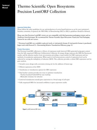
  • 1. Technical Manual Thermo Scientific Open Biosystems Precision LentiORF Collection Important Safety Note Please follow the safety guidelines for use and production of vector-based lentivirus as set by your institution’s biosafety committee. In general, the NIH Office of Biotechnology BSL2 or BSL2+ guidelines should be followed. Please note that Precision LentiORF vectors are not compatible with third generation packaging systems such as ViraPower from Invitrogen. We recommend the Thermo Scientific Open Biosystems TransLenti Viral Packaging System for use with our vectors. **Precision LentiORFs are available in glycerol stock or viral particle format. If viral particle format is purchased, begin work with Protocol X – Determining Relative Transduction Efficiency, page 11. Product Description The Precision LentiORF collection is a library of expression ready lentiviral ORFs generated using gene content from the fully sequenced ORFeome Collaboration Collection. Its unique design expresses the ORF, the fluorescent marker and the selection marker from one promoter, providing a visual marker for ORF expression and two methods of transduction optimization. When used in viral format precise control of expression level can be achieved by varying the multiplicity of infection (MOI). This collection provides a robust ORF expression tool for gene analysis. • Versatile vector design with convenient cloning site for the addition of fusion tags • Efficient expression at low MOI • Transfection or transduction options for ORF expression • Dual marker cassette provides two ways to optimize transduction - Nuclear localized TurboGFP for easy tracking - Blasticidin resistance for selection • Lentiviral transduction extends gene expression to a broad range of cell types • Fully sequenced ORFs for increased confidence in gene expression results CMV ORF IRES tGFP(nuc) -2a- Blastr cPPT WRE Multi Tag Cloning Site pLOC Ampr pUC Sv40 Ori Figure 1. pLOC lentiviral ORF clone 1
  • 2. Table 1. Features Of The pLOC Vector ORF Cassette Elements CMV Promoter RNA Polymerase II promoter ORF Custom MultiTag Cloning site Restriction site for purification or localization tag TurboGFP(nuc) Marker to track ORF expression 2a-peptide signal Allows dual translation of BlastR and TurboGFP(nuc) Blastr Mammalian selectable marker Lentiviral Elements SIN-LTR Increases safety - 3' Self inactivating long terminal repeat 5'LTR 5' long terminal repeat WRE Enhances the stability and translation of transcripts cPPT (Central Polypurine Tract) Increases translocation into the nucleus of non-dividing cells RRE (Rev Response Element) Increases efficient packaging full-length viral genomes Plasmid Backbone Elements pUC ori High copy replication and maintenance in E.coli AMPr Ampicillin bacterial selectable marker Note: The specific sequence of the att site may vary between clones. Refer to the data file online for the specific sequence. Figure 2. Detailed vector map of pLOC lentiviral vector. Antibiotic Resistance pLOC contains 2 resistance markers (Table 2). Table 2. Antibiotic resistances conveyed by pLOC Antibiotic Concentration Utility Ampicillin (carbenicillin) 100 μg/ml Bacterial selection marker (outside LTRs) Blasticidin S variable Mammalian selectable marker Quality Control Each ORF identity has been sequence validated following the cloning process. The att site, MultiTag cloning site and secondary stop codons have also been sequence validated. 2
  • 3. Protocol I - Replication Table 3. Materials for plate replication Item Vendor Catalog # LB-Lennox Broth (low salt) VWR EM1.00547.0500 Peptone, granulated, 2 kg - Difco VWR 90000-368 Yeast Extract, 500 g, granulated VWR EM1.03753.0500 NaCl Sigma S-3014 Glycerol VWR EM-2200 or 80030-956 Carbenicillin Novagen 69101-3 Blasticidin S Invivogen ant-zn-5p 96-well microplates Nunc 260860 Aluminum seals Nunc 276014 Disposable replicators Genetix X5054 Disposable replicators Scinomix SCI-5010-OS Vector Sumary Replication From Individual Glycerol Stock Prepare media with 8% glycerol* and the appropriate antibiotics, and inoculate from glycerol stock. For archive replication, grow all pLOC clones at 37°C in LB-Lennox (low salt) media plus 100 μg/ml carbenicillin in order to provide maximum stability of the clones. Replication Of Plates Prepare target plates by dispensing ~160 μl of LB-Lennox (low salt) media supplemented with 8% glycerol* and appropriate antibiotic (100 μg/ml carbenicillin). Prepare Source Plates 1. Remove foil seals while the source plates are still frozen. This minimizes cross-contamination. 2. Thaw the source plates with the lid on. Wipe any condensation underneath the lid with a paper wipe soaked in ethanol. Replicate 1. Gently place a disposable replicator in the thawed source plate and lightly move the replicator around inside the well to mix the culture. Make sure to scrape the bottom of the plate of the well. 2. Gently remove the replicator from the source plate and gently place in the target plate and mix in the same manner to transfer cells. 3. Dispose of the replicator. 4. Place the lids back on the source plates and target plates. 5. Repeat steps 1-4 until all plates have been replicated. 6. Return the source plates to the -80°C freezer. 7. Place the inoculated target plates in a 37°C incubator for 18-19 hours. Freeze at –80°C for long term storage. Avoid long periods of storage at room temperature or higher in order to control background recombination products. Note: Due to the tendency of all viral vectors to recombine, we recommend keeping the incubation times as short as possible and avoid subculturing. Return to your glycerol stock for each plasmid preparation. *Glycerol can be omitted from the media if you are culturing for plasmid preparation. If making copies of the constructs for long term storage at –80°C, 8% glycerol is required. Protocol II - Plasmid Preparation Culture Conditions For Individual Plasmid Preparations For plasmid preparation, grow all pGIPZ clones at 37°C in LB broth (low salt) media plus 100 μg/ml carbenicillin only. 3
  • 4. 2X LB broth (low salt) media preparation LB-Broth-Lennox 20 g/l Peptone 10 g/l Yeast Extract 5 g/l Appropriate antibiotic(s) at recommended concentration(s) Most plasmid mini-prep kits recommend a culture volume of 1–10 ml for good yield. 1. Upon receiving your glycerol stock(s) containing the shRNAmir of interest store at -80°C until ready to begin. 2. To prepare plasmid DNA first thaw your glycerol stock culture and pulse vortex to resuspend any E. coli that may have settled to the bottom of the tube. 3. Take a 10 μl inoculum from the glycerol stock into 3-5 ml of 2X LB (low salt) with 100 μg/ml carbenicillin. Return the glycerol stock(s) to -80°C. *Note: If a larger culture volume is desired, incubate the 3-5 ml culture for 8 hours at 37˚C with shaking and use as a starter inoculum. Dilute the starter culture 1:500-1:1000 into the larger volume. 4. Incubate at 37°C for 18-19 hours with vigorous shaking. 5. Pellet the 3-5 ml culture and begin preparation of plasmid DNA. 6. Run 3-5 μl of the plasmid DNA on a 1% agarose gel. The size of the pLOC vector containing your ORF will vary depending on the length of the ORF. It can range from <500 nt to >4 kb. Note: Due to the tendency of all viral vectors to recombine, we recommend keeping the incubation times as short as possible and avoid subculturing. Return to your original glycerol stock for each plasmid preparation. Culture Conditions For 96-Well Bio-Block Plasmid Preparation Inoculate a 96-well bio-block containing 1 ml per well of 2X-LB (low salt) media with 100 μg/ml carbenicillin with 1 µl of the glycerol stock culture. Incubate at 37°C with shaking (~170-200 rpm). We have observed that incubation times between 18-19 hours produce good plasmid yield. For plasmid preparation, follow the protocols recommended by the plasmid isolation kit manufacturer. Note: 96-well bio-block plasmid preparation protocol in conjunction with a Qiagen Turbo kit (Catalog # 27191) are used to purify plasmid DNA. 2 bio-blocks are combined, the optional wash step is not performed and water is used for the elution. Protocol III - Precision Lentiorf Plasmid Dna Analysis By Restriction Digest The following is a sample protocol for restriction analysis of Precision LentiORF plasmid DNA using EcoRI restriction enzyme. EcoRI cuts 15 bases upstream of attB1 site flanking the ORF at 5’ (which corresponds to 30-50 nt upstream of ORF start codon, depending on the clone configuration –see specific clone information). A second EcoRI site is located 1348 bp downstream of attB2 site, approximately 1.37 kb downstream of the last sense codon of the ORF. Alternatively, SpeI or PacI flanking attB1 site at the 5’ and NheI or AscI flanking attB2 at 3’ in the vector may be used to cut out the fragment corresponding to the ORF. Use of EcoRI is preferable for smaller ORFs. Table 4. PCR Components Component Amount Sterile, nuclease-free water X μl NEBuffer EcoRI (10X) 2.0 μl DNA sample (500 ng) in water X μl EcoRI, 10 U/ul 0.5 μl Final volume 20.0 μl 1. Set up EcoRI restriction reaction as follows (Table 4). 2. Mix gently by pipetting. 3. Incubate at 37°C for 1 hour to digest 4. Run cut and uncut plasmid in 1% agarose gel (Figure 3). Protocol IV – Tagging Lentiorf With C-Terminal Tag pLOC-ORF LentiORFs are available in “open” (the native stop codon has been removed) and “closed” (the ORF 4
  • 5. Figure 3. Restriction analysis of 2 Precision LentiORF clones with different sized ORFs. Lanes 1 & 10 are a 10 kb molecular weight ladder (10 kb, 7 kb, 5 kb, 4 kb, 3 kb, 2.5 kb, 2 kb, 1.5 kb, 1 kb). Lanes 2 and 4 show uncut plasmid. Lanes 3 and 5 are EcoRI digests. The white arrow points to the 7.8 kb band common to the back bone of all Precision LentiORF clones. The lower bands (ORF1 producing a 2.8 kb band and ORF2 producing two bands, 2.5 kb and 1.3 kb) are specific to each ORF. ORF2 has an additional EcoRI site within it producing two ORF specific bands noted with the asterisks. retains its native stop codon) configurations (see clone information). Translation of the “closed” configuration ORFs terminates at its natural stop codon immediately after last sense codon of the ORF reference sequence. “Open” configuration of the ORFs was created to allow for C-terminal fusions of the ORFs with the tag of interest. In these ORFs translation terminates at the stop codon in the vector immediately downstream of the attB2 site adding 11-16 amino acids to its C-terminus (see figure 4 below). If desired the stop codons in the vector can be ORF attB2.1 site Stop condons ATG GAC GAG CTG TAC AAG TAC CCA ACT TTC TTG TAC AAA GTG GTT GCT AGC TAA TGA Frame of translation NheI * * ACCGGGCGCGCC CCGCCCCTCTC AscI Figure 4. Example of 3’ end of ORF and flanking sequence. Highlighted gray are NheI and AscI sites flanking ORF-attB2 site at 3’. Highlighted yellow is the attB2 site (version attB2.1) Underlined is an invariable part of the attB2 site and the adjacent vector sequence. The secondary stop codons have asterisks beneath them. replaced by the fusion tag at Nhe/AscI sites flanking the stop codon in the vector. Care should be taken to place the fusion tag in the same reading frame as the Stop codon. Table 5. Suggested concentration of Blasticidin S to use for Protocol V - Blasticidin S Selection generation of stable cell lines expressing pLOC-ORF. Blasticidin S Kill Curve and Blasticidin S Selection Cell Line Blasticidin S, ug/ml pLOC-vector confers resistance to Blasticidin S in transduced HEK 293T 7-15 or transfected cells. Blasticidin S selection can be used to HeLa 5-7 eliminate non-transduced or non-transfected cells. In order HepG2 5-7 to generate stable cell lines, it is important to determine MCF7 9-12 the minimum amount of Blasticidin S required to kill non- OVCAR8 9-12 transfected or non-transduced cells. We have tested several A549 9-12 cell lines for their sensitivity to Blasticidin S (see Table 5). These data can be used as a general guidelines. However, we recommend testing your cell line of by generating a Blasticidin S kill curve following the protocol below. Blasticidin S Kill Curve 1. On day 0, plate 5 x 10⁴ cells per well in a 24-well plate in enough wells to carry out your Blasticidin S dilutions. Incubate overnight. 2. Prepare media specifically for your cells containing a range of antibiotic, for example: 0 - 20 μg/ml Blasticidin S. 3. The next day (day 1) replace the growth media with the media containing the dilutions of the antibiotic into the appropriate wells. 4. Incubate at 37°C. 5. Approximately every 2-3 days replace with freshly prepared selective media. 6. Monitor the cells daily and observe the percentage of surviving cells. Optimum effectiveness should be reached in 3-6 days under Blasticidin S selection. 7. The minimum antibiotic concentration to use is the lowest concentration that kills 100% of the cells in 3-6 days from the start of antibiotic selection. 5
  • 6. Protocol VI - Transfection The protocol below is optimized for transfection of the pLOC-ORF plasmid DNA into HEK293T cells in a 24- well plate using serum-free media. If a different culture dish is used, adjust the number of cells, volumes and reagent quantities in proportion to the change in surface area (Table 6). It is preferable that transfections be carried out in medium that is serum-free and antibiotic-free. A reduction in transfection efficiency occurs in the presence of serum, however it is possible to carry out successful transfections with serum present (see Transfection Optimization). Warm Express-In to ambient temperature (approximately 20 minutes at room temperature) prior to use. Always mix well by vortex or inversion prior to use. Table 6. Suggested amounts of DNA, medium and Express-In reagent for transfection of shRNA plasmid DNA into adherent cells. Tissue Culture Dish Surface Area per Total Serum-Free Media Plasmid DNA (μg)* Arrest-In (μg)** Plate or Well (cm2) Volume per Well (ml) 60 mm 20.0 2.0 4.0 21.0 35 mm 8.0 1.0 2.0 10.0 6-well 9.4 1.0 2.0 10.0 12-well 3.8 0.5 1.0 5.0 24-well 1.9 0.25 0.5 2.5 96-well 0.3 0.1 0.1 - 0.2 0.5 - 1.0 *Recommended starting amount of DNA. May need to be optimized for the highest efficiency. **Recommended starting amount of Express-In reagent. See Transfection Optimization. Maintain sterile working conditions with the DNA and Express-In mixtures as they will be added to the cells. 1. The day before transfection (day 0), plate the cells at a density of 5 x 10⁴ cells per well of a 24-well plate. Full medium (i.e. with serum and antibiotics) will be used at this stage. 2. On the day of transfection, form the DNA/Express-In transfection complexes. a. For each well to be transfected, dilute 500 ng shRNA plasmid DNA into 50 µl (total volume) of serum-free medium in a microfuge tube. b. For each well to be transfected, dilute 2.5 µg (2.5 µl) of Express-In into 50 µl (total volume) serum-free medium into a separate microfuge tube. c. Add the diluted DNA (step a) to the diluted Express-In reagent (step b), mix rapidly then incubate for 20 minutes at room temperature. This will give a 1:5 DNA:Express-In ratio which is recommended for optimal transfection into HEK293T cells. Your total volume will be 100 µl at this stage. 3. Aspirate the growth medium from the cells. Add an additional 150 µl of serum-free medium to each of the tubes containing transfection complexes and mix gently. Add the 250 µl DNA/Express-In complex mixture to the cells and incubate for 5-6 hours in a CO2 incubator at 37˚C. Your total volume will be 250 µl at this stage (150 μl serum-free medium + 100 μl DNA:Express-In mixture). 4. Following the 5-6 hour incubation, add an equal volume of growth medium (250 µl) containing twice the amount of normal serum to the cells (i.e. to bring the overall concentration of serum to what is typical for your cell line). Alternatively, the transfection medium can be aspirated and replaced with the standard culture medium (see note). Return the cells to the CO2 incubator at 37˚C. Note – Express-In has displayed low toxicity in the cell lines tested, therefore removal of transfection reagent is not required for many cell lines. In our experience higher transfection efficiencies have been achieved if the transfection medium is not removed. However, if toxicity is a problem, aspirate the transfection mixture after 5-6 hours and replace with fresh growth medium. Additionally, fresh growth medium should be replenished as required for continued cell growth. 5. After 24-72 hours of incubation, examine the cells microscopically for the TurboGFP expression. Note: If desired, Blasticidin S selection can be applied 24 h post transfection. The working concentration of Blasticidin S varies between cell lines. We recommend you determine the optimal concentration of Blasticidin S required to kill your host cell line prior to selection for pLOC-ORF transfectants (see Blasticidin S kill curve protocol). Typically, the working concentration ranges from 5-20 μg/ml. We recommend that you change 6
  • 7. Figure 5. pLOC -ORF transfected into HEK293T cells 24 hours post transfection. a) Phase b) TurboGFP fluorescence of the expression marker. Note the high percentage of cells successfully transfected. Factors affecting transfection efficiency are not limited to but include purity of plasmid DNA, health of transduced cells, and inconsistencies in number of cells plated. selection media every 2-3 days. You should use the lowest concentration that kills 100% of the cells in 3-6 days from the start of Blasticidin S selection. Transfection Optimization using Express-In It is essential to optimize transfection conditions to achieve the highest transfection efficiencies and lowest toxicity with your cells. The most important parameters for optimization are DNA to transfection reagent ratio, DNA concentrations and cell confluency. We recommend that you initially begin with the Thermo Scientific Open Biosystems Express-In and DNA amount indicated in Table 6 and extrapolate the number of cells needed for your vessel size from the number of cells used in a well of a 24-well plate as listed in step 1 of the protocol for delivery of plasmid DNA. Cells Grown In Suspension Transfection of cells in suspension would follow all the above principles and the protocol would largely remain the same, except that the DNA/Open Biosystems Express-In™ mixture should be added to cells (post 20 minute incubation for complex formation) to a total volume of 250 μl serum-free medium or to a total volume of 250 μl of medium with serum (no antibiotics). Protocol VII -Packaging Lentivirus The pLOC vector is tat dependant, so you must use a packaging system that expresses the tat gene. For packaging lentiviral constructs, we recommend the TransLenti Viral Packaging System. The TransLenti Viral Packaging System allows creation of a replication-incompetent (Shimada, et al. 1995), HIV-1-based lentivirus which can be used to deliver and express your gene of interest in either dividing or non-dividing mammalian cells. The TransLenti Viral Packaging System uses a replication-incompetent lentivirus based on the translentiviral system developed by Kappes (Kappes and Wu 2001). For protocols and information on packaging pLOC with our TransLenti Viral GIPZ Packaging System, please see the product manual available on our website. Protocol VIII - Titering Viral Titering Follow the procedure below to determine the titer of your lentiviral stock using the mammalian cell line of choice IF YOU HAVE PRODUCED VIRAL PARTICLES YOURSELF. This protocol uses the TLA-HEK293T cell line that is available as part of our TransLenti Viral shRNA Packaging System. You can use a standard HEK293T cell line as an alternative. Note: If you have generated a lentiviral stock of the expression control (e.g. pLOC Non-Silencing), we recommend titering this stock as well. 1. The day before transduction, seed a 24-well tissue culture plate with TLA-HEK293T cells at 5 x 10⁴ cells per well in DMEM (10% FBS, 1% pen-strep). The following day, the well should be no more than 40-50% confluent. TLA-HEK293T (Thermo Scientific Open Biosystems Catalog # HCL4517). 2. Make dilutions of the viral stock in a round bottom 96-well plate using serum-free media. Utilize the plate as 7
  • 8. Five - fold dilutions 1 2 3 4 5 6 7 8 9 10 11 12 Virus stock 1 A Virus stock 2 B Virus stock 3 C Virus stock 4 D E F G H Figure 6. Five-fold serial dilutions of virus stock. shown in Figure 6 using one row for each virus stock to be tested. Use the procedure below (starting at step 4) for dilution of the viral stocks. The goal is to produce a series of 5-fold dilutions to reach a final dilution of 390625-fold. 3. To each well add 80 μl of serum-free media. 4. Add 20 μl of thawed virus stock to each corresponding well in column 1 (5 fold dilution). Pipette contents of well up and down 10-15 times. Discard pipette tip. 5. With new pipette tips, transfer 20 μl from each well of column 1 to the corresponding well in column 2. Pipette 10-15 times and discard pipette tips. 6. With new pipette tips, transfer 20 μl from each well of column 2 to the corresponding well in column 3. Pipette 10-15 times and discard pipette tip. 7. Repeat transfers of 20 μl from columns 3 through 8, pipetting up and down 10-15 times and changing pipette tips between each dilution. It is strongly recommended that you use a high quality multichannel pipettor when performing multiple dilutions. Pre-incubate the dilutions of the virus stock for 5 minutes at room temperature. 8. Label 24-well plate as shown in Figure 7 using one row for each virus stock to be tested. 1 2 3 4 5 6 Virus stock 1 A Virus stock 2 B Virus stock 3 C Virus stock 4 D Figure 7. Twenty-four well tissue culture plate, seeded with TLA-HEK293T cells, used to titer the virus. 9. Remove culture media from the cells in the 24-well plate. 10. Add 225 μl of serum-free media to each well. 11. Transduce cells by adding 25 μl of diluted virus from the original 96-well plate (Figure 6) to a well on the 8
  • 9. 24-well destination plate (Figure 7) containing the cells. For example, transfer 25 μl from well A2 of the 96-well plate into well A1 in the 24-well plate (Table 7). Table 7. Example of set up for dilutions Well (Row A, B, C, or D) Volume Diluted Virus Used Dilution Factor Originating Destination (96-well plate) (24-well plate) A1 25 μl 5* A2 A1 25 μl 25 A3 A2 25 μl 125 A4 A3 25 μl 625 A5 A4 25 μl 3125 A6 A5 25 μl 15625 A7 A6 25 μl 78125 A8 25 μl 390625 * *Please note that when expecting very high or very low titers, it would be advisable to include either well 8 or well 1 respectively. 12. Incubate transduced cultures at 37°C for 4 hours. 13. Remove transduction mix from cultures and add 1 ml of DMEM (10% FBS, 1% pen-strep). 14. Culture cells for 48 hours. 15. Count the TurboGFP expressing cells or colonies of cells (Figure 8). Count each multi-cell colony as 1 transduced cell, as the cells will be dividing over the 48 hour culture period. Figure 8 illustrates this principle of counting. Note: The intensity of TurboGFP may vary between LentiORF clones. If the TurboGFP intensity in your cells is low, we recommend counting colonies at a higher magnification. 16. Transducing units per ml (TU/ml) can be determined using the following formula: # of TurboGFP positive colonies counted x dilution factor x 40 = #TU/ml Example: 55 TurboGFP positive colonies counted in well A3. (TurboGFP positive colonies) x 625 (dilution factor) x 40 = 1.38x10⁶ TU/ml Once you have generated a lentiviral stock with a suitable titer, you are ready to transduce the lentiviral vector into the mammalian cell line of choice and assay for expression of your recombinant protein. Multiplicity Of Infection (MOI) To obtain optimal expression of your gene of interest, you will need to transduce the lentiviral vector into your mammalian cell line of Figure 8. Example of individual colony 72 h post choice using a suitable MOI. MOI is defined as the number of transduction. Imaged at 40x magnification. transducing units per cell. Although this is cell line dependent, this generally correlates with the number of integration events per cell and as a result, level of expression. Determining The Optimal MOI A number of factors can influence determination of an optimal MOI including the nature of your mammalian cell (actively- versus non-dividing), its transduction efficiency, your application of interest, and the nature of your gene of interest. If you are transducing your lentiviral construct into the mammalian cell line of choice for the first time, after you have titered it, we recommend using a range of MOIs (e.g. 0, 0.5, 1, 2, 5, 10, 20) to determine the MOI required to obtain optimal expression for your particular application. If minimal expression is preferred, it should be noted that to achieve single copy knockdown, an MOI of 0.3 is generally used, as less than 4% of your cells will have more than one insert. Protocol IX - Transduction Transduction Of Target Cells The protocol below is optimized for transduction of the lentiviral particles into TLA-HEK293T, OVCAR8 or MCF7 cells in a 24-well plate using serum-free media. If a different culture dish is used, adjust the number of 9
  • 10. cells, volumes and reagent quantities in proportion to the change in surface area (see Table 8). It is strongly recommended that you optimize transduction conditions to suit your target cell line to provide for the highest transduction efficiency possible. It is preferable that transduction be carried out in medium that is serum-free and antibiotic-free. A reduction in transduction efficiency occurs in the presence of serum, however it is possible to carry out successful transductions with serum present; you will have to optimize the protocol according to your needs. 1. On day 0 plate 5 x 10⁴ cells per well in a 24-well plate. Incubate overnight. You will be using full medium (i.e. with serum) at this stage. 2. The next day (day 1), remove the medium and add the virus to the MOI you wish to use. Set up all desired experiments and controls in a similar fashion. Bring the total volume of liquid up so that it just covers the cells efficiently with serum-free media (See Table 8 for guidelines). If you are using concentrated virus you are likely to use very little virus volume and a lot of serum-free media; if you are using unconcentrated virus you will find you need much more virus volume. Table 8. Suggested volumes of media per surface area per well of adherent cells. Tissue Culture Dish Surface Area per Well (cm2) Suggested total serum-free medium volume per well (ml) 100 mm 56.0 5.0 60 mm 20.0 2.0 35 mm 8.0 1.0 6-well 9.4 1.0 12-well 3.8 0.5 24-well 1.9 0.25 96-well 0.3 0.1 3. Approximately 4-6 hours post-transduction, add an additional 1ml of full media (serum plus pen-strep if you are using it) to your cells and incubate overnight. We have experienced low toxicity with transduction in the cell lines tested, therefore removal of virus is not required for many cell lines. In our hands higher transduction efficiencies have been achieved if the virus is not removed after 6 hours. However, if toxicity is a problem, aspirate the mixture after 3-6 hours and replace with fresh growth medium. Additionally, fresh growth medium should be replenished as required for continued cell growth. 4. At 48 hours post-transduction examine the cells microscopically for the presence of reporter expression as this will be your first indication as to the efficiency of your transduction. Note: When visualizing TurboGFP expression, if less than 90% of all cells are green, it is recommended in these cases to utilize Blasticidin S selection in order to increase the percentage of cells expressing your gene of interest. a. If adding Blasticidin S, use the appropriate concentration as determined based on the above kill curve. If the cell density is close to or above 50% confluency, it is recommended that you split the cells at the time of Blasticidin S addition. Incubate. b. Approximately every 2-3 days replace with freshly prepared selective media. c. Monitor the cells daily and observe the percentage of surviving cells. At some time point almost all of the cells surviving selection will be harboring the gene of interest. Optimum effectiveness should be reached in 3-6 days with Blasticidin S. Please note that the higher the MOI you have chosen the more copies of the gene of interest and Blasticidin S resistance gene you will have per cell. When selecting on Blasticidin S, it is worth remembering that at higher MOIs, cells containing multiple copies of the resistance gene can withstand higher Blasticidin S concentrations than those at lower MOIs. Even at single copy the marker in pLOC lentiORFs provides significant resistance to Blasticidin in transduced cells. Cells with very low transgene expression level may still survive the selection even if TurboGFP is expressed at an undetectable level (see Figure 9). We recommend using untransduced control cells to monitor the selection process. Longer selection and higher concentrations of Blasticidin S are recommended if high level of transgene expression is desired. Adjust the concentration of Blasticidin S to a level that will select for the population of transduced cells you wish to select for, without going below the minimum antibiotic concentration you have established in your kill curve. 5. Once your transduction efficiency is at an acceptable level (with or without Blasticidin S selection), you can proceed to assay cells for gene expression by quantitative/real-time PCR (QPCR), western blot or other appropriate functional assay. The TurboRFP expression construct may be used as a control. 10
  • 11. a Figure 9. HEK293T cells after 4 days of selection on Blasticidin S at concentration 10ug/ml. a) untransduced control. The rounded cell morphology compared to the transduced cells is an indication of cell death for HEK293T cells, b) and c) cells transduced with pLOC-ORF viral particles (MOI=0.3) and placed on selection 24 hours post transduction. Imaged 5 days post transduction. Optimal length of incubation from the start of transfection/transduction to analysis is dependent on cell type and the gene of interest. Protocol X – Determining Relative Transduction Efficiency Follow the procedure below to determine the relative transduction efficiency of purchased LentiORF viral particles (Thermo Scientific Open Biosystems Catalog # OHS5831). This protocol should be used with purchased LentiORF RFP in viral particle format. Prior to transducing with purchased LentiORF individual clones in viral particle format, we recommend determining the relative transduction efficiency of your cell type. Lentiviral titers provided with purchased LentiORF viral particles have been calculated by transducing TLAHEK293T cells. Transduction efficiencies vary significantly by cell type. The relative transduction efficiency of your cells may be estimated by determining the functional titer of a control virus such as LentiORF TurboRFP control viral particles (Thermo Scientific Open Biosystems Catalog # OHS5831) in your cell type of choice. Follow the procedure below to determine the functional titer of the LentiORF viral stock in the mammalian cell line of your choice. The following conditions have been optimized for transduction of TLA-HEK293T cells. When determining the relative transduction efficiency of your cell type, use the transduction conditions that have been optimized for your cell type of choice. 1. The day before transduction, seed a 24-well tissue culture plate with your cells at 5x10⁴ cells per well in their respective media. The following day, the well should be no more than 40-50% confluent. 2. Make dilutions of the LentiORF control viral stock in a round bottom 96-well plate using serum-free medium. Five - fold dilutions 1 2 3 4 5 6 7 8 9 10 11 12 Replicate 1 A Replicate 2 B Replicate 3 C Replicate 4 D E F G H Figure 10. Five-fold serial dilutions of virus stock. 11
  • 12. Utilize the plate as shown in Figure 10 with one row for each replicate (we recommend performing at least two replicates). Use the procedure below for dilution of the viral stock. The goal is to produce a series of 5-fold dilutions to reach a final dilution of 390625-fold. 3. Add 40 ul of serum-free media to each well in column 1. Add 80 ul of serum-free media to each well of columns 2-8. If desired, include 8 μg/mL polybrene in the dilution media 4. Add 10 µl of thawed control virus stock to each well in column 1 (5-fold dilution). Pipette contents of well up and down 10-15 times. Discard pipette tip. 5. With new pipette tips, transfer 20 µl from each well of column 1 to the corresponding well in column 2. Pipette 10-15 times and discard pipette tips. 6. With new pipette tips, transfer 20 µl from each well of column 2 to the corresponding well in column 3. Pipette 10-15 times and discard pipette tip. 7. Repeat transfers of 20 µl from columns 3 through 8, pipetting up and down 10-15 times and changing pipette tips between each dilution. It is strongly recommended that you use a high quality multichannel pipettor when performing multiple dilutions. Pre-incubate the dilutions of the virus stock for 5 minutes at room temperature. 8. Label a 24-well plate as shown in Figure 11 using one row for each replicate. 1 2 3 4 5 6 Replicate 1 A Replicate 2 B Replicate 3 C Replicate 4 D Figure 11. Twenty-four well tissue culture plate, seeded with TLA-HEK293T cells, used to titer the virus. 9. Remove culture medium from the cells in the 24-well plate. 10. Add 225 µl of serum-free medium to each well. 11. Transduce cells by adding 25 μl of diluted control LentiORF lentivirus from the original 96-well plate (Figure 10) to a well on the 24-well destination plate (Figure 11) containing the cells. Table 9. Example of set up for dilutions Well (Row A, B, C, or D) Volume Diluted Virus Used Dilution Factor Originating Destination (96-well plate) (24-well plate) A1 25 μl 5* A2 A1 25 μl 25 A3 A2 25 μl 125 A4 A3 25 μl 625 A5 A4 25 μl 3125 A6 A5 25 μl 15625 A7 A6 25 μl 78125 A8 25 μl 390625 * *Please note that when expecting very high or very low transduction efficiency, it would be advisable to include either higher or lower dilution factors. 12
  • 13. For example, transfer 25 μl from well A2 of the 96-well plate into well A1 in the 24-well plate (Table 9). 12. Incubate transduced cultures at 37°C for 4-6 hours. 13. Add 1 ml of your medium (normal serum concentration). 14. Culture cells for 72 hours. 15. Count the TurboGFP expressing cells or colonies of cells. Count each multi-cell colony as 1 transduced cell, as the cells will be dividing over the 72 hour culture period. Figure 6 illustrates this principle of counting. Count the number of TurboGFP expressing colonies in wells corresponding to at least two viral dilutions. Note: The intensity of TurboGFP may vary between LentiORF clones. If the TurboGFP intensity in your cells is low, we recommend counting colonies at a higher magnification. 16. Transducing units per ml (TU/ml) can be determined using the following formula: # of TurboGFP positive colonies counted x dilution factor x 40 = # TU/mL (Note: 25 μl of diluted virus was added to the cells. This is 1/40th of a mL) Example: 55 TurboGFP positive colonies counted in well A3. 55 (TurboGFP positive colonies) x 625 (dilution factor) x 40 = 1.38x10⁶ TU/ml 17. The functional titer calculated for your cell line under your experimental conditions can be used to determine the relative transduction efficiency of your cell type by using the following formula: Functional titer of Non-silencing control virus stock in your cell line ÷ Titer of Non-silencing control shRNAmir virus stock as calculated by Thermo Scientific Open Biosystems in TLA-HEK293T = Relative transduction efficiency For example, if the titer of the non-silencing control shRNAmir virus stock in TLA-HEK293T (as provided on the product specification sheet) is 6.9x10⁶ TU/mL and the functional titer of the control shRNAmir virus stock in your cell line is 1.38x10⁶ TU/mL, the relative transduction efficiency of your cell type is 0.2. To extrapolate the average functional titer of the provided GIPZ viral particles, multiply the average titer of each plate as provided on the product specification sheet by the relative transduction efficiency of your cell type. In our example, if the titer of the GIPZ viral partilces in TLA-HEK293T cells is 2x10⁶ TU/mL and the relative transduction efficiency of your cell type is 0.2, the extrapolated average functional titer of that plate your cell type is 4x10⁵ TU/mL. Once the relative transduction efficiency of the GIPZ virus has been established in your cell line, use the optimized transduction conditions determined in Protocol IX to transduce your cell line with the purchased GIPZ shRNAmir individual clones in viral particle format. If the titer of the non-silencing control shRNAmir virus is not satisfactory in your cell line you might consider choosing a different cell line more permissive to transduction by lentivirus before proceeding. Controls And Validation Precision LentiORF Starter Kits The use of vector-based ORF expression is a powerful and versatile tool. Successful gene silencing in vitro is dependent on several variables including 1) The target cell line being studied 2) Transfection and transduction efficiency 3) Abundance of the mRNA or protein of interest in the target cell line 4) Half life of the protein 5) Robust experimental protocols. For all these reasons it is very important to run controlled experiments where the transfection and transduction efficiencies are as high as possible and measurable. Controls are a critical part of a gene silencing experiment. They enable accurate representation of knockdown data and provide confidence in the specificity of the response. Changes in the mRNA or protein levels in cells treated with negative or non-silencing controls reflect nonspecific responses in cells and can be used as a baseline against which specific knockdown can be measured. Positive controls are useful to demonstrate that your experimental system is functional and your shRNA construct is successfully activating the RNAi pathway. Controls The pLOCTM TurboRFP lentiviral expression construct has been validated as control for ORF expression experiments. This control has been tested in transfection and transduction based experiments for TurboRFP expression and selection on Blasticidin S. Additional cell lines to the one below have been tested and are listed in Table 5. 13
  • 14. Figure 12. Cells transduced with pLOC-TurboRFP control particles 72 hours post transduction (MOI=3, titered in HEK293T cells. a) Cell line A549 b) HepG2 c) OVCAR8. TurboRFP is shown in red. The nuclear localized TurboGFP is overlaid resulting in yellow nuclei. What Packaging Cell Line Should I Use For Making Lentivirus? The pLOC vector is tat dependant, so you must use a packaging system that expresses the tat gene. For packaging our lentiviral shRNAmir constructs, we recommend the TransLenti Viral Packaging System. The TransLenti Viral shRNA Packaging System allows creation of a replication-incompetent (Shimada, et al. 1995), HIV-1-based lentivirus which can be used to deliver and express your gene or shRNAmir of interest in either dividing or non- dividing mammalian cells. The TransLenti Viral shRNA Packaging System uses a replication-incompetent lentivirus based on the trans-lentiviral system developed by Kappes (Kappes and Wu 2001). For protocols and information on packaging pLOC with our TransLenti Viral shRNA Packaging System, please see the product manual available on our website. Can I Use Any 2nd Generation Packaging System With The GIPZ Vector? The pLOC vector is tat dependant, so you must use a packaging system that expresses the tat gene. What does the number 40 refer to in the formula for the calculation of titer? The titer units are given in transducing units (TU) per ml, so the number 40 is used to convert the 25 μl used in the titration ("volume of diluted virus used", Table 8) to one milliliter. How Can I Make A Stable Cell Line? In order to generate stable cell lines, it is important to determine the minimum amount of Blasticidin S required to kill non-transfected/transduced cells. This can be done by generating a Blasticidin S kill curve. After you have determined the appropriate concentration of Blasticidin Sto use, you can transfect or transduce your cells with the construct and culture with Blasticidin S in order to select for those cells that have a stable integrant. Cells not containing a stable integrant will not be selected for. Where do you purchase Blasticidin S? We purchase Blasticidin S from Cellgro™ (Catalog # 61-385-RA). How Many Transfections Are Available In Each Volume Size Of Express-In? The number of transfections that can be performed depends on the size of the culture dish used and the volume size of Express-In purchased. Refer to Table 10 below for the approximate number of transfections.. Table 10. Number of transfections depending on culture dish size and volume of Express-In purchased. Tissue Culture Dish Surface Area Arrest-In 0.5 ml qty 1.0 ml qty 5.0 ml qty 10 ml qty per Well (cm2) (1 mg/ml) (µg)* (rxns)** (rxns)** (rxns)** (rxns)** 60 mm 20.0 21.0 47-50 100 500 1000 35 mm 8.0 10.0 100 200 1000 2000 6-well 9.4 10.0 100 200 1000 2000 12-well 3.8 5.0 200 400 2000 4000 24-well 1.9 2.5 400 800 4000 8000 96-well 0.3 0.5-1.0 1000 2000 10000 20000 **Recommended starting amounts of Arrest-In reagent as defined in Table 1. **Approximate number of transfections based on recommended starting amount of Express-In. Individual results may vary depending on amounts of Express-In used. 1. How much time elapsed from transfection/transduction to Blasticidin S selection? 14
  • 15. If Transfection Into Your Cell Line Is Unsuccessful, You May Need To Consider The Following List Of Factors Influencing Successful Transfection 1. Concentration and purity of plasmid DNA and nucleic acids-determine the concentration and purity of your DNA using 260/280 nm absorbance. Avoid cytotoxic effects by using pure preparations of nucleic acids. 2. Insufficient mixing of transfection reagent or transfection complexes. 3. Transfection in serum containing or serum free media-our studies indicate that Express-In/DNA complexes should preferably be formed in the absence of serum. In the cell lines tested we found that the highest transfection efficiencies can be obtained if the cells are exposed to the transfection complexes in serum free conditions followed by the addition of medium containing twice the amount of normal serum to the complex medium 3-6 hours post transfection (leaving the complexes on the cells). However, the serum-free transfection medium can be replaced with normal growth medium if high toxicity is observed. 4. Presence of antibiotics in transfection medium-the presence of antibiotics can adversely affect the transfection efficiency and lead to increased toxicity levels in some cell types. It is recommended that antibiotics be excluded until transfection has mostly occurred (3-6 hours) and then be added together with the full medium. 5. High protein expression levels–some proteins when expressed at high levels can be cytotoxic; this effect can also be cell line specific. 6. Cell history, density, and passage number-it is very important to use healthy cells that are regularly passaged and in growth phase. The highest transfection efficiencies are achieved if cells are plated the day before, however, adequate time should be given to allow the cells to recover from the passaging (generally >12 hours). Plate cells at a consistent density to minimize experimental variation. If transfection efficiencies are low or reduction occurs over time, thawing a new batch of cells or using cells with a lower passage number may improve the results. If Transduction Into Your Cell Line Is Unsuccessful, You May Need To Consider The Following List Of Factors Influencing Successful Transduction 1. Transduction efficiency is integrally related to the quality and the quantity of the virus you have produced. Factors to bear in mind when transducing include MOI (related to accurate titer), the presence of serum in the media, the use of polybrene in the media, length of expose to virus, and viral toxicity to your particular cells. 2. High quality transfer vector DNA and the appropriate and efficient viral packaging are required to make high quality virus able to transduce cells effectively. See also suggestions 3-6 for factors influencing successful transfection (above). 3. All cell lines are not equally permissible to transduction by lentivirus. You may consider testing additional cell lines to find one more suitable for your experiments. If Express-In seems to be toxic to a particular cell line, try reducing the DNA:Express-In ratio. FAQS/Troubleshooting For answers to questions that are not addressed here, please email technical support at openbiosystems@thermofisher.com with your question, your sales order or purchase order number and the catalog number or clone ID of the construct or collection with which you are having trouble. 13
  • 16. Limited Use Licenses Limited Use Label License: Evrogen Fluorescent Proteins This product contains a proprietary nucleic acid(s) coding for a proprietary fluorescent protein being, including its derivatives or modifications, the subject of pending U.S. and foreign patent applications and/or patents owned by Evrogen JSC (hereinafter “Evrogen Fluorescent Proteins”). The purchase of this product conveys to the buyer the non-transferable right to use Evrogen Fluorescent Proteins for (i) not-for-profit internal Research conducted by the buyer (whether the buyer is an academic or for-profit entity), where “Research” means non-commercial uses or activities which (or the results of which) do not generate revenue, and (ii) evaluation of Evrogen Fluorescent Proteins for the purpose of testing its appropriateness for development of a therapeutic, clinical diagnostic, vaccine or prophylactic product, provided that Evrogen Fluorescent Proteins are not used in the development or manufacture of such product. Offer of Evrogen Fluorescent Proteins for resale; distribution, transfer, or otherwise providing access to Evrogen Fluorescent Proteins to any third party for any purpose, or any use of Evrogen Fluorescent Proteins other than for Research is strictly prohibited. The buyer cannot sell or otherwise transfer materials made by the employment of Evrogen Fluorescent Proteins to a third party or otherwise use Evrogen Fluorescent Proteins for Commercial Purposes. The buyer may transfer information made through the use of this product solely for research and not for Commercial Purposes. Commercial Purposes means any activity by a party for consideration and may include, but is not limited to: (1) use of Evrogen Fluorescent Proteins in manufacturing; (2) use of Evrogen Fluorescent Proteins to provide a service, information, or data; (3) use of Evrogen Fluorescent Proteins for therapeutic, diagnostic or prophylactic purposes. The purchase of this product does not convey a license under any method claims in the foregoing patents or patent applications. For information on the foregoing patents or patent applications or on purchasing a license to use Evrogen Fluorescent Proteins for purposes other than Contact Information those permitted above, contact Licensing Department, Evrogen JSC, Miklukho-Maklaya street 16/10, Moscow, 117997, Russian Federation license@ Technical Support evrogen.com. Tel: 1.888.412.2225 Fax: 1.256.704.4849 Limited Use Label License: Benitec This product is sold solely for use for research purposes in fields other than plants.  This product is not transferable.  If the purchaser is not willing openbiosystems@ to accept the conditions of this label license, supplier is willing to accept the return of the unopened product and provide the purchaser with a full thermofisher.com refund. However, if the product is opened, then the purchaser agrees to be bound by the conditions of this limited use statement.  This product is sold by supplier under license from Benitec Australia Ltd and CSIRO as co-owners of U.S. Patent No. 6,573,099 and foreign counterparts.  For information © 2009 Thermo Fisher regarding licenses to these patents for use of ddRNAi as a therapeutic agent or as a method to treat/prevent human disease, please contact Benitec at Scientific Inc. All rights licensing@benitec.com.  For the use of ddRNAi in other fields, please contact CSIRO at www.pi.csiro.au/RNAi. reserved. Trademarks are the property of their shRNA Limited Use Agreement respective owners. All Permitted Use. Portions of this product are covered by published patent applications (the "shRNA IP Rights") owned by Cold Spring Harbor other trademarks are Laboratory, referred to herein as “CSHL.” This product, and/or any components or derivatives of this product, and/or any materials made using this the property of Thermo product or its components, are referred to herein as the “Product.” Subject to the terms of this Limited Use Agreement, sale of the Product by Open Fisher Scientific Inc. Biosystems (“OBS”) conveys the limited, nonexclusive, nontransferable right to any company or other entity that orders, pays for and takes delivery and its subsidiaries. For of the Product (the “Customer”) from OBS to use the Product solely for its internal research and only at such Customer’s facility where the Product is Research Use Only. delivered by OBS. Restrictions. Customer obtains no right to transfer, assign, or sublicense its use rights, or to transfer, resell, package, or otherwise distribute the Product, or to use the Product for the benefit of any third party or for any commercial purpose. The Product or components of the Product may not be used in vitro or in vivo for any diagnostic, preventative, therapeutic or vaccine application, or in the manufacture or testing of a product therefore, or used (directly or indirectly) in humans for any purpose. Compliance. Customer may only use the Product in compliance with applicable local, state and federal laws, regulations and rules, including without limitation (for uses in the United States) EPA, FDA, USDA and NIH guidelines. Customer may not (directly or indirectly) use the Product, or allow the transfer, transmission, export or re-export of all or any part of the Product, in violation of any export control law or regulation of the United States or any other relevant jurisdiction. Other Uses. Except for the permitted use expressly specified above and subject to the restrictions set forth above, no other use is permitted. CSHL shall retain all right, title and interest in and to the shRNA IP Rights. Nothing herein confers to Customer (by implication, estoppel or otherwise) any right or license under any patent of CSHL, including, for avoidance of doubt, under any patent that may issue from or relate to the shRNA IP Rights. For information on obtaining an agreement to use this Product for purposes other than those expressly permitted by this Limited Use Agreement, or to practice under CSHL patent rights, please contact the CSHL Office of Technology Transfer at (516) 367-8301. Termination of Rights. Upon issuance of any patent covering any portion of the Product, any and all rights conveyed to any Customer (other than a research or educational institution that is a tax-exempt organization under section 501(c)(3) of the U.S. IRS code) under this Limited Use Agreement shall terminate and such Customer shall need a license from CSHL to continue to use any Product that was previously purchased, or to purchase from OBS or any other vendor or use any product claimed by such issued patent. At such time, such Customer may contact the CSHL Office of Technology Transfer at (516) 367-8301 and seek a license to use this Product under CSHL’s patent rights. YOUR USE OF THIS PRODUCT CONSTITUTES ACCEPTANCE OF THE TERMS OF THIS LIMITED USE AGREEMENT. If you are not willing to accept the limitations of this agreement, OBS is willing to accept return of the Product for a full refund.