2. 1384 • J. Neurosci., January 25, 2012 • 32(4):1383–1394 Gerstner et al. • Diurnal Fabp7 Post-Transcriptional Processing
cytic cellular and molecular processes contribute significantly to min with 0.1% Triton X-100 in PBS followed by three 10 min rinses in
linking behavioral and physiological processes. PBS. The slices were then blocked for nonspecific antibody binding with
Fabp7 is enriched near synapses. Its abundance in synap- Aurion goat-blocking agent (Cat. #25596, containing 5% BSA, 0.1% cold
toneurosomal fractions declines with age (Pu et al., 1999). We water fish gelatin, and 5% normal goat serum in PBS, pH 7.4) for 30 min.
The slices were then rinsed three times for 10 min each in incubation
found Fabp7 abundance cycles in synaptoneurosomes, suggesting
buffer containing PBS ϩ 0.1% BSA-C (Aurion Immuno Gold Reagents,
daily variations in trafficking machinery. One potential mechanism Cat. #25558). Finally, the samples were incubated in a mixture of incu-
is through mRNA-binding proteins, such as cytoplasmic polyade- bation buffer and rabbit primary Fabp7 antibody (Millipore) at a dilu-
nylation element (CPE)-binding protein (CPEB). One such protein, tion of 1:1000 overnight at 4°C. The following day, the slices were rinsed
CPEB1, is present in astrocytes (Jones et al., 2008), but its role in in incubation buffer every 10 min for 2 h (12 rinses total), and then
targeting mRNAs to perisynaptic processes in astrocytes has not incubated overnight in a 1:100 dilution of ultrasmall gold conjugate
been explored. CPE-mediated mechanisms exist for trafficking and [F(abЈ) 2 fragment of goat-anti-rabbit IgG (heavy and light chains), Au-
local translation of neuronal mRNAs, so we conducted a bioinfor- rion Immuno Gold Reagents, Cat. #25360] at 4°C. After the overnight
matic search that revealed phylogenetically conserved CPE se- incubation, the slices were rinsed in incubation buffer 6ϫ for 10 min
quences in Fabp7 mRNA. We hypothesized that Fabp7 CPEs are each, and then rinsed in PBS 6ϫ for 5 min each. The slices were postfixed
in 2% glutaraldehyde in 0.1 M PB for 30 min and rinsed 2ϫ for 5 min each
functional and able to mediate translation, predicting Fabp7 mRNA
in PB.
could undergo time-of-day subcellular translocation and polyade- The use of ultrasmall gold conjugates requires a silver-enhancing step
nylation. Our novel findings confirm that, in addition to neurons, to increase the size of the subnanometer gold particles to more easily
cytoplasmic polyadenylation processes exist for astrocytic CPE- visualize the particles by transmission electron microscopy. After post-
containing mRNA (Jones et al., 2008) and establish the existence of fixation, the slices were rinsed 3ϫ for 10 min each in Enhancing Condi-
coordinated control for mRNA trafficking and subsequent transla- tioning Solution (ECS) (Aurion). Next, the slices were developed in
tion at the perisynaptic astrocytic process compartment across the Silver Enhancement Solution (Aurion) for 1.25 h. To terminate silver
day. enhancement, the slices were exposed to a solution of 0.3 M sodium
thiosulfate in ECS for 5 min. The slices were then rinsed twice in 10 min
intervals in ECS and finally processed routinely for electron microscopy.
Materials and Methods After silver enhancing, the slices were fixed in a solution of 0.5% os-
Subjects and handling mium tetroxide in 0.1 M PB for 30 min. Following OsO4 postfixation, the
Male mice (C57BL/6) were either purchased directly from Harlan samples were dehydrated in a graded ethanol series, then further dehy-
Sprague Dawley (adult) or taken at specific ages from a breeding colony. drated in propylene oxide and flat-embedded in Epon epoxy resin. The
Animals were entrained to a 12 h light, 12 h dark (12:12) schedule for a specific regions used for the analysis were trimmed from the flat-
minimum of 2 weeks. Zeitgeber time (ZT) 0 was lights-on, and ZT12 was embedded sections and flat-mounted on blank epoxy resin stubs. Excess
lights-off, on this diurnal schedule. For tissue collection, animals were epoxy resin was then sectioned away in 1 m increments with a Diatome
killed by decapitation, their brains dissected, flash frozen at Ϫ30°C in Histo diamond knife using a Reichert–Jung Ultracut-E ultramicrotome
2-methylbutane, and stored at Ϫ80°C. For tissue punches of specific until the surface of the tissue was exposed. Samples were then sectioned
regions, brains were embedded in optimal cutting temperature media using a Diatome Ultra 45 diamond knife with the same microtome and
and sectioned on a Leica (CM3050) cryostat at Ϫ25°C as previously contrasted with Reynolds lead citrate and 8% uranyl acetate in 50%
described (Gerstner et al., 2006, 2008). Punches from the same brain EtOH. Ultrathin sections were observed with a Philips CM120 electron
region were pooled from each of the animals within the same group, and microscope and images were captured with a MegaView III side-
stored at Ϫ80°C until RNA isolation. All animal care and use procedures mounted digital camera.
were in strict accordance with University of Wisconsin Institutional An-
imal Care and Use Committee and National Institutes of Health guide-
Northern blot analysis
lines for the humane treatment of laboratory animals. Northern blotting was performed as described previously (Gerstner et al.,
Synaptoneurosome preparation 2006). Briefly, brains were dissected and total RNA was isolated using
Synaptoneurosomes (SNs) were prepared from mouse whole brain as TRIzol (Invitrogen), according to the manufacturer’s specifications, and
described previously (Westmark and Malter, 2007; Westmark et al., stored at Ϫ80°C. Before loading, each sample was incubated with sample
2011). Briefly, mice were asphyxiated with carbon dioxide followed by buffer [7.5% formaldehyde, 43% formamide, 12% 10ϫ 3-(N-
removal of the brain. Brains were washed in ice-cold gradient medium morpholino)propanesulfonic acid, 0.12% ethidium bromide] for 5 min
(GM) buffer (0.25 M sucrose, 5 mM Tris, pH 7.5, and 0.1 mM EDTA), at 56°C, and then cooled on ice for 5 min. Northern blots consisted of 1
transferred to a glass Dounce homogenizer containing ice-cold GM buf- g of total RNA per lane and were electrophoresed on a 1.2% agarose gel.
fer, and gently homogenized with five strokes of the loose pestle followed RNA was transferred overnight onto a sheet of GeneScreen Plus nitrocel-
by five strokes of the tight pestle. The homogenate was spun at 1000 g for lulose (NEN Life Science Products). DNA templates generated by PCR
10 min at 4°C in round-bottom tubes to pellet cellular debris and nuclei. were labeled with 32P using the Megaprime DNA labeling system (GE
The supernatant (2 ml of aliquots) was applied to Percoll gradients (lay- Healthcare). The following primers were used: Fabp7 forward primer,
ers, 2 ml each of 23%, 15%, 10%, and 3% isomotic Percoll) and spun at AGACCCGAGTTCCTCCAGTT, reverse primer, CCTCCACACCGA
speed (32,500 g) for 5 min at 4°C. The third band from the top of the AGACAAAC, Cpeb1 forward primer, CATCTTGGGACCTTCTTGGA,
gradient (23%–15% interface) containing intact SNs was removed and Cpeb1 reverse primer, AAGACCCAAGGGATTACC, Cpeb2 forward
pooled for the experiments. primer, GGTCGCTCTTCCCTATTTCC, Cpeb2 reverse primer, TCCA
AAGGCTGAGAACCATC, Cpeb3 forward primer, CCCTTCTCCAGC
Immunocytochemistry and electron microscopy AATGTGAT, Cpeb3 reverse primer, CTCGTTCCCCATTTTGACAT,
Animals were perfused through the heart with 0.1% heparin in 0.01 M Cpeb4 forward primer, CCTCACTGCTTCACTCACCA, Cpeb4 reverse
PBS, pH 7.4, until liver cleared, followed by 2% paraformaldehyde, 1% primer, AACAAAGCGGGCACTGATAG. Probes were mixed with hy-
glutaraldehyde in 0.1 M phosphate buffer (PB), pH 7.4. Sections were cut brisol and incubated overnight at 42°C. Blots were washed 3ϫ with 100
at 50 m on a vibratome and stored in 0.01 M PBS and 0.01% azide ml of 2ϫ SSC, 1% SDS, at room temperature, then twice for 30 min each
solutions until processing. The hippocampal section was prepared from with 100 ml of 2 ϫ SSC, 1% SDS, at 50°C, and finally once for 30 min with
a mouse killed at ZT12. 100 ml of 0.5 ϫ SSC, 1% SDS, at 60°C. Following washes, blots were
Following fixation, the slices were rinsed in 0.1 M PB and then treated exposed to a phosphoscreen for 3 d and image analysis was performed
with freshly prepared 0.1% sodium borohydride in 0.1 M PB for 10 min to using the Storm 860 and ImageQuant 5.2 software (Molecular Dynam-
quench unbound aldehydes. Next, the slices were permeabilized for 30 ics). A 983 bp PCR product for glyceraldehyde-3-phosphate dehydroge-
3. Gerstner et al. • Diurnal Fabp7 Post-Transcriptional Processing J. Neurosci., January 25, 2012 • 32(4):1383–1394 • 1385
nase (GAPDH) was generated as described previously (Gerstner et al., In situ hybridization
2006). In situ hybridization was performed as previously described (Gerstner
and Landry, 2007). Briefly, postfixed, cryostat sections (20 m) were
RNase H and poly(A) analysis pretreated with proteinase K (0.2 g/ml; Promega) and hybridized over-
The RNase H digestion procedure was modified from previously pub- night at 55°C in 150 l of 35S-labeled antisense riboprobe (10,000 cpm/
lished methods (Goodwin and Rottman, 1992). Briefly, 5 g of total l). Following posthybridization washes, sections were exposed to a
RNA was incubated at 80°C for 10 min, rapid cooled to 20°C, and com- phosphoscreen for 6 d. Image analysis was performed using the Storm
bined with or without 0.15 g of Oligo-dT(12–18) in 1 M EDTA for 15 860 and ImageQuant 5.2 software (Molecular Dynamics). For densito-
min. The addition of 50 U RNase H (Promega) was added to RNase H metric analysis of in situ hybridization data, specific regions from a min-
buffer (20 mM HEPES-KOH, pH 7.8, 50 mM KCl, 10 mM MgCl2) at a final imum of four sections were averaged per animal per time point, and
volume of 50 l, and digested at 37°C for 30 min. RNA was isolated from normalized to background as described previously (Gerstner and Lan-
the reactions by phenol-chloroform extraction, ethanol was precipitated, dry, 2007).
and Northern blotting was performed as above. Antisense ( 35S-labeled) was made from Fabp7 PCR templates derived
Densitometric analysis of the poly(A) tail was performed using Im- from T7-tagged reverse or forward primers, respectively. The following
ageQuant software. Briefly, a 1 ϫ 3 pixel window (i.e., the width of a lane primers were used: Forward primer: AGACCCGAGTTCCTCCAGTT,
in the Northern blot ϫ 3 pixel window) was generated, and the starting reverse primer, CCTCCACACCGAAGACAAAC. Fabp7 template used
spot was determined at ZT 12 by the lane that contained the time point for in vitro transcription was generated using standard PCR conditions.
with the lowest poly(A) tail size. The starting spot within this lane was T7 sequence tag extending the reverse primer was GGCCAGTGAATTGT
located above the resolved band with a signal intensity value nearest the AATACGACTCACTATAGGGAGGCGG. In vitro transcription reac-
background value. This height on the gel was determined to be the start- tions were performed as described by Promega Biotech.
ing spot across all lanes. Identical boxes were placed at this level in adja- For emulsion autoradiography, sections processed for in situ hybrid-
cent lanes for all ZT time points, and a signal intensity value was ization (ISH) were dipped in NTB 3 liquid emulsion (Eastman Kodak)
determined within each box. The boxes were then moved up a single under safelight conditions and stored at 4°C for 6 weeks. Processing was
pixel, and the new signal intensities tabulated (single rung). Signal inten- as described by the manufacturer. For analysis of diurnal time points, all
sity values for each rung were done repeatedly until the lane with the last sections within a series were processed under identical conditions in the
value closest to the background value was tabulated (equal to longest same run (N ϭ 2–5 animals per group). Images of ISH emulsion-dipped
poly(A) tail). Tabulated values for each rung were averaged, and this hippocampal sections were taken using darkfield microscopy, and saved
value was used to generate a normalized value across all time points as Tagged Image File Format (TIFF) files for analysis. The region of
within a given single rung. These normalized values within a given time hippocampus was analyzed using ImageJ software (National Institutes of
point were averaged across all rungs to control for relative signal inten- Health) extending from interaural plane 1.62 to 2.22 mm as defined by
sity. This normalization controls for time-of-day changes in transcript Paxinos and Franklin (2003). Counts were tabulated, background was
intensity of the band, but retains a signal above noise/background for subtracted, and averaged. Briefly, a standardized ϳ230 ϫ 230 m box
calculating band length. was made that covered the precursor layer and extended through the
granular cell layer and into the molecular layer of the dentate gyrus of the
Quantitative reverse transcriptase PCR hippocampus. Each individual animal had ϳ4 sections that were ana-
All quantitative reverse transcriptase PCR (qRT-PCR) was performed at
lyzed per time point and these were then averaged to generate a single
the University of Wisconsin-Madison Genome Center. Complementary
number that was used as an N ϭ 1. Final counts for each time point were
DNA was generated using Superscript II Reverse Transcriptase (SSRII;
calculated by averaging across all animals in each group. Density mea-
Invitrogen) as follows: 5 g of total RNA was added to 100 pmol of T7
sures were used to calculate distribution of silver grains by normaliz-
oligo-dT primer [GGCCAGTGAATTGTAATACGACTCACTATAGG
ing signal intensity values of the molecular layer against the highest
GAGGCGG-d(T)24] and the reaction volume was brought to 12 l of
average background-subtracted value within the 230 ϫ 230 m box.
total volume with PCR grade water. The reaction was incubated at 70°C
These silver grain distribution values (background-subtracted pixel
for 10 min in a Mastercycler ep thermocycler (Eppendorf) and placed on
counts per maximal pixel count) were then averaged and divided by
ice for 2 min. Five times SSRII buffer (4 l), 2 l of 0.1 M DTT, and 1 l
the square area (m 2) analyzed. Individual samples were averaged
of 10 mM deoxyribonucleotide triphosphates were added and the reac-
within groups, plotted with SEM bars, and subjected to ANOVA sta-
tion was incubated in a thermocycler for 2 min at 42°C. After incubation,
tistical analysis.
1 l of SSRII was added and the reaction was incubated at 42°C for 1 h.
CDNA was then quantified using an ND-1000 Spectrophotometer
(NanoDrop Technologies) and normalized to 20 ng/l with PCR grade Western blotting
water. Samples prepared from whole brain were subjected to SDS-PAGE by
separating 10 g of protein per lane on a 10 –20% gradient precast gel
PCR methods (Biorad). Protein was transferred to 0.2 m Protran nitrocellulose mem-
Individual reactions contained 1ϫ Probes MasterMix (F. Hoffmann-La branes (PerkinElmer), blocked in 5% dried milk powder in 50 mM Tris-
Roche), 0.05 U uracil-N-glycosylase, 1ϫ LCGreen Plus (Idaho Technol- HCl, pH 7.5, 15 mM NaCl, 0.5% Tween-20 (TBST), washed briefly in
ogy), 200 nM each primer, and 20 ng of cDNA. The following primers TBST and incubated in primary antibody in TBST overnight at 4°C.
were used: Fabp7 forward primer, CTCTGGGCGTGGGCTTT, reverse Fabp7 antibody (Millipore) was used at 1:1000 dilution in TBST; anti-
primer, TTCCTGACTGATAATCACAGTTGGTT, 2M forward body to -actin (Imgenex) was used at 1:10,000 dilution; and anti-
primer, CATGGCTCGCTCGGTGACC, 2M reverse primer, AATGTG glyceraldehyde-3-phosphate dehydrogenase (Imgenex) was used at
AGGCGGGTGGAACTG, -actin forward primer, AGAGGGAAATCG 1:5000 dilution. Blots were then washed three times in TBST, incubated
TGCGTGAC, -actin reverse primer, CAATAGTGATGACCTGGCCG for 1 h in antirabbit horseradish peroxidase-conjugated secondary anti-
T, GAPDH forward primer, CATGGCCTTCCGTGTTCCTA, GAPDH body (1:7500 in TBST; KPL), washed three times in TBST and incubated
reverse primer, ATGCCTGCTTCACCACCTTCT. The total reaction in ECL plus Western blotting detection reagent for 5 min based on the
volume of 5 l was overlaid with 10 of Chillout Wax (Bio-Rad Labo- manufacturer’s instructions (GE Healthcare). For time-of-day synap-
ratories). All reactions were run on a LightCycler 480 Real-Time PCR toneurosomal blots, Fabp7 antibody was used at 1:2000 dilution, PSD-95
System (F. Hoffmann-La Roche). The cycling parameters were 50°C for 5 (MAB1598, Millipore) was used at 1:1000, and normalization to total
min, 95°C for 10 min, and then 50 cycles of 95°C for 15 s and 60°C for protein was evaluated on the same blots using India ink (Higgins Black
30 s. Fluorescence data were collected at a wavelength setting of 450 –500 Magic, No. 44011). Visualization was performed by chemoluminescence
nm. Analysis was performed using both LightCycler 480 proprietary soft- scanning (medium sensitivity, photomultiplier voltage, 600 V) on a Ty-
ware and qBASE v1.3.5 (Jan Hellemans, Center for Medical Genetics, phoon 9410 PhosphorImager (GE Healthcare). Densitometric analysis
Ghent University, Belgium). was performed using ImageQuant version 5.2.
4. 1386 • J. Neurosci., January 25, 2012 • 32(4):1383–1394 Gerstner et al. • Diurnal Fabp7 Post-Transcriptional Processing
Tissue culture
Dissociated primary cortical astrocyte culture tissue was a gift from
the laboratory of Dr. Dandan Sun (University of Wisconsin-
Madison) and prepared as described previously (Su et al., 2002).
Briefly, cerebral cortices were removed from 1-d-old mice. The cor-
tices were incubated in a solution of 0.25 mg of trypsin per milliliter of
HBSS for 25 min at 37°C. The tissue was then mechanically triturated
and filtered through nylon meshes. The dissociated cells were rinsed
and resuspended in Eagle’s minimal essential medium (EMEM) con-
taining 10% fetal bovine serum. Viable cells (1 ϫ 104 cells per well)
were plated in 24-well plates coated with collagen type 1. Cultures
were maintained in a 5% CO2 atmosphere at 37°C. To obtain mor-
phologically differentiated astrocytes, confluent cultures (days 12–15
in culture) were treated with EMEM containing 0.25 mM dibutyryl
cAMP (DBcAMP) for 7 d to induce differentiation.
Immunoprecipitation
To assess the relative binding of Fabp7 mRNA to CPEB, coimmunopre-
cipitation experiments were done according to those previously de-
scribed by Brown et al. (2001). Briefly, mouse brain astrocyte culture was
lyzed in 1 ml of ice-cold buffer (10 mM HEPES, pH 7.4, 200 mM NaCl, 30
mM EDTA, and 0.5% Triton X-100) with 2ϫ complete protease inhibi-
tors (Boehringer Mannheim) and 400 U/ml RNasin (Promega). Nuclei
were pelleted, the supernatant was precleared, and an aliquot (20%) of
this precleared supernatant (“input”) was saved for RNA extraction. The
remaining lysate was immunoprecipitated with anti-CPEB antibody (Af-
finity BioReagents). The immunoprecipitate was resuspended in DEPC-
treated water for protein analysis and RNA extraction.
Xenopus oocyte maturation assay
Reporter constructs. The 277 nt long Fabp7 3ЈUTR cDNA sequence (as in
NCBI accession no. BC055280) was directionally cloned downstream of
the luciferase open reading frame in pLuci-XlGLD-2A(S) (Rouhana and
Wickens, 2007) using XhoI and SpeI restriction endonucleases. For mu-
tational analysis of the Fabp7 3ЈUTR, sense and antisense oligonucleo- Figure 1. Fabp7 is enriched in the perisynaptic process compartment. A, Western blot anal-
tides corresponding to the last 100 nt of the Fabp7 3ЈUTR wild-type ysis of protein isolated from mouse brain total homogenate (H) or synaptoneurosome (S) frac-
sequence and mutations (3XCPE mutant and hexanucleotide mutant) tions shows an abundance of Fabp7 protein compared with -actin. B, C, Electron microscopy
were synthesized (Integrated DNA Technologies), annealed, and ligated of nerve terminals in (B) hippocampus and (C) cerebellum revealed the presence of nanogold-
downstream of the luciferase ORF as above. Control constructs for Gld-2 labeled Fabp7 immunoreactive particles (filled arrowheads) in the perisynapstic compartment
have been previously described (Rouhana and Wickens, 2007), as have of astrocyte processes (a). White arrows point to the synaptic density; dendrites (d). Scale bars,
the construct used for transcription of -galatosidase mRNA (Gray et al., 500 nm. D, Electron microscopic image of background labeling of nanogold-treated sections
2000). lacking Fabp7 primary antibody. Scale bar, 2 m; astrocyte (a). Inset: electron microscopic
In vitro transcription of capped reporter mRNAs. DNA templates were image of astrocyte-specific labeling of nanogold-treated sections with Fabp7 primary antibody
linearized with SpeI or BamHI and used as substrate for transcription of for comparison to background. Note the concentration of nanogold particles (filled arrowheads)
capped mRNAs using Megascript T7 RNA polymerase (Ambion) in the in astrocytes (a). Scale bar, 1 m.
presence of 6 mM m7GpppG (New England Biolabs). Transcripts were
DNase treated, ethanol precipitated, and resuspended in RNase-free
water.
Injection of mRNA and manipulation of Xenopus oocytes. Oocyte re- dependent physiology and memory behavior (Owada et al.,
moval, injection, and the induction of meiotic maturation were per- 2006), we decided to examine Fabp7 synaptic localization in fur-
formed as previously described (Ballantyne et al., 1997) with the ther detail. A significant enrichment of Fabp7 protein was ob-
following minor modifications. MRNAs were injected at ϳ2–3 fmol of served in whole brain synaptoneurosomal fractions compared
the luciferase reporter mRNA, along with 12 fmol of -gal reporter with -actin (t test, p Ͻ 0.05, N ϭ 3) as measured by Western blot
mRNA in 50 nL of RNase-free water. Oocytes were then allowed to
(Fig. 1 A), confirming its presence in synaptic preparations as
recover for 2 h and left intact or stimulated for meiotic maturation with
10 g/ml of progesterone overnight at room temperature, except for
previously reported (Pu et al., 1999). Since Fabp7 protein expres-
time points indicated in Figure 5C. sion is confined to astrocytes and neural progenitors in adult
Luciferase and -galactosidase measuring assay. Groups of five oocytes mammalian brain, this suggests that it is enriched within the
were pooled and analyzed for luciferase and -galactosidase activity as perisynaptic compartment of fine astrocytic processes that sur-
previously described (Gray et al., 2000). round synapses, which together comprise the tripartite synapse
(Halassa et al., 2007). We validated the presence of Fabp7 protein
Results in the tripartite synapse using electron microscopy (EM) follow-
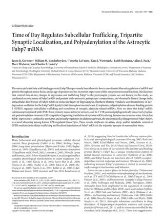
Location ing immunocytochemistry on mouse brain tissue sections. Analysis
Fabp7 tripartite synapse enrichment of nanogold-labeled anti-Fabp7 immunoreactive (IR) protein
The localization of Fabp7 in the mammalian adult CNS is not with EM showed a clear localization of the IR product in the fine
well characterized. One previous study described the decline of glial processes of hippocampal (Fig. 1 B) and cerebellellar tripar-
both Fabp7 and Fabp3 protein abundance within brain synaps- tite synapses (Fig. 1C). Fabp7 IR was not observed in neuronal
tosomal fractions of aged mice (Pu et al., 1999). Because Fabp7 compartments and nanogold labeling was nonspecific in control
knock-out mice have been shown to influence NMDA-receptor- sections lacking primary Fabp7 antibody (Fig. 1 D).
5. Gerstner et al. • Diurnal Fabp7 Post-Transcriptional Processing J. Neurosci., January 25, 2012 • 32(4):1383–1394 • 1387
homogenate was similar to the ratio of Fabp7 at ZT0 and ZT18.
Together, these data show that there are dynamic time-of-day-
regulated changes in the relative levels of Fabp7 mRNA and pro-
tein in synaptic compartments.
Fabp7 mRNA is trafficked in adult hippocampus
Previously, we have shown that time-of-day oscillations in Fabp7
mRNA abundance occur broadly throughout murine brain, in-
cluding various layers of adult murine hippocampus (Gerstner et
al., 2008). Given that Fabp7 protein is enriched in perisynaptic
processes of astrocytes, and that Fabp7 mRNA is increased in the
hippocampus following intense neuronal stimulation (Owada et
al., 2006), we more closely examined circadian changes of Fabp7
mRNA in hippocampus. We specifically determined whether
there is a time-of-day change on the distribution pattern of
mRNA in molecular layers of the hippocampus. A more focal
distribution of Fabp7 mRNA near astroglial somata would sug-
gest recently synthesized transcript from the nucleus, whereas a
more scattered distribution would reflect mRNA trafficking away
from the cell body into the cytoplasm of astrocyte processes. Pre-
viously we reported significant time-of-day changes in Fabp7
mRNA levels in various brain regions of mouse brain (Gerstner et
al., 2006, 2008). Quantification of Fabp7 mRNA by in situ hybrid-
ization using radio-labeled antisense riboprobes on this tissue
Figure 2. Fabp7 mRNA and protein enrichment cycles in the tripartite synapse. A, Analysis of subjected to liquid emulsion in the dentate gyrus of hippocampus
mouse whole-brain mRNA by qPCR shows a dramatic change in the ratio of synaptoneurosomal revealed a significant time-of-day change in mRNA levels with
(S) to total homogenate (H) Fabp7 mRNA compared with GAPDH and -actin mRNA across the peak levels of expression observed at ZT0 and ZT6 (Gerstner et
day. B, Analysis of mouse brain protein also shows a time-of-day cycling of Fabp7 enrichment in al., 2008), in phase with other brain regions (Gerstner et al., 2006,
the ratio of synaptoneurosomal (S) to total homogenate (H). The cycling of the synaptic protein
2008). We next examined the density of silver grains over the
PSD-95 is also observed, but in a different phase to Fabp7. Light bars, light period; dark bars,
dark period. light/dark cycle to determine whether diurnal changes exist in
histological localization for Fabp7 mRNA. Dispersal of silver
grains is greater at the light/dark transition (ZT12), compared
Fabp7 mRNA and protein enrichment cycles in tripartite synapses with a less distributed pattern between ZT0 and ZT6 (Fig. 3 A, B).
Circadian cycling of core clock components and clock-controlled Analysis of silver grain density shows statistically significant dif-
genes are well known in terms of transcriptional autoregulatory ference in mRNA distribution based on time of day (one-way
processing in the nuclear compartment of cells. In contrast, very ANOVA, p ϭ 0.0156; Fig. 3B). The more somatic and punctate
little is known about time-of-day changes of cycling molecules in distribution observed at the ZT0 – 6 time period (Fig. 3A) corre-
other cellular locations, including the synapses in brain. We sponds with times of higher mRNA levels in the hippocampus
therefore examined the localization of Fabp7 mRNA in synap- (Gerstner et al., 2008), likely reflecting localization of recently
toneurosomal fractions over the light/dark cycle. We observed transcribed Fabp7 mRNA in the cell bodies of astrocytes (Gerst-
dramatic time-of-day changes in Fabp7 mRNA in synaptoneuro- ner et al., 2008). Changes in the temporal distribution of Fabp7
somal fractions compared with total homogenate, with higher mRNA were not specific to hippocampus. Similar patterns of
levels in the synaptoneurosome during the dark phase compared Fabp7 mRNA localization were observed in other brain regions,
with the light phase (Fig. 2 A). We also observed diurnal changes such as the cortex, thalamus, amygdala, and hypothalamus (data
in GAPDH and -actin mRNA expression in synaptoneurosomal not shown). Together, these data indicate a coordinated process
fractions compared with total homogenate. However, these dif- in directed Fabp7 mRNA trafficking throughout brain.
ferences did not compare with the ϳ1000-fold time-of-day dif-
ferences observed for Fabp7. We next examined whether time of CPEB machinery
day changes Fabp7 protein levels in synaptneurosomes. We ob- Phylogenetic conservation of CPE sequences in the Fabp7 3ЈUTR
served Ͼ2-fold changes in the relative abundance of Fabp7 pro- CPEs represent one mechanism that controls trafficking of mRNA
tein in synaptoneurosomal fractions compared with total in neurons. The CPEs interact with CPEB1 within neurons follow-
homogenate (Fig. 2 B). Since -actin protein is not as enriched in ing increased activity to regulate experience-dependent local
the synaptoneurosomal fraction as Fabp7 (Fig. 1), and since translation (Wu et al., 1998). CPEB1 has also been shown to
-actin mRNA abundance cycles in the opposite direction as operate on -catenin mRNA in astrocytes to regulate transla-
Fabp7 mRNA (Fig. 2 A), we compared relative Fabp7 protein tion and subsequent migration (Jones et al., 2008). Given the
levels in synaptoneurosomes to the postsynaptic density protein diurnal changes in tripartite synaptic enrichment and traffick-
95 (PSD-95), a member of the membrane-associated guanylate ing observed in Fabp7 mRNA, we first bioinformatically
kinase family that aids in receptor and ion channel clustering at searched for cis-acting elements within the 3ЈUTR that are
synapses. We observed Ͼ4-fold changes in the relative abun- involved in signaling pathways known to influence traffick-
dance of PSD-95 protein in synaptoneurosome versus total ho- ing and synaptic targeting. The consensus CPE sequence
mogenate (Fig. 2 B), with lower levels at lights on (ZT0). PSD-95 UUUUAU and likely similar elements were therefore bioin-
was more enriched in synaptic fractions at ZT6 and ZT12, and the formatically searched in the Fabp7 mRNA 3ЈUTR of various
ratio of its relative abundance in synaptoneurosomal versus total species. Multiple phylogenetically conserved CPEs were ob-
6. 1388 • J. Neurosci., January 25, 2012 • 32(4):1383–1394 Gerstner et al. • Diurnal Fabp7 Post-Transcriptional Processing
Figure 4. CPEB1 binds Fabp7 mRNA in astrocytes. A, Northern blot analysis of various CPEBs
reveals expression from whole mouse brain total RNA (T) or CPEB1 and CPEB4 from cultured
primary astrocyte RNA (A). Enrichment of Fabp7 mRNA in astrocytes is shown as a positive
control. B, Coimmunoprecipitation of Fabp7 mRNA with CPEB1 from astrocyte lysate. Enrich-
ment of Fabp7 and the positive control -2 microglobulin mRNA (IP vs input) compared with
GAPDH mRNA (IP vs input) (t test, ***p Ͻ 0.001). Fabp7 vs GAPDH; -2M vs GAPDH. Data are
mean Ϯ SEM. N ϭ 3/group).
CPEB machinery exists in astrocytes and binds Fabp7 mRNA
Four CPEB proteins have been characterized in adult mamma-
lian brain (Theis et al., 2003), and CPEB1 was previously shown
to be expressed in astrocytes (Jones et al., 2008). To confirm the
presence of CPEB1, and determine whether the other CPEBs exist
in astrocytes, Northern blot analysis of mRNA from cultured
Figure 3. Fabp7 mRNA distribution cycles diurnally in hippocampus. A, Representative cor- murine primary astrocytes was performed. The presence of
onal in situ hybridization hippocampal tissue sections probed for Fabp7 mRNA, subjected to
CPEB1 and CPEB4, but not CPEB2 or CPEB3 mRNA, was iden-
emulsion autoradiography and dark-field microscopy at four diurnal time points. Note the
dispersion in signal observed at ZT12 compared with the other time points. ML, molecular layer; tified in astrocyte culture homogenates (Fig. 4 A). All CPEBs were
GL, granular layer (between large and small dashed lines), PL, precursor layer (on edge of small observed in total RNA from whole brain (Fig. 4 A), confirming
dashed line). B, Analysis of relative mRNA distribution over the light/dark cycle in the molecular previous studies (Theis et al., 2003; Rouhana et al., 2005).
layers of hippocampus reveals a significant time-of-day effect. One-way ANOVA, p ϭ 0.0156; Given murine Fabp7 mRNA contains CPEs, we performed
*p Ͻ 0.05 ZT12 vs ZT0 and 6, post hoc Tukey. Each value represents the mean Ϯ SEM. Light coimmunoprecipitation experiments on lysate from mouse pri-
bars, light period; dark bars, dark period. Emulsion image contrast was inverted for clarity. mary astrocyte culture to assess whether CPEB1 binds to Fabp7
mRNA. This method selectively restricts interactions of CPEB
Table 1. Phylogenetic conservation of CPEs in the 3UTR of Fabp7 mRNA protein with Fabp7 mRNA expressed specifically in astrocytes,
Sequence Nucleotide number Location eliminating the possibility for false-positive interactions from
whole-brain lysate. There was a ϳ12-fold enrichment of Fabp7
CPE-1
DR 590 UGUUUUUUAAUUAU 603 ϭ 56 nt Upstream of hex mRNA in the eluate of anti-CPEB1-conjugated protein-A beads
GG 630 UUUUUUUUAACAGU 643 ϭ 57 nt Upstream of hex compared with input (Fig. 4 B). Previously the CPE-containing
MM 691 UACUUUUUAUAGAA 704 ϭ 56 nt Upstream of hex 2 microglobulin mRNA was identified in a microarray screen
RN 679 UUUUUUUUAUAAAG 692 ϭ 57 nt Upstream of hex search for CPEB1-bound mRNAs from mammalian brain (Du
SS 715 UUUUUUUUAAAUAU 728 ϭ 59 nt Upstream of hex and Richter, 2005). 2 microglobulin is also present in astrocytes
HS 919 UUUUUUUUAUAAAC 932 ϭ 57 nt Upstream of hex (Diedrich et al., 1993; Vanguri, 1995), and therefore we used it as
CPE-2ϩ a positive control for a CPEB-interacting mRNA. QRT-PCR
DR 603 UUAUUUUUAUUUGU 616 ϭ 43 nt Upstream of hex analysis of CPEB1 co-IP showed a ϳ3-fold enrichment of 2
GG 537 UUUUUUUUAAGAAA 550 ϭ 153 nt Upstream of hex microglobulin mRNA compared with input, confirming previ-
MM 680 AUAAUUUUAACAAU 693 ϭ 66 nt Upstream of hex
ous findings of binding to CPEB1 (Du and Richter, 2005). A
MM 669 UUUUUUUUAAAUAA 682 ϭ 77 nt Upstream of hex
RN 655 UUUUUUUUAAUAAC 668 ϭ 81 nt Upstream of hex statistically significant elevation in Fabp7 mRNA compared with
SS 621 CUUGUUUUAAAAAC 634 ϭ 152 nt Upstream of hex 2 microglobulin mRNA co-IP was observed (t test, p Ͻ 0.01),
HS 940 UACAUUUUAUAAUU 953 ϭ 35 nt Upstream of hex suggesting that Fabp7 mRNA binds to CPEB with greater affinity.
HS 879 GUACUUUUAUAAUU 892 ϭ 97 nt Upstream of hex
CPE-1 and CPE-2ϩ are shown within proximity to their nucleotide number and location relative to the polyadenyl- CPE function
ation hexanucleotide (hex) sequence. DR, Danio rerio; GG, Gallus gallus; MM, Mus musculus; RN, Rattus norvigicus; SS, CPE mediates cytoplasmic regulation of Fabp7 mRNA translation
Sus scrofa; HS, Homo sapiens. Since Fabp7 mRNA binds CPEB1 in cultured astrocytes (Fig. 4 B),
we were interested in determining whether the CPEs are func-
tional. To do so, we tested whether the Fabp7 mRNA 3ЈUTR
served in all species examined (Table 1). The relative distance would control translation during Xenopus oocyte maturation.
and loci of the CPEs from the hexanuculeotide (AAUAAA) are Legitimate CPEs from neuronal mRNAs are commonly active in
also conserved in multiple species (Table 1), suggesting func- this assay, enhancing translation and promoting polyadenylation
tional regulatory mechanisms underlying Fabp7 mRNA trans- during oocyte maturation (Wu et al., 1998; Kim and Richter,
lational control conserved across species. 2008; Novoa et al., 2010; Igea and Mendez, 2010).
7. Gerstner et al. • Diurnal Fabp7 Post-Transcriptional Processing J. Neurosci., January 25, 2012 • 32(4):1383–1394 • 1389
RNase H enzyme hydrolizes phosphodiester bonds of RNA hy-
bridized to DNA, and is used for determining polyadenylation
(Goodwin and Rottman, 1992). Using an oligo-dT DNA primer
and hybridizing it to total brain poly(A) RNA, a consistent mo-
bility of digested Fabp7 mRNA products were observed following
digestion in the presence of the RNase H enzyme at both ZT12
and 24 time points in whole-brain homogenates examined by
Northern blotting (data not shown). Closer examination of
RNase H digested poly(A) mRNA products using oligo-dT at
multiple time points confirmed a single predicted Fabp7 mRNA
band at 762 bp (Fig. 7A). Synchronization of this banding pattern
was observed in multiple regions throughout murine brain (Ger-
Figure 5. Translational stimulation by the Fabp7 3ЈUTR. Luciferase activity of progesterone- stner et al., 2006, 2008), including regions involved in circadian
stimulated Xenopus oocytes shows a statistically significant increase in translation for mouse rhythms (suprachiasmatic nucleus) and sleep/wake regulation
Fabp7 3ЈUTR (t test, **p Ͻ 0.01). Cyclin B1 3ЈUTR (positive control) shows a typical activation (ventrolateral preoptic nucleus, tuberomammalary nucleus, lat-
response following progesterone stimulation (t test, ***p Ͻ 0.001). Data are mean Ϯ SEM, eral hypothalamus, locus ceruleus, pons) (Saper et al., 2010). A
normalized to coinjected -gal for control (N ϭ 3/group). comparison of the changes in mRNA level and polyadenylation
shows an increased length in poly(A) tail when the Fabp7 mRNA
is low, during the dark period, at ZT18 (Fig. 7B).
The murine Fabp7 3ЈUTR was subcloned downstream of the
luciferase open-reading frame, and the resulting mRNA in-
jected into Xenopus oocytes, half of which were then induced
Discussion
The results presented here are the first to show that circadian
to mature using progesterone (Rouhana and Wickens, 2007). A
changes in polyadenylation occur in a non-neuronal mRNA in
-galactosidase reporter without the Fabp7 3ЈUTR was coinjected
mammalian brain, and that a diurnally controlled mRNA is post-
for normalization. A 2.4-fold induction of luciferase activity was
transcriptionally regulated by CPEB-mediated cytoplasmic poly-
observed following progesterone stimulation, demonstrating
adenylation. Fabp7 mRNA polyadenylation is mediated, at least
that translational stimulation elements operate on the Fabp7 3Ј
in part, through cis-regulatory elements in the Fabp7 mRNA’s
UTR (Fig. 5). An mRNA carrying the cyclin B1 3ЈUTR served as a
3ЈUTR that are phylogenetically conserved. Further, CPEs in the
positive control, and exhibited translational repression in resting
Fabp7 3ЈUTR are necessary for repression of translation in the
oocytes and translational activation after maturation, as expected
Xenopus oocyte maturation assay and Fabp7 transcript is bound by
(de Moor and Richter, 1999; Barkoff et al., 2000).
CPEB1 in mouse astrocytes. These data provide a novel mechanism
A time course analysis showed a temporal window of lu-
for coordinated CPEB-mediated translational processing of mRNA
ciferase/Fabp7 mRNA 3ЈUTR translation peaking at 7 h follow-
in astrocytes over the circadian cycle in mammalian brain.
ing progesterone stimulation (Fig. 6). Mutations in either the
An aging-dependent decline in Fabp7 protein in synaptoneuro-
CPEs or the cleavage and polyadenylation specificity hexanucle-
somal fractions of mouse brain was previously observed (Pu et al.,
otide, both of which are required for cytoplasmic polyadenyla-
1999). We confirmed that the Fabp7 was present in the fine perisyn-
tion (Fox et al., 1989), blocked the translational stimulation
aptic processes of astrocytes using electron microscopy, and ob-
mediated by the Fabp7 3ЈUTR (Fig. 6). Mutations in the CPEs
served a time-of-day-dependent alteration in Fabp7 mRNA
release repression of translation, leading to an increase in lu-
localization in synaptoneurosomal fractions. Exactly whether an
ciferase activity in unstimulated oocytes compared with wild
aging-dependent decrease in Fabp7 protein in synaptoneurosomal
type. These observations are similar to other validated CPE-
fractions is due to an overall decrease in Fabp7 levels, or some deficit
containing 3ЈUTRs using this assay, such as in the 3ЈUTR of
in subcellular localization and/or polyadenylation mechanisms re-
CPEB4 mRNA (Igea and Mendez, 2010). We conclude that the
mains to be determined. However, since Fabp7 binds the -3 poly-
Fabp7 3ЈUTR possesses functional CPEs.
unsaturated fatty-acid, docosahexaenoic acid (DHA), to high
Fabp7 mRNA poly(A) tail is diurnally regulated in mouse brain affinity (Xu et al., 1996) and DHA has been implicated in Alzhei-
CPE-containing mRNAs are known to regulate polyadenylation mer’s disease and cognitive function (Morley and Banks, 2010), it is
and subsequent translation (Wu et al., 1998; Wells et al., 2001; Du possible that alterations in Fabp7 processing or targeting contributes
and Richter, 2005; Igea and Mendez, 2010). Since Fabp7 mRNA to age-related cognitive decline. This is an intriguing possibility since
binds to CPEB1, is translationally regulated by existing CPEs amyloid-precursor proteins, which are believed to contribute to Alz-
in its 3ЈUTR, and trafficked in astrocytes in a time-of-day- heimer’s disease etiology (Tanzi and Bertram, 2005; Haass and Sel-
dependent manner, a corresponding diurnal change in the koe, 2007), have been shown to act as membrane anchors for the
poly(A) tail can be predicted. To determine whether Fabp7 cytoplasmic translational activity of CPEB (Cao et al., 2005). It is
mRNA has a corresponding change in polyadenylation, we exam- possible that misregulation of CPEB-mediated Fabp7 mRNA post-
ined the pattern of mRNA length over the light/dark cycle. We transcriptional events could contribute to an aging-dependent de-
previously reported that the abundance of Fabp7 mRNA varies cline in cognitive processing and/or neurodegenerative disease
with time of day in multiple brain regions (Gerstner et al., 2006, related to fatty-acid signaling (Di Paolo and Kim, 2011).
2008). Reanalysis of that data revealed that the lengths of the CPEBs are mRNA-binding proteins involved in subcellular
Fabp7 mRNA, and not merely its abundance, vary in each of these trafficking and local translation of targeted mRNAs at synapses
regions of the brain. This is a specific effect, since Fabp5 mRNA (Huang et al., 2003). We confirmed that CPEB1 also exists in
length (data not shown), another Fabp of similar size expressed in astrocytes as described previously (Jones et al., 2008), and char-
CNS glia (Veerkamp and Zimmerman, 2001), does not vary. To acterized functional CPE-sites in the Fabp7 3ЈUTR. However, the
confirm that this change in Fabp7-banding pattern was a result of physiological signals that direct Fabp7 mRNA trafficking and
alterations in poly(A) tail length, we used RNase H analysis. The polyadenylation remain unknown. Since Fabp7 has been shown
8. 1390 • J. Neurosci., January 25, 2012 • 32(4):1383–1394 Gerstner et al. • Diurnal Fabp7 Post-Transcriptional Processing
to regulate cellular outgrowth (Feng et al.,
1994; Arai et al., 2005) and motility (Mita
et al., 2010), CPE-mediated polyadenyla-
tion and targeting of Fabp7 may be in-
volved in regulating astrocytic process
extension in plasticity-related contexts.
It is not yet known, however, whether
changes in neuronal activity and/or syn-
aptic plasticity affect trafficking and
polyadenylation of Fabp7 mRNA, lead-
ing to morphological changes in astro-
cytic processes.
Our results show that Fabp7 mRNA un-
dergoes longer poly(A) extension during a
time window when the base levels of mRNA
are lower. Interestingly, the shortest poly(A)
tail was observed at ZT12, the time point of
greatest mRNA dispersal, suggesting either
dormant or degraded Fabp7 mRNA is di-
rected away from cell bodies at the light-to-
dark transition. Dramatic increases in levels
of Fabp7 heteronuclear RNA transcripts
were previously observed at the dark-to-
light transition (Gerstner et al., 2008), sug-
gesting a major transcriptional event and
reflected by the concentrated somatic local-
ization of silver grain density observed in as-
trocytes during the light period (Fig. 3A).
This suggests that increases in Fabp7
poly(A) tail length at times of lower mRNA
levels are likely not derived from nuclear
transcriptional events per se, but instead are
controlled post-transcriptionally through
mechanisms involving CPEB-mediated
translational activation (see Fig. 8). Protein
kinase activity is thought to direct phos-
phorylation of CPEB and to regulate poly-
adenylation of target transcripts (Richter,
2007). One prediction is that similar kinase
activation of CPEB exists in astrocytes, gated
by time-of-day alterations in neuronal ac- Figure 6. Fabp7 mRNA translation is regulated by CPEs. A, The 3ЈUTR sequence of the murine Fabp7 mRNA is depicted with the
tivity, metabolism, or behavioral state. This CPEs in bold, and the hexanucleotide underlined and in bold. B, Schematic diagram of the Fabp7 mRNA structure, with the
might be expected at light/dark transition sequence and relative locations of wild type (WT), 3XCPE mutant (3XCPEmt), and hexanucleotide mutant (HEXmt). Mutated
times, when animals switch between dor- nucleotides are shown in lower case. C, Firefly luciferase ORF fused to 3ЈUTR of Fabp7 3ЈUTR wild type (Fabp7); Fabp7 3ЈUTR
mant and active states, where sleep/wake 3XCPEmt; and Fabp7 3ЈUTR HEXmt was injected into oocytes, and the percentage of induced luciferase activity is shown at
neurophysiology could signal changes in indicated time points following progesterone stimulation (normalized to baseline unstimulated oocyte controls). Two-way
Fabp7 mRNA perisynaptic localization, ANOVA; effect of genotype with time p Ͻ 0.001; post hoc Bonferroni Fabp7 vs 3XCPEmt and vs HEXmt at 7 h, ***p Ͻ 0.001. Data
polyadenylation, and subsequent transla- are mean Ϯ SEM (N ϭ 4). D, Firefly luciferase ORF fused to 3ЈUTR of wild-type Gld2 (positive control), Fabp7, 3XCPEmt, and HEXmt
tion. Therefore, one prediction would be were injected into oocytes and incubated in the absence (repression) or presence (activation) of progesterone. Luciferase activities
that activity state at specific times of day were determined after 7 h. One-way ANOVA, p Ͻ 0.001; post hoc Bonferroni Fabp7, ***p Ͻ 0.001, and Gld2, **p Ͻ 0.01,
contributes to Fabp7 mRNA trafficking, untreated to progesterone treatment. Data are mean Ϯ SEM (N ϭ 4).
polyadenylation, and protein accumulation
in perisynaptic astroglial processes. Whether CPEB activation fits activity. Fabp7 mRNA increases following neuronal activity
this model (Fig. 8) or state-dependent signaling pathways and ki- within hippocampus following kainate stimulation (Owada et al.,
nases mediate this process remains to be determined. 1996). While mechanisms underlying post-transcriptional pro-
The oscillation in trafficking observed within the hippocam- cessing of clock and clock-controlled genes are beginning to be
pus introduces an additional level of complexity for Fabp7 post- understood (Kojima et al., 2011), to the best of our knowledge, a
transcriptional regulation in astrocytes. Since CPEB is thought to role for CPEB in the regulation of post-transcriptional processing
stimulate trafficking and translation of plasticity-related mRNAs, of diurnally regulated mRNA has not yet been examined. Our
such as ␣CaMKII, to recently activated synapses (Wu et al., 1998; data presented here indicate that astrocytic Fabp7 transcription
Bramham and Wells, 2007; Richter, 2007), it is tempting to spec- and subsequent trafficking to perisynaptic process compart-
ulate that the diurnal trafficking observed for Fabp7 mRNA may ments oscillate based on time of day through a putative CPEB-
be mediated by CPEB through astrocytic responses to neuronal mediated mechanism (Fig. 8).
9. Gerstner et al. • Diurnal Fabp7 Post-Transcriptional Processing J. Neurosci., January 25, 2012 • 32(4):1383–1394 • 1391
Figure 8. Model for time-of-day regulation of Fabp7 mRNA processing in astrocytes. Top,
Figure 7. Diurnal changes in Fabp7 mRNA polyadenylation. A, Northern blot analysis of Summary of findings showing diurnal changes in levels of Fabp7 mRNA, poly(A) tail length, and
RNase H digestion of Fabp7 mRNA in the presence of Oligo-dT shows a single band at various trafficking. Bottom, Peak in Fabp7 transcription occurs near dawn, with moderate polyadenyl-
time points over the light/dark cycle. RNase H in the absence of Oligo-dT reveals the time-of-day ation signal and a somatic distribution, reflecting recently transcribed RNA in the nucleus. A
regulation of Fabp7 mRNA polyadenylation, which is lowest near the light-to-dark transition at gradual decline in transcription and polyadenylation signal continues over the light phase.
ZT12. B, In situ hybridization of Fabp7 mRNA on sagittal sections of mouse brain over the Lowest levels of poly(A) are observed at dusk, at a time of greatest mRNA dispersal. Targeting of
light/dark cycle (top) show broad changes in expression, with lower levels during the dark mRNA is initiated in the dark phase to perisynaptic processes (S) and corresponding induction of
period. Relative changes in Fabp7 mRNA and poly(A) tail are observed out of phase, with mRNA polyadenylation via CPEB (C). CPEB translational activation via phosphorylation (p) may occur
levels highest early in the light period, and poly(A) tail longest toward the end of the dark during this period. Localization at S is higher during this time. Fabp7 mRNA returns to higher
period. OB, olfactory bulb; Ct, cortex; MB, midbrain; HB, hind-brain; Cb, cerebellum. Light bars, levels the following morning, following new transcription. Light bars, light period; dark bars,
light period; dark bars, dark period. dark period.
In addition to the presynaptic and postsynaptic neuronal ter- changes in Fabp7 post-transcriptional processing. Future work
minals, an astroglial process, which surrounds the synapse, is an will determine whether neuronal activity is required for the ob-
active participant in neural transmission and defines a unique served effects on Fabp7 mRNA regulation.
compartment called the tripartite synapse (Halassa et al., 2007, Since we show a time-of-day change in Fabp7 mRNA local-
2009b). Astrocytes, through these perisynaptic processes, are be- ization to synapses and subcellular trafficking in hippocampus,
lieved to both sequester neurotransmitters and release gliotrans- another intriguing possibility is that Fabp7 function at synapses
mitters and factors to modulate synaptic activity (Halassa and may be related to time-of-day-dependent changes observed in
Haydon, 2010). Previously, the glial fibrillary acidic protein synaptic plasticity and memory formation (Gerstner et al., 2009;
(GFAP) mRNA was identified in SN fractions (Rao and Steward, Gerstner and Yin, 2010). DHA is an -3 polyunsaturated fatty-
1993), molecular evidence for the presence of astroglial processes acid known to bind Fabp7 to high affinity (Xu et al., 1996), is
of the tripartite synapse. Glial cells also respond to neuronal ac- abundant in the synaptic compartment (Breckenridge et al.,
tivity. For example, an increase in GFAP expression has been 1972; Salem et al., 2001), regulates synaptic plasticity (Young et
observed in rat hippocampus following electrically induced sei- al., 1998; Itokazu et al., 2000; Fujita et al., 2001; Mirnikjoo et al.,
zures (Steward et al., 1991). A fivefold increase of GFAP mRNA 2001), and influences learning and memory behavior (Moriguchi
was also observed at 24 h following seizure stimulation before et al., 2000; Moriguchi and Salem, 2003; Fedorova et al., 2007,
returning to baseline levels 4 d later (Torre et al., 1993). Interest- 2009). DHA-stimulated NMDA-receptor-dependent current is
ingly, Fabp7 mRNA has a similar hippocampal expression profile blocked in neurons derived from Fabp7 KO mice (Owada et al.,
following kainate injection, where it is elevated 24 h later, reach- 2006), suggesting that Fabp7 localization in tripartite synapses
ing peak expression after 48 h (Owada et al., 1996), a time that influences neurophysiological function. Such Fabp7-dependent
corresponds to peak GFAP protein expression (Torre et al., modulation of synaptic efficacy may in turn regulate memory
1993). Since Fabp7 mRNA is enriched in dendritic layers of processes. This is evident from observations of significant altera-
hippocampus (Zhong et al., 2006), and present in synaptoneuro- tions in later periods of memory behavior in a passive avoidance-
somes (Pu et al., 1999) (Figs. 1, 2), these data suggest that Fabp7- conditioning task in Fabp7 KO mice compared with wild-type
targeted perisynaptic translocation and subsequent translation mice (Owada et al., 2006). Recently we have shown that Fabp7
within astrocytes may have some function related to changes in overexpression in Drosophila leads to changes in daytime sleep
neuronal activation or synaptic homeostasis. We did not observe behavior and increases in memory consolidation (Gerstner et al.,
time-of-day-dependent changes in polyadenylation of the Fabp7 2011a,b). These data implicate a functional role for subcellular
mRNA in primary astrocyte culture (data not shown), suggesting localization of Fabp7 in mediating neural plasticity and time-of-
that the presence of nonastroglial factors are responsible for day-dependent changes in complex behavior.
10. 1392 • J. Neurosci., January 25, 2012 • 32(4):1383–1394 Gerstner et al. • Diurnal Fabp7 Post-Transcriptional Processing
In conclusion, we provide the first evidence of circadian n-3 fatty acid deficiency impairs rat spatial learning in the barnes maze.
changes in polyadenylation for the astrocyte Fabp7 mRNA in Behav Neurosci 123:196 –205.
Feillet CA, Ripperger JA, Magnone MC, Dulloo A, Albrecht U, Challet E
mammalian brain. We characterize poly(A) tail processing and
(2006) Lack of food anticipation in Per2 mutant mice. Curr Biol
translational regulation of Fabp7 mRNA through CPE-sites in its 16:2016 –2022.
3ЈUTR, and describe an interaction with CPEB1 in astrocytes. We Feng L, Hatten ME, Heintz N (1994) Brain lipid-binding protein
show the time-of-day-dependent trafficking of Fabp7 mRNA in (BLBP): a novel signaling system in the developing mammalian CNS.
the mouse hippocampus. We also provide biochemical and ultra- Neuron 12:895–908.
structural evidence of Fabp7 protein in perisynaptic processes of Florian C, Vecsey CG, Halassa MM, Haydon PG, Abel T (2011) Astrocyte-
the tripartite synapse, where mRNA abundance changes logarith- derived adenosine and A1 receptor activity contribute to sleep loss-
induced deficits in hippocampal synaptic plasticity and memory in mice.
mically based on time of day. These changes are novel for known
J Neurosci 31:6956 – 6962.
circadian genes and implicate Fabp7 post-transcriptional pro- Fox CA, Sheets MD, Wickens MP (1989) Poly(A) addition during matura-
cessing in the temporal regulation of the astrocytic component of tion of frog oocytes: Distinct nuclear and cytoplasmic activities and reg-
the tripartite synapse. ulation by the sequence UUUUUAU. Genes Dev 3:2151–2162.
Fujita S, Ikegaya Y, Nishikawa M, Nishiyama N, Matsuki N (2001) Docosa-
hexaenoic acid improves long-term potentiation attenuated by phospho-
References lipase A(2) inhibitor in rat hippocampal slices. Br J Pharmacol
Abarca C, Albrecht U, Spanagel R (2002) Cocaine sensitization and reward 132:1417–1422.
are under the influence of circadian genes and rhythm. Proc Natl Acad Sci Fuller PM, Gooley JJ, Saper CB (2006) Neurobiology of the sleep-wake
U S A 99:9026 –9030. cycle: sleep architecture, circadian regulation, and regulatory feed-
Andretic R, Chaney S, Hirsh J (1999) Requirement of circadian genes for back. J Biol Rhythms 21:482– 493.
cocaine sensitization in Drosophila. Science 285:1066 –1068. Garcia JA, Zhang D, Estill SJ, Michnoff C, Rutter J, Reick M, Scott K, Diaz-
Arai Y, Funatsu N, Numayama-Tsuruta K, Nomura T, Nakamura S, Osumi N Arrastia R, McKnight SL (2000) Impaired cued and contextual memory
(2005) Role of Fabp7, a downstream gene of Pax6, in the maintenance of in NPAS2-deficient mice. Science 288:2226 –2230.
neuroepithelial cells during early embryonic development of the rat cor- Gerstner JR, Landry CF (2007) The zinc-binding protein chordc1 under-
tex. J Neurosci 25:9752–9761. goes complex diurnal changes in mRNA expression during mouse brain
Ballantyne S, Daniel DL Jr, Wickens M (1997) A dependent pathway of development. Neurochem Res 32:241–250.
cytoplasmic polyadenylation reactions linked to cell cycle control by
Gerstner JR, Lyons LC, Wright KP Jr, Loh DH, Rawashdeh O, Eckel-Mahan
c-mos and CDK1 activation. Mol Biol Cell 8:1633–1648.
KL, Roman GW (2009) Cycling behavior and memory formation.
Barkoff AF, Dickson KS, Gray NK, Wickens M (2000) Translational control
J Neurosci 29:12824 –12830.
of cyclin B1 mRNA during meiotic maturation: coordinated repression
Gerstner JR, Yin JC (2010) Circadian rhythms and memory formation. Nat
and cytoplasmic polyadenylation. Dev Biol 220:97–109.
Rev Neurosci 11:577–588.
Bracchi-Ricard V, Brambilla R, Levenson J, Hu WH, Bramwell A, Sweatt JD,
Gerstner JR, Vander Heyden WM, Lavaute TM, Landry CF (2006) Profiles
Green EJ, Bethea JR (2008) Astroglial nuclear factor-kappaB regulates
of novel diurnally regulated genes in mouse hypothalamus: expression
learning and memory and synaptic plasticity in female mice. J Neurochem
analysis of the cysteine and histidine-rich domain-containing, zinc-
104:611– 623.
binding protein 1, the fatty acid-binding protein 7 and the GTPase, ras-
Bramham CR, Wells DG (2007) Dendritic mRNA: transport, translation
like family member 11b. Neuroscience 139:1435–1448.
and function. Nat Rev Neurosci 8:776 –789.
Gerstner JR, Bremer QZ, Vander Heyden WM, Lavaute TM, Yin JC, Landry
Breckenridge WC, Gombos G, Morgan IG (1972) The lipid composition of
CF (2008) Brain fatty acid binding protein (Fabp7) is diurnally regu-
adult rat brain synaptosomal plasma membranes. Biochim Biophys Acta
266:695–707. lated in astrocytes and hippocampal granule cell precursors in adult ro-
Brown V, Jin P, Ceman S, Darnell JC, O’Donnell WT, Tenenbaum SA, Jin X, dent brain. PLoS One 3:e1631.
Feng Y, Wilkinson KD, Keene JD, Darnell RB, Warren ST (2001) Mi- Gerstner JR, Vanderheyden WM, Shaw PJ, Landry CF, Yin JC (2011a) Fatty-
croarray identification of FMRP-associated brain mRNAs and altered acid binding proteins modulate sleep and enhance long-term memory
mRNA translational profiles in fragile X syndrome. Cell 107:477– 487. consolidation in Drosophila. PLoS One 6:e15890.
Cao Q, Huang YS, Kan MC, Richter JD (2005) Amyloid precursor proteins Gerstner JR, Vanderheyden WM, Shaw PJ, Landry CF, Yin JC (2011b) Cy-
anchor CPEB to membranes and promote polyadenylation-induced toplasmic to nuclear localization of fatty-acid binding protein correlates
translation. Mol Cell Biol 25:10930 –10939. with specific forms of long-term memory in Drosophila. Commun Integr
Chaudhury D, Colwell CS (2002) Circadian modulation of learning and Biol 4:623– 626.
memory in fear-conditioned mice. Behav Brain Res 133:95–108. Gibbs ME, Hertz L (2008) Inhibition of astrocytic energy metabolism by
Chaudhury D, Wang LM, Colwell CS (2005) Circadian regulation of hip- D-lactate exposure impairs memory. Neurochem Int 52:1012–1018.
pocampal long-term potentiation. J Biol Rhythms 20:225–236. Gibbs ME, Anderson DG, Hertz L (2006) Inhibition of glycogenolysis in
de Moor CH, Richter JD (1999) Cytoplasmic polyadenylation elements me- astrocytes interrupts memory consolidation in young chickens. Glia
diate masking and unmasking of cyclin B1 mRNA. EMBO J 54:214 –222.
18:2294 –2303. Goodwin EC, Rottman FM (1992) The use of RNase H and poly(A) junc-
Diedrich JF, Carp RI, Haase AT (1993) Increased expression of heat shock tion oligonucleotides in the analysis of in vitro polyadenylation reaction
protein, transferrin, and beta 2-microglobulin in astrocytes during products. Nucleic Acids Res 20:916.
scrapie. Microb Pathog 15:1– 6. Gray NK, Coller JM, Dickson KS, Wickens M (2000) Multiple portions of
Di Paolo G, Kim TW (2011) Linking lipids to Alzheimer’s disease: choles- poly(A)-binding protein stimulate translation in vivo. EMBO J
terol and beyond. Nat Rev Neurosci 12:284 –296. 19:4723– 4733.
Du L, Richter JD (2005) Activity-dependent polyadenylation in neurons. Haass C, Selkoe DJ (2007) Soluble protein oligomers in neurodegeneration:
RNA 11:1340 –1347. Lessons from the Alzheimer’s amyloid beta-peptide. Nat Rev Mol Cell
Dudley CA, Erbel-Sieler C, Estill SJ, Reick M, Franken P, Pitts S, McKnight SL Biol 8:101–112.
(2003) Altered patterns of sleep and behavioral adaptability in NPAS2- Halassa MM, Haydon PG (2010) Integrated brain circuits: astrocytic net-
deficient mice. Science 301:379 –383. works modulate neuronal activity and behavior. Annu Rev Physiol
Eckel-Mahan KL, Storm DR (2009) Circadian rhythms and memory: not so 72:335–355.
simple as cogs and gears. EMBO Rep 10:584 –591. Halassa MM, Fellin T, Haydon PG (2007) The tripartite synapse: roles for
Fedorova I, Hussein N, Di Martino C, Moriguchi T, Hoshiba J, Majchrzak S, gliotransmission in health and disease. Trends Mol Med 13:54 – 63.
Salem N Jr (2007) An n-3 fatty acid deficient diet affects mouse spatial Halassa MM, Florian C, Fellin T, Munoz JR, Lee SY, Abel T, Haydon PG,
learning in the Barnes circular maze. Prostaglandins Leukot Essent Fatty Frank MG (2009a) Astrocytic modulation of sleep homeostasis and cog-
Acids 77:269 –277. nitive consequences of sleep loss. Neuron 61:213–219.
Fedorova I, Hussein N, Baumann MH, Di Martino C, Salem N Jr (2009) An Halassa MM, Fellin T, Haydon PG (2009b) Tripartite synapses: roles for
11. Gerstner et al. • Diurnal Fabp7 Post-Transcriptional Processing J. Neurosci., January 25, 2012 • 32(4):1383–1394 • 1393
astrocytic purines in the control of synaptic physiology and behavior. Sasaki H, Kato H, Saino-Saito S, Matsumoto N, Akaike N, Noda T, Kondo
Neuropharmacology 57:343–346. H (2006) Altered emotional behavioral responses in mice lacking brain-
Harris KM, Teyler TJ (1983) Age differences in a circadian influence on type fatty acid-binding protein gene. Eur J Neurosci 24:175–187.
hippocampal LTP. Brain Res 261:69 –73. Panatier A, Theodosis DT, Mothet JP, Touquet B, Pollegioni L, Poulain DA,
Herzog ED (2007) Neurons and networks in daily rhythms. Nat Rev Neu- Oliet SH (2006) Glia-derived D-serine controls NMDA receptor activity
rosci 8:790 – 802. and synaptic memory. Cell 125:775–784.
Huang YS, Carson JH, Barbarese E, Richter JD (2003) Facilitation of den- Pascual O, Casper KB, Kubera C, Zhang J, Revilla-Sanchez R, Sul JY, Takano
dritic mRNA transport by CPEB. Genes Dev 17:638 – 653. H, Moss SJ, McCarthy K, Haydon PG (2005) Astrocytic purinergic sig-
Igea A, Mendez R (2010) Meiosis requires a translational positive loop
´ naling coordinates synaptic networks. Science 310:113–116.
where CPEB1 ensues its replacement by CPEB4. EMBO J 29:2182–2193. Paxinos G, Franklin KBJ (2003) The mouse brain in stereotaxic coordinates,
Itokazu N, Ikegaya Y, Nishikawa M, Matsuki N (2000) Bidirectional actions compact Ed 2. Oxford: Elsevier.
of docosahexaenoic acid on hippocampal neurotransmissions in vivo. Pu L, Igbavboa U, Wood WG, Roths JB, Kier AB, Spener F, Schroeder F
Brain Res 862:211–216. (1999) Expression of fatty acid binding proteins is altered in aged mouse
Jones KJ, Korb E, Kundel MA, Kochanek AR, Kabraji S, McEvoy M, Shin CY, brain. Mol Cell Biochem 198:69 –78.
Wells DG (2008) CPEB1 regulates beta-catenin mRNA translation and Raghavan AV, Horowitz JM, Fuller CA (1999) Diurnal modulation of
cell migration in astrocytes. Glia 56:1401–1413. long-term potentiation in the hamster hippocampal slice. Brain Res
Kim JH, Richter JD (2008) Measuring CPEB-mediated cytoplasmic 833:311–314.
polyadenylation-deadenylation in xenopus laevis oocytes and egg ex- Rao A, Steward O (1993) Evaluation of RNAs present in synaptodendro-
tracts. Methods Enzymol 448:119 –138. somes: dendritic, glial, and neuronal cell body contribution. J Neurochem
Kojima S, Shingle DL, Green CB (2011) Post-transcriptional control of cir- 61:835– 844.
cadian rhythms. J Cell Sci 124:311–320. Richter JD (2007) CPEB: a life in translation. Trends Biochem Sci
Kondratova AA, Dubrovsky YV, Antoch MP, Kondratov RV (2010) Circa- 32:279 –285.
dian clock proteins control adaptation to novel environment and mem- Roth TL, Sweatt JD (2008) Rhythms of memory. Nat Neurosci 11:993–994.
ory formation. Aging 2:285–297. Rouhana L, Wickens M (2007) Autoregulation of GLD-2 cytoplasmic
Li X, Sankrithi N, Davis FC (2002) Transforming growth factor-alpha is poly(A) polymerase. RNA 13:188 –199.
expressed in astrocytes of the suprachiasmatic nucleus in hamster: Role of Rouhana L, Wang L, Buter N, Kwak JE, Schiltz CA, Gonzalez T, Kelley AE,
glial cells in circadian clocks. NeuroReport 13:2143–2147. Landry CF, Wickens M (2005) Vertebrate GLD2 poly(A) polymerases in
Lyons LC, Roman G (2009) Circadian modulation of short-term memory the germline and the brain. RNA 11:1117–1130.
in Drosophila. Learn Mem 16:19 –27. Sakai T, Tamura T, Kitamoto T, Kidokoro Y (2004) A clock gene, period,
Lyons LC, Rawashdeh O, Katzoff A, Susswein AJ, Eskin A (2005) Circadian plays a key role in long-term memory formation in Drosophila. Proc Natl
modulation of complex learning in diurnal and nocturnal aplysia. Proc Acad Sci U S A 101:16058 –16063.
Natl Acad Sci U S A 102:12589 –12594. Salem N Jr, Litman B, Kim HY, Gawrisch K (2001) Mechanisms of action of
Lyons LC, Rawashdeh O, Eskin A (2006a) Non-ocular circadian oscillators docosahexaenoic acid in the nervous system. Lipids 36:945–959.
and photoreceptors modulate long term memory formation in aplysia. Saper CB (2006) Staying awake for dinner: hypothalamic integration of
J Biol Rhythms 21:245–255. sleep, feeding, and circadian rhythms. Prog Brain Res 153:243–252.
Lyons LC, Collado MS, Khabour O, Green CL, Eskin A (2006b) The circa- Saper CB, Fuller PM, Pedersen NP, Lu J, Scammell TE (2010) Sleep state
dian clock modulates core steps in long-term memory formation in aply- switching. Neuron 68:1023–1042.
sia. J Neurosci 26:8662– 8671. Sarov-Blat L, So WV, Liu L, Rosbash M (2000) The Drosophila takeout gene
Marpegan L, Swanstrom AE, Chung K, Simon T, Haydon PG, Khan SK, Liu is a novel molecular link between circadian rhythms and feeding behav-
AC, Herzog ED, Beaule C (2011) Circadian regulation of ATP release in
´ ior. Cell 101:647– 656.
astrocytes. J Neurosci 31:8342– 8350. Stellwagen D, Malenka RC (2006) Synaptic scaling mediated by glial TNF-
Masri S, Sassone-Corsi P (2010) Plasticity and specificity of the circadian alpha. Nature 440:1054 –1059.
epigenome. Nat Neurosci 13:1324 –1329. Steward O, Torre ER, Tomasulo R, Lothman E (1991) Neuronal activity
McClung CA, Sidiropoulou K, Vitaterna M, Takahashi JS, White FJ, Cooper up-regulates astroglial gene expression. Proc Natl Acad Sci U S A
DC, Nestler EJ (2005) Regulation of dopaminergic transmission and co- 88:6819 – 6823.
caine reward by the clock gene. Proc Natl Acad Sci U S A 102:9377–9381. Su G, Kintner DB, Flagella M, Shull GE, Sun D (2002) Astrocytes from
Mirnikjoo B, Brown SE, Kim HF, Marangell LB, Sweatt JD, Weeber EJ (2001) Na(ϩ)-K(ϩ)-Cl(Ϫ) cotransporter-null mice exhibit absence of swelling
Protein kinase inhibition by omega-3 fatty acids. J Biol Chem and decrease in EAA release. Am J Physiol Cell Physiol 282:C1147– 60.
276:10888 –10896. Suh J, Jackson FR (2007) Drosophila ebony activity is required in glia for the
Mita R, Beaulieu MJ, Field C, Godbout R (2010) Brain fatty acid-binding circadian regulation of locomotor activity. Neuron 55:435– 447.
protein and omega-3/omega-6 fatty acids: mechanistic insight into ma- Suzuki A, Stern SA, Bozdagi O, Huntley GW, Walker RH, Magistretti PJ,
lignant glioma cell migration. J Biol Chem 285:37005–37015. Alberini CM (2011) Astrocyte-neuron lactate transport is required for
Moriguchi T, Greiner RS, Salem N Jr (2000) Behavioral deficits associated long-term memory formation. Cell 144:810 – 823.
with dietary induction of decreased brain docosahexaenoic acid concen- Tanzi RE, Bertram L (2005) Twenty years of the Alzheimer’s disease amy-
tration. J Neurochem 75:2563–2573. loid hypothesis: a genetic perspective. Cell 120:545–555.
Moriguchi T, Salem N Jr (2003) Recovery of brain docosahexaenoate leads Theis M, Si K, Kandel ER (2003) Two previously undescribed members of
to recovery of spatial task performance. J Neurochem 87:297–309. the mouse CPEB family of genes and their inducible expression in the
Morley JE, Banks WA (2010) Lipids and cognition. J Alzheimers Dis principal cell layers of the hippocampus. Proc Natl Acad Sci U S A
20:737–747. 100:9602–9607.
Nishiyama H, Knopfel T, Endo S, Itohara S (2002) Glial protein S100B Torre ER, Lothman E, Steward O (1993) Glial response to neuronal activity:
modulates long-term neuronal synaptic plasticity. Proc Natl Acad Sci GFAP-mRNA and protein levels are transiently increased in the hip-
U S A 99:4037– 4042. pocampus after seizures. Brain Res 631:256 –264.
Novoa I, Gallego J, Ferreira PG, Mendez R (2010) Mitotic cell-cycle progres- Turek FW, Joshu C, Kohsaka A, Lin E, Ivanova G, McDearmon E, Laposky A,
sion is regulated by CPEB1 and CPEB4-dependent translational control. Losee-Olson S, Easton A, Jensen DR, Eckel RH, Takahashi JS, Bass J
Nat Cell Biol 12:447– 456. (2005) Obesity and metabolic syndrome in circadian clock mutant mice.
Owada Y, Yoshimoto T, Kondo H (1996) Increased expression of the Science 308:1043–1045.
mRNA for brain- and skin-type but not heart-type fatty acid binding Turrigiano GG (2006) More than a sidekick: glia and homeostatic synaptic
proteins following kainic acid systemic administration in the hippocam- plasticity. Trends Mol Med 12:458 – 460.
pal glia of adult rats. Brain Res Mol Brain Res 42:156 –160. Vanguri P (1995) Interferon-gamma-inducible genes in primary glial cells
Owada Y, Abdelwahab SA, Kitanaka N, Sakagami H, Takano H, Sugitani Y, of the central nervous system: comparisons of astrocytes with microglia
Sugawara M, Kawashima H, Kiso Y, Mobarakeh JI, Yanai K, Kaneko K, and Lewis with brown Norway rats. J Neuroimmunol 56:35– 43.
![1384 • J. Neurosci., January 25, 2012 • 32(4):1383–1394 Gerstner et al. • Diurnal Fabp7 Post-Transcriptional Processing
cytic cellular and molecular processes contribute significantly to min with 0.1% Triton X-100 in PBS followed by three 10 min rinses in
linking behavioral and physiological processes. PBS. The slices were then blocked for nonspecific antibody binding with
Fabp7 is enriched near synapses. Its abundance in synap- Aurion goat-blocking agent (Cat. #25596, containing 5% BSA, 0.1% cold
toneurosomal fractions declines with age (Pu et al., 1999). We water fish gelatin, and 5% normal goat serum in PBS, pH 7.4) for 30 min.
The slices were then rinsed three times for 10 min each in incubation
found Fabp7 abundance cycles in synaptoneurosomes, suggesting
buffer containing PBS ϩ 0.1% BSA-C (Aurion Immuno Gold Reagents,
daily variations in trafficking machinery. One potential mechanism Cat. #25558). Finally, the samples were incubated in a mixture of incu-
is through mRNA-binding proteins, such as cytoplasmic polyade- bation buffer and rabbit primary Fabp7 antibody (Millipore) at a dilu-
nylation element (CPE)-binding protein (CPEB). One such protein, tion of 1:1000 overnight at 4°C. The following day, the slices were rinsed
CPEB1, is present in astrocytes (Jones et al., 2008), but its role in in incubation buffer every 10 min for 2 h (12 rinses total), and then
targeting mRNAs to perisynaptic processes in astrocytes has not incubated overnight in a 1:100 dilution of ultrasmall gold conjugate
been explored. CPE-mediated mechanisms exist for trafficking and [F(abЈ) 2 fragment of goat-anti-rabbit IgG (heavy and light chains), Au-
local translation of neuronal mRNAs, so we conducted a bioinfor- rion Immuno Gold Reagents, Cat. #25360] at 4°C. After the overnight
matic search that revealed phylogenetically conserved CPE se- incubation, the slices were rinsed in incubation buffer 6ϫ for 10 min
quences in Fabp7 mRNA. We hypothesized that Fabp7 CPEs are each, and then rinsed in PBS 6ϫ for 5 min each. The slices were postfixed
in 2% glutaraldehyde in 0.1 M PB for 30 min and rinsed 2ϫ for 5 min each
functional and able to mediate translation, predicting Fabp7 mRNA
in PB.
could undergo time-of-day subcellular translocation and polyade- The use of ultrasmall gold conjugates requires a silver-enhancing step
nylation. Our novel findings confirm that, in addition to neurons, to increase the size of the subnanometer gold particles to more easily
cytoplasmic polyadenylation processes exist for astrocytic CPE- visualize the particles by transmission electron microscopy. After post-
containing mRNA (Jones et al., 2008) and establish the existence of fixation, the slices were rinsed 3ϫ for 10 min each in Enhancing Condi-
coordinated control for mRNA trafficking and subsequent transla- tioning Solution (ECS) (Aurion). Next, the slices were developed in
tion at the perisynaptic astrocytic process compartment across the Silver Enhancement Solution (Aurion) for 1.25 h. To terminate silver
day. enhancement, the slices were exposed to a solution of 0.3 M sodium
thiosulfate in ECS for 5 min. The slices were then rinsed twice in 10 min
intervals in ECS and finally processed routinely for electron microscopy.
Materials and Methods After silver enhancing, the slices were fixed in a solution of 0.5% os-
Subjects and handling mium tetroxide in 0.1 M PB for 30 min. Following OsO4 postfixation, the
Male mice (C57BL/6) were either purchased directly from Harlan samples were dehydrated in a graded ethanol series, then further dehy-
Sprague Dawley (adult) or taken at specific ages from a breeding colony. drated in propylene oxide and flat-embedded in Epon epoxy resin. The
Animals were entrained to a 12 h light, 12 h dark (12:12) schedule for a specific regions used for the analysis were trimmed from the flat-
minimum of 2 weeks. Zeitgeber time (ZT) 0 was lights-on, and ZT12 was embedded sections and flat-mounted on blank epoxy resin stubs. Excess
lights-off, on this diurnal schedule. For tissue collection, animals were epoxy resin was then sectioned away in 1 m increments with a Diatome
killed by decapitation, their brains dissected, flash frozen at Ϫ30°C in Histo diamond knife using a Reichert–Jung Ultracut-E ultramicrotome
2-methylbutane, and stored at Ϫ80°C. For tissue punches of specific until the surface of the tissue was exposed. Samples were then sectioned
regions, brains were embedded in optimal cutting temperature media using a Diatome Ultra 45 diamond knife with the same microtome and
and sectioned on a Leica (CM3050) cryostat at Ϫ25°C as previously contrasted with Reynolds lead citrate and 8% uranyl acetate in 50%
described (Gerstner et al., 2006, 2008). Punches from the same brain EtOH. Ultrathin sections were observed with a Philips CM120 electron
region were pooled from each of the animals within the same group, and microscope and images were captured with a MegaView III side-
stored at Ϫ80°C until RNA isolation. All animal care and use procedures mounted digital camera.
were in strict accordance with University of Wisconsin Institutional An-
imal Care and Use Committee and National Institutes of Health guide-
Northern blot analysis
lines for the humane treatment of laboratory animals. Northern blotting was performed as described previously (Gerstner et al.,
Synaptoneurosome preparation 2006). Briefly, brains were dissected and total RNA was isolated using
Synaptoneurosomes (SNs) were prepared from mouse whole brain as TRIzol (Invitrogen), according to the manufacturer’s specifications, and
described previously (Westmark and Malter, 2007; Westmark et al., stored at Ϫ80°C. Before loading, each sample was incubated with sample
2011). Briefly, mice were asphyxiated with carbon dioxide followed by buffer [7.5% formaldehyde, 43% formamide, 12% 10ϫ 3-(N-
removal of the brain. Brains were washed in ice-cold gradient medium morpholino)propanesulfonic acid, 0.12% ethidium bromide] for 5 min
(GM) buffer (0.25 M sucrose, 5 mM Tris, pH 7.5, and 0.1 mM EDTA), at 56°C, and then cooled on ice for 5 min. Northern blots consisted of 1
transferred to a glass Dounce homogenizer containing ice-cold GM buf- g of total RNA per lane and were electrophoresed on a 1.2% agarose gel.
fer, and gently homogenized with five strokes of the loose pestle followed RNA was transferred overnight onto a sheet of GeneScreen Plus nitrocel-
by five strokes of the tight pestle. The homogenate was spun at 1000 g for lulose (NEN Life Science Products). DNA templates generated by PCR
10 min at 4°C in round-bottom tubes to pellet cellular debris and nuclei. were labeled with 32P using the Megaprime DNA labeling system (GE
The supernatant (2 ml of aliquots) was applied to Percoll gradients (lay- Healthcare). The following primers were used: Fabp7 forward primer,
ers, 2 ml each of 23%, 15%, 10%, and 3% isomotic Percoll) and spun at AGACCCGAGTTCCTCCAGTT, reverse primer, CCTCCACACCGA
speed (32,500 g) for 5 min at 4°C. The third band from the top of the AGACAAAC, Cpeb1 forward primer, CATCTTGGGACCTTCTTGGA,
gradient (23%–15% interface) containing intact SNs was removed and Cpeb1 reverse primer, AAGACCCAAGGGATTACC, Cpeb2 forward
pooled for the experiments. primer, GGTCGCTCTTCCCTATTTCC, Cpeb2 reverse primer, TCCA
AAGGCTGAGAACCATC, Cpeb3 forward primer, CCCTTCTCCAGC
Immunocytochemistry and electron microscopy AATGTGAT, Cpeb3 reverse primer, CTCGTTCCCCATTTTGACAT,
Animals were perfused through the heart with 0.1% heparin in 0.01 M Cpeb4 forward primer, CCTCACTGCTTCACTCACCA, Cpeb4 reverse
PBS, pH 7.4, until liver cleared, followed by 2% paraformaldehyde, 1% primer, AACAAAGCGGGCACTGATAG. Probes were mixed with hy-
glutaraldehyde in 0.1 M phosphate buffer (PB), pH 7.4. Sections were cut brisol and incubated overnight at 42°C. Blots were washed 3ϫ with 100
at 50 m on a vibratome and stored in 0.01 M PBS and 0.01% azide ml of 2ϫ SSC, 1% SDS, at room temperature, then twice for 30 min each
solutions until processing. The hippocampal section was prepared from with 100 ml of 2 ϫ SSC, 1% SDS, at 50°C, and finally once for 30 min with
a mouse killed at ZT12. 100 ml of 0.5 ϫ SSC, 1% SDS, at 60°C. Following washes, blots were
Following fixation, the slices were rinsed in 0.1 M PB and then treated exposed to a phosphoscreen for 3 d and image analysis was performed
with freshly prepared 0.1% sodium borohydride in 0.1 M PB for 10 min to using the Storm 860 and ImageQuant 5.2 software (Molecular Dynam-
quench unbound aldehydes. Next, the slices were permeabilized for 30 ics). A 983 bp PCR product for glyceraldehyde-3-phosphate dehydroge-](data:image/gif;base64,R0lGODlhAQABAIAAAAAAAP///yH5BAEAAAAALAAAAAABAAEAAAIBRAA7)