

Recomendados
Más contenido relacionado
La actualidad más candente
La actualidad más candente (20)
Similar a Medoralv9supplip58
Similar a Medoralv9supplip58 (20)
Medoralv9supplip58
- 1. 52 Infecciones orofaciales / Orofacial infections Patología pulpar y periapical / Pulp and periapical disease RESUMEN En la actualidad, gran parte de los tratamientos que se realizan en la clínica son debidos a patologías que afectan a la pulpa y al periápice. La pulpa es un tejido ricamente vascularizado e inervado, de- limitado por un entorno inextensible como es la dentina, con una circulación sanguínea terminal y con una zona de acceso circulatorio –periápice– de pequeño calibre. Todo ello, hace que la capacidad defensiva del tejido pulpar sea muy limitada ante las diversas agresiones que pueda sufrir. El tejido pulpar también puede ser afectado por una infección retrógrada (1), a partir de los canalículos secundarios, desde el ligamento periodontal o desde el ápice durante un proceso de periodontitis. Debido a que la patología periapical va casi siempre precedida de una afectación de la pulpa, describiremos en primer lugar las causas (2) de enfermedad pulpar y a continuación las causas de la patología periapical. De dicha etiología dependerá la evolución y la clasificación de estas patologías. Analizaremos la necrosis pulpar y la degeneración pulpar que pueden desencadenar una periodontitis apical reversible o una periodontitis apical irreversible. Palabras clave: Pulpitis, periodontitis, patología periapical, clasificación de pulpitis, clasificación de patología periapical, necrosis pulpar. AFECTACION PULPAR Etiología Infecciones Las infecciones producidas por microorganismos anaerobios y bacterias gramnegativas (3) son una de las causas más impor- tantes que pueden afectar a la pulpa. Kakehashi y cols. (4) confirmaron la importancia de estos microorganismos como causantes de dichas patologías. Esta infección puede llegar a la pulpa a través de la corona o de la raíz del diente. Las caries, las fisuras o fracturas y los defectos del desarrollo dentario son las causas más frecuentes de infec- ción a través de la corona. Por la raíz son las caries del cuello, las bolsas periodontales y las bacteriemias. Algunos autores (5) citan la pulpitis por anacoresis y explican que las bacterias pueden circular a través del torrente sanguíneo y colonizar zonas donde, gracias a un irritante físico o mecánico, está facilitada la inflamación pulpar. Otras causas Traumatismos: agudos, como las luxaciones, fisuras y fracturas; crónicos como el bruxismo y la abrasión, o bien iatrogénicos como los movimientos ortodóncicos, preparación de cavidades o tallados dentarios. Cambios bruscos de temperatura con generación de calor. El uso de instrumental rotatorio sin refrigeración adecuada, materiales como la godiva, gutapercha caliente o el fraguado de acrílicos, generan un calor excesivo que puede producir daño pulpar. Las grandes restauraciones metálicas, que transmiten intensamente los cambios de temperatura, sobre todo el frío, pueden llegar fácilmente a la pulpa sin una protección entre la obturación y la misma produciendo dolor; si el estímulo es prolongado e intenso, provoca una pulpitis; los cambios térmicos moderados pueden estimular la formación de dentina de reparación. Electrogalvanismo: la presencia en el medio bucal de restaura- ciones con distintos metales puede producir descargas eléctricas con la consiguiente afectación de la pulpa. Variaciones bruscas de presión: en las que se produce una libe- ración de burbujas de gas nitrógeno de la sangre, dando lugar a las barodontalgias. Radiaciones: en pacientes bajo tratamiento de radioterapia por tumoraciones de cabeza y cuello. Toxicidad de los materiales de obturación: cada vez menos frecuente debido a su mayor biocompatibilidad. Cuando se produce daño pulpar por los materiales de obturación es debido a un mal sellado o a la filtración marginal. Intoxicaciones: en ciertas enfermedades como la diabetes, gota Etiología, clasificación y patogenia de la patología pulpar y periapical Joaquín F. López Marcos (1) (1) Profesor Asociado. Facultad de Medicina y Odontología. Universidad de Salamanca Correspondencia: Dr. D. Joaquín Francisco López Marcos Facultad de Medicina y Odontología Universidad de Salamanca Avda. Campo Charro s/n 37007 Salamanca 923-29 44 00 E-mail: jflmarcos@usal.es López-Marcos JF. Etiología, clasificación y patogenia de la patología pulpar y periapical. Med Oral Patol Oral Cir Bucal 2004;9 Suppl:S52-62. © Medicina Oral S. L. C.I.F. B 96689336 - ISSN 1137 - 2834 Indexed in: -Index Medicus / MEDLINE / PubMed -EMBASE, Excerpta Medica -Indice Médico Español -IBECS
- 2. 53 Med Oral Patol Oral Cir Bucal 2004;9 Suppl:52-62. Patología pulpar y periapical / Pulp and periapical disease o nefropatías se puede producir intoxicaciones endógenas que pueden afectar a la pulpa.Algo similar sucede en las intoxicacio- nes de carácter exógeno producidas por mercurio o plomo. Fisiológicas: las que ocurren con el envejecimiento. Idiopáticas: en las cuales no se encuentra causa conocida. Estas dos últimas causas son las fundamentales en los procesos degenerativos pulpares. AFECTACION PERIAPICAL Etiología Marcada por traumatismos dentarios que afecten tanto a la corona como a la raíz del diente. Asimismo, las alteraciones oclusales como bruxismo, sobrecarga oclusal y maloclusiones pueden desencadenar daño periapical. También la patología pulpar en forma de pulpitis y necrosis produce alteración periapical, además de la etiología iatrogénica debido a la so- breinstrumentación o sobreobturación en los tratamientos de conductos radiculares. CLASIFICACION Se han preconizado muchas y distintas clasificaciones por dis- tintos autores (6-9) para las patologías pulpar y periapical, según su etiología, anatomía patológica o sus manifestaciones clínicas, pero, como afirma Lasala (10), casi todas eran clasificaciones histopatológicas, que no son prácticas para la aplicación clínica y el establecimiento de una terapéutica racional. Cohen y Burns (11) opinan que desde una visión más global, la pulpa se clasifica fundamentalmente como enferma o sana y, atendiendo a criterios de tratamiento adecuados, debe decidirse si ha de extirparse o no. Estableceremos una clasificación siguiendo a Pumarola y Ca- nalda (12), basada en la de Walton y Torabinejad (13), en la que diferenciaremos entre pulpitis, necrosis y procesos degenerativos pulpares (Tabla 1). Los procesos patológicos periapicales se clasifican en la Tabla 2. PATOGENIA Debido a las diversas causas que producen una patología pulpar y periapical, el proceso patogénico básico que se desarrolla es el de la respuesta inflamatoria. La pulpa va a reaccionar originando una pulpitis, inflamación que ocurre como respuesta a mecanismos directos e inmunitarios. Los mecanismos directos son los microorganismos, los cuáles llegan a la pulpa a través de los túbulos dentinarios expuestos, ya sea por caries, traumatismos o factores irritantes (productos bacterianos, bacterias, endotoxinas, etc.), que al penetrar a tra- vés de los túbulos dentinarios, destruyen el odontoblasto y las células subyacentes (14). En los mecanismos inmunitarios actúan factores del comple- mento e inmunoglobulinas. El resultado final, ya sea inducido por irritación directa o por el sistema inmunitario, hace que se liberen mediadores químicos que inician la inflamación. La respuesta inicial a nivel vascular va a ser una rápida vaso- constricción seguida de una vasodilatación casi inmediata con enlentecimiento del flujo sanguíneo, acúmulo de hematíes en el centro del vaso y emigración de los leucocitos a la periferia (Gráfico 1), pegándose a la pared del vaso (15). Esto hace que aparezcan pequeñas fisuras en el endotelio de los vasos, a través de las cuales se produce una extravasación plasmática hacia los espacios de tejido conectivo, dando lugar a un edema que pro- duce una elevación en la presión local y que es el responsable de la compresión de las terminaciones nerviosas originando el dolor (Gráfico 2). El resultado final de la inflamación va a ser un infiltrado de linfocitos, macrófagos y células plasmáticas. En la fase aguda de la inflamación, se produce una exudación como respuesta de los tejidos pulpar y periapical ante cualquier agresión, con predominio de los PMN neutrófilos. Al llegar a la fase crónica la respuesta del huésped es proliferativa, en un intento del tejido pulpar y periapical de reparar la lesión, con la formación de nuevas células, vasos y fibras, que sería lo que se denomina tejido de granulación. EVOLUCION DE LA PULPITIS Y LA PATOLO- GIA PERIAPICAL Según el tipo, la pulpitis y la patología periapical evolucionan de distinta manera (Tabla 1 y 2). Pulpitis reversibles En las pulpitis reversibles, en los casos de hipersensibilidad (16), la pulpa se encuentra vital pero inflamada (con predominio crónico), y con capacidad de repararse una vez que se elimine el factor irritante. Los cambios inflamatorios que ocurren (17) son: vasodilatación, congestión, estasia, trombosis, aglomeración de leucocitos dentro de los vasos sanguíneos, edema, ruptura de los vasos y hemorragia local. Ocurre por factores externos que pueden dar lugar a un proceso inflamatorio reversible de la pulpa, siempre y cuando eliminemos estos factores de agresión. Entre ellos encontramos tallados dentarios generalmente con finalidad protésica, túbulos dentinarios expuestos, heridas pulpares produ- cidas por maniobras iatrogénicas, microfiltración por mal sellado de los materiales de obturación y caries poco profundas. Pulpitis irreversibles En las pulpitis irreversibles la pulpa se encuentra vital, inflama- da, pero sin capacidad de recuperación, aún cuando se hayan eliminado los estímulos externos que provocan el estado infla- matorio. Generalmente son debidas a una pulpitis reversible no tratada. Las bacterias alcanzan la pulpa (1) y allí se asientan, estableciendo formas sintomáticas y asintomáticas. La reacción inicial de la pulpa es la liberación de mediadores químicos de la inflamación. Se forma entonces un edema intersticial que va a incrementar la presión intrapulpar, comprimiendo las fibras nerviosas, y dando lugar a un dolor muy intenso, espontáneo y provocado. Si el edema encuentra salida a través de los tú- bulos dentinarios, da lugar a formas asintomáticas, que serán sintomáticas en el momento en que ocurra la obstrucción de la cavidad, ya sea por impacto de alimentos, o por una restauración realizada sin un correcto diagnóstico. En las formas serosas prevalece el exudado inflamatorio, mien- tras que en las formas purulentas hay aumento de pus, debido a los leucocitos que han llegado a resolver la inflamación (1) Pulpitis irreversible asintomática Se desarrolla a partir de una pulpitis sintomática no tratada en la que ha cedido la fase aguda o en la que los estímulos externos
- 3. 54 Infecciones orofaciales / Orofacial infections Patología pulpar y periapical / Pulp and periapical disease Periodontitis Apical Reversible HIPEREMIAAPICAL Periodontitis Sintomática Serosa PURULENTA Apical irreversible Asintomática GRANULOMATOSA SUPURADA OSTEOESCLEROSIS APICAL Tabla 2. Clasificación de la patología periapical PULPITIS REVERSIBLES HIPERSENSIBILIDAD HERIDA PULPAR (Iatrogénica) IRREVERSIBLES SINTOMÁTICAS SEROSA PURULENTA ASINTOMÁTICAS HIPERPLÁSICA (Pólipo pulpar) ULCERADA NECROSIS PARCIAL ASÉPTICA SÉPTICA TOTAL ASÉPTICA SÉPTICA DEGENERACIONES PULPARES ATRÓFICA CALCIFICACIÓN REABSORCIÓN DENTINARIA INTERNA OTRAS GRASA HIALINA FIBROSA METAPLASIA Tabla 1. Clasificación de la patología pulpar
- 4. 55 Med Oral Patol Oral Cir Bucal 2004;9 Suppl:52-62. Patología pulpar y periapical / Pulp and periapical disease múltiples focos de calcificación, y una degeneración laminar o pulpolitos (Figura 3) perfectamente delimitados que se desarro- llan con mayor frecuencia en la cámara pulpar. Reabsorción dentinaria interna Es producida por la acción de los odontoclastos, existiendo una pulpa vital generalmente inflamada. Puede aparecer a cualquier nivel de la cámara pulpar o de la pulpa radicular. Las causas pueden ser idiopáticas, infecciosas y traumáticas (especialmente pulpotomía), siendo los traumatismos o la pulpitis crónica per- sistente los responsables de la formación de odontoclastos, los cuales van reabsorbiendo la dentina que rodea la pulpa dando una imagen radiolúcida en la radiografía. Degeneraciones grasa, hialina y fibrosa En las degeneraciones grasa, hialina y fibrosa hay depósitos de tejido adiposo, amiloideo y fibroso en la pulpa debido a alteraciones del metabolismo y la permeabilidad del tejido pulpar. En algunas avitaminosis pueden aparecer procesos de metaplasia con aparición de tejido osteoide en el interior del tejido pulpar. Una vez que se ha llegado a la fase de necrosis pulpar, las bac- terias y sus productos de degradación pueden llegar a través del ápice a los tejidos periapicales (18), en donde mientras no haya absceso o periodontitis serosa, el proceso puede permanecer asintomático durante largos periodos de tiempo (1). Periodontitis apical serosa En la periodontitis apical serosa (aguda) se produce una infla- mación de los tejidos periapicales dando lugar a una hiperemia y vasodilatación con exudado de líquido e infiltración leucocitaria, que aumenta la presión tisular estimulando a los osteoclastos que producenunareabsorciónósea.Elexudadoylainfiltracióncelular distienden las fibras del ligamento periodontal originando dolor. Periodontitis apical purulenta Si la enfermedad continúa, las bacterias y sus productos de degradación llegan al periápice y se produce una periodontitis apical purulenta, dando lugar a un absceso primario en primera instancia y secundario cuando se debe a una exacerbación del mismo por fallo en las defensas o la llegada de nuevos gérmenes (1). Si el proceso se mantiene, hay un equilibrio con las defensas del huésped, estableciéndose las formas crónicas. En la forma granulomatosa (Figuras 4 y 5) existen células inflamatorias cró- nicas, tejido de granulación y restos de tejido epitelial recubiertos de una cápsula periférica. Hay actividad osteoclástica que da lugar a una reabsorción ósea apical. A partir del tejido de granulación se puede desarrollar una cavidad llena de líquido, revestida de epitelio y con una cápsula fibrosa que sería el denominado quiste apical. En la forma supurada hay un acúmulo purulento que busca una vía de drenaje, a través de un trayecto fistuloso, a los tejidos blandos. Si en algún momento se produce la obliteración de la fístula, se va a producir la agudización del proceso, apareciendo el denominado absceso fénix. Osteosclerosis apical La osteosclerosis apical es una periodontitis apical irreversible, asintomática y crónica en la que se produce un aumento de la densidad ósea por estimulación osteoclástica, siendo frecuente en pacientes jóvenes. son leves o moderados, pero mantenidos en el tiempo, debido a un equilibrio entre las bacterias y las defensas, dado que las células de defensa son capaces de neutralizar la agresión bacte- riana y hacer que permanezca asintomática. A veces se abre un drenaje hacia el exterior por una comunicación entre la cavidad pulpar y la lesión cariosa (Figura 1), produciéndose un drenaje espontáneo del exudado seroso y evitando así la formación de edema intrapulpar. La forma ulcerada se observa en el fondo de una caries abierta al exterior y está caracterizada por la formación de una ulce- ración en la superficie de una pulpa expuesta. Puede ocurrir a cualquier edad y es capaz de resistir un proceso infeccioso de escasa intensidad, pero que de un modo crónico grave (16) puede progresar sin síntomas clínicos a una necrosis. La pulpitis hiperplásica también denominada pólipo pulpar (Figu- ra 2), puede ocurrir en pacientes jóvenes, con una cámara pulpar grande y una amplia cavidad de caries. La pulpa crece a través del orificio carioso, dando lugar a una masa exofítica, granuloma- tosa, de color rosa-rojizo y de consistencia fibrosa (12). El tejido hiperplásico es tejido de granulación formado por fibras de tejido conectivo entremezcladas con numerosos capilares. Necrosis pulpar Es la descomposición séptica o no (aséptica), del tejido con- juntivo pulpar que cursa con la destrucción del sistema micro- vascular y linfático de las células y, en última instancia, de las fibras nerviosas (12). Se observa un drenaje insuficiente de los líquidos inflamatorios debido a la falta de circulación colateral y la rigidez de las paredes de la dentina, originando un aumento de la presión de los tejidos y dando lugar a una destrucción progresiva hasta que toda la pulpa se necrosa. La necrosis pulpar se puede originar por cualquier causa que dañe la pulpa. La flora microbiana presente en las pulpitis irreversibles asintomáticas, de respiración aerobia y anaerobia facultativa, se va transformando en un medio de respiración anaerobia estricta a medida que disminuye el potencial de óxido- reducción hístico lo que, al dificultar los procesos fagocíticos, facilita el desarrollo y multiplicación microbiana, especialmente de bacterias anaerobias. Las bacterias gramnegativas anaerobias estrictas tienen una elevada capacidad proteolítica y colagenolíti- ca, por lo que contribuyen en gran medida a la desestructuración del tejido conjuntivo pulpar. En los procesos degenerativos pulpares, la atrofia pulpar (dege- neración atrófica) se produce lentamente con el avance de los años, considerándose fisiológica en la edad senil (10), aunque también pueden ser secundarias a traumatismos, alteraciones oclusales, caries e inflamaciones pulpares y periodontales. Hay un incremento en la cantidad de fibras colágenas pulpares y una disminución en el número de células. Calcificación pulpar La calcificación pulpar (degeneración cálcica) se produce por el depósito de sales cálcicas, acumuladas con mayor frecuencia en el tercio apical, aumentando su incidencia con la edad. Hay que distinguir entre la calcificación fisiológica que progresivamente va disminuyendo el volumen pulpar con la edad dental, de la calcificación patológica como respuesta reactiva pulpar ante un traumatismo o ante un proceso destructivo como la caries o la abrasión. Hay una degeneración cálcica difusa, si existen
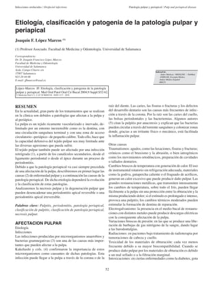
- 5. 56 Infecciones orofaciales / Orofacial infections Patología pulpar y periapical / Pulp and periapical disease Fig. 2. Pulpitis hiperplásica o pólipo pulpar. Hyperplastic pulpitis or polyp of the pulp. Fig. 3. Pulpolito en conducto radicular distal de un molar inferior. Pulp stone in distal root canal in a mandibular molar. Fig. 1. Comunicación de la cámara pulpar con el exterior. Pulp chamber communication with the outside.
- 6. 57 Med Oral Patol Oral Cir Bucal 2004;9 Suppl:52-62. Patología pulpar y periapical / Pulp and periapical disease Fig. 5. Granuloma periapical. Periapical granuloma. Fig. 4. Imagen radiolúcida periapical. Periapical radiolucent image.
- 7. 58 Infecciones orofaciales / Orofacial infections Patología pulpar y periapical / Pulp and periapical disease Aetiology, classification and pathogenesis of pulp and pe- riapical disease LÓPEZ-MARCOS JF.AETIOLOGY, CLASSIFICATION AND PATHOGENESIS OF PULP AND PERIAPICAL DISEASE. MED ORAL PATOL ORAL CIR BUCAL 2004;9 SUPPL:S52-62. ABSTRACT At present, the majority of the treatments that are performed in the clinic are due to disease entities involving the dental pulp and periapex. Dental pulp is a richly vascularized and innervated tissue, enclo- sed by surrounding tissues that are incapable of expanding, such as dentin. It has terminal blood flow and small-gauge circula- tory access – the periapex. All of these characteristics severely constrain the defensive capacity of the pulp tissue when faced with the different aggressions it may be subjected to. Pulp tissue can also be affected by a retrograde infection (1), arising from the secondary canaliculi, from the periodontal liga- ment or from the apex during the course of periodontitis. Due to the fact that periapical disease is almost inevitably preceded by pulp disease, we shall begin by describing the causes (2) of pulp disease and will then proceed to a discussion of the causes of periapical disease. The course of illness and classification of these pathological entities will depend on the aetiology involved. We will analyse pulp necrosis and pulp degeneration that are capable of triggering reversible apical periodontitis or irrever- sible apical periodontitis. Keywords: Pulpitis, periodontitis, periapical pathology, clas- sification of pulpitis, classification of periapical disease, pulp necrosis. PULP INVOLVEMENT Aetiology Infections Infections that are caused by anaerobic microorganisms and gram-negative bacteria (3) are one of the leading causes that affect the dental pulp. Kakehashi et al. (4) confirmed the importance of these microor- ganisms as causative agents in these disease entities. These types of infection can reach the pulp through the crown or by means of the root of the tooth. Caries, cracks or fractures and dental developmental defects are the most common causes of infection entering through the crown. If we are referring to the root as the point of entry, we must make specific mention of periodontal pockets and bacteremia. Some authors (5) cite anachoresis-indu- ced pulpitis and explain that the bacteria can circulate throughout the bloodstream and colonise areas where, thanks to a physical or mechanical irritant, pulp inflammation occurs. Other Causes of Pulp Disease Trauma:Acute trauma, such as dislocations, cracks and fractures, chronic trauma, for instance bruxism and abrasion, or iatrogenic injury, that can occur while performing orthodontic manoeuvres, while preparing dental cavities or carvings can produce dental pulp disease. Abrupt changes of temperature with heat generation are ano- ther important cause. The use of rotary instrumentation without proper cooling, materials such as godiva, hot gutta-percha or the setting of acrylic material all generate excessive heat that can cause pulp injury. Large metal restorations that are intense transmitters of temperature changes, especially cold, can easily reach the pulp if there is no protection placed between the ob- turation and the pulp; this in turn, causes pain. If the stimulus is prolonged and intense, pulpitis results; moderate temperature changes can promote the formation of dentin repair tissue. Electrogalvanism: The presence of restorations made using different metals in the oral environment can produce electrical charges with the consequent pulp involvement. Abrupt pressure variations: Pressure variations in which ni- trogen gas bubbles are released into the bloodstream lead to barodontalgia. Radiation: Radiation can cause pulp disease in patients under- going radiation therapy for tumours located in the head and neck. Toxicity of filling materials: This cause of pulp pathology is less and less frequent due to the higher degree of biocompatibility of the materials used in obturation. Whenever pulp damage occurs as a result of these materials, it is due to a poor seal or marginal filtration. Poisoning: Certain diseases such as diabetes, gout or kidney disease can cause endogenous intoxications that can affect the pulp. Something similar occurs in exogenous mercury or lead poisoning. Physiological causes: Physiological causes in this case are the ones that occur during the aging process. Idiopathic causes: These are the cases in which no known cause can be determined. The last two aetiological classes are the leading causes of pulp degenerative illnesses. PERIAPICAL INVOLVEMENT Aetiology The aetiology of periapical involvement is marked by dental trauma that affects both the crown and the root of the tooth. Likewise, occlusal alterations such as bruxism, excessive oc- clusal loading and malocclusions can trigger periapical injury. Pulp pathology in the form of pulpitis and necrosis also provokes periapical alteration, in addition to iatrogenic aetiology due to the excessive use of dental instrumentation or to overfilling during root canal treatments. CLASSIFICATION Different authors (6-9) have advocated many different classi- fication systems for pulp and periapical pathological entities based on aetiology, pathology or clinical manifestations, but, as Lasala has stated (10), almost all of them were histopathological classifications, which are not convenient for clinical practice or for when establishing a rational treatment plan.
- 8. 59 Med Oral Patol Oral Cir Bucal 2004;9 Suppl:52-62. Patología pulpar y periapical / Pulp and periapical disease Cohen and Burns (11) are of the opinion that from more global standpoint, pulp is basically classified as diseased or healthy and, by applying proper therapeutic criteria, the decision is made to excise or not. We will elaborate a classification system following Pumarola and Canaldaʼs work (12) that is based on Walton and Torabinejadʼs classification (13), in which we will distinguish between pulpitis, necrosis and degenerative pulp conditions (Table 1). Periapical pathological conditions are classified in Table 2. PATHOGENESIS Because of the numerous causes that produce pulp and periapical disease, the fundamental pathogenic condition that develops is an inflammatory response. The dental pulp reacts by developing pulpitis, an inflammation that occurs in response to direct and immune mechanisms. The direct mechanisms are the microorganisms that gain access to the pulp by means of the dentinary tubules that have become exposed, as a result of dental caries, trauma or irritants (bacte Reversible Apical Periodontitis APICAL HYPEREMIA Periodontitis Symptomatic Serous PURULENT IRREVERSIBLE APICAL ASYMPTOMATIC GRANULOMATOUS SUPPURATED APICAL OSTEOESCLEROSIS Table 2. Classification of periapical pathological conditions Table I. Classification of pulp pathological conditions PULPITIS REVERSIBLE HYPERSENSITIVITY PULP (Iatrogenic) IRREVERSIBLE SYMPTOMATIC SEROUS PURULENT ASYMTOMATIC HYPERPLASTIC (Pulp polyps) ULCERATED NECROSIS PARTIAL ASEPTIC SEPTIC TOTAL ASEPTIC SEPTIC PULP DEGENERATION ATROPHIC CALCIFICATION INTERNAL DENTIN RESORPTION OTHER FATTY HYALINE FIBROUS METAPLASIA
- 9. 60 Infecciones orofaciales / Orofacial infections Patología pulpar y periapical / Pulp and periapical disease . © L.Dguez Extravasación plasmática Plasma extravasation DOLOR PAIN NERVIO NERVE Edema Oedema Gráfico 2. Debido a la extravasación plasmática, el plasma escapa a los espacios tisulares donde produce edema que incrementa la presión y la compresión de las fibras nerviosas originando dolor. Plasma extravasation lets plasma into the surrounding tissue where resulting oedema increases pressure on nerve fibres and causes pain. Leucocitos Leucocytes Hematies Erythrocyte © L.Dguez Gráfico 1. Respuesta inicial a nivel vascular, durante el proceso de inflamación. Contracción transitoria de la microcirculación, seguida de una inmediata vasodilatación. Los hematíes migran al centro del vaso y los leucocitos a la periferia. Se produce un agrietamiento de las paredes del vaso debido a la contracción de las células endo- teliales bajo la influencia de la histamina. Vascular initial response, during inflammation. Temporary contraction of the microcirculation system, followed by an immediate vasodilatation. Erythrocytes migrate to the center of the vessel and leucocytes to the perifery. Hystamine induced contraction of the endothelium causes the apparition of cracks on the vessel walls.
- 10. 61 Med Oral Patol Oral Cir Bucal 2004;9 Suppl:52-62. Patología pulpar y periapical / Pulp and periapical disease rial products, bacteria, endotoxins, etc.) that penetrate through the dentinary tubules, destroying odontoblasts and underlying cells (14). On the other hand, complement factors and immunoglobulins act as immune mechanisms. The end result, whether induced by direct irritation or by the immune system, is that chemical mediating factors are released that set off the inflammation. The first response on a vascular level will be rapid vasocons- triction followed almost immediately by vasodilatation, slowing down the local blood flow, leading in turn, to an accumulation of red blood cells in the centre of the vessel and leukocyte emigration to peripheral areas (graph 1), adhering to the blood vessel wall (15). This leads to the formation of small cracks in the endothelium of the vessels, by means of which plasma is extravasated towards the connective tissue spaces; in turn, this leads to oedema which increases local pressure and compres- ses nerve endings, thereby causing pain (graph 2). The final consequence of the inflammatory process will be an infiltrate containing lymphocytes, macrophages and plasma cells. In the acute stage of the inflammation, exudation occurs as a response of the pulp and periapical tissues to any kind of aggression, with a predominance of PMN neutrophils. Once the inflammation has reached the chronic stage, the host responds by means of proliferation in an attempt made by the dental pulp and periapical tissue to repair the lesion, forming new cells, vessels and fibres. This is known as granulation tissue. EVOLUTION OF PULPITIS AND PERIAPICAL PATHOLOGY Depending on the type, pulpitis and periapical pathology evolve in different ways (Table 1 and 2). Reversible Pulpitis In reversible pulpitis, in cases of hypersensitivity (16), the pulp is still vital, albeit inflamed (predominantly chronic pulpitis) and has the capacity to self-repair once the irritant has been eliminated. The inflammatory changes that occur are (17): vessel dilatation, congestion, stasis, thrombosis, and agglomeration of leukocytes inside the blood vessels, oedema, blood vessel rupture and local haemorrhage. This occurs due to external factors that trigger an inflammatory condition in the pulp that is reversible, provided that we eliminate these factors of aggression. These factors of aggression include dental carving, generally for prosthetic purposes, exposed dentinary tubules, pulp injury caused by iatrogenic manipulations, microfiltration resulting from improper sealing of the materials of obturation and shallow caries. Irreversible Pulpitis In irreversible pulpitis, the pulp is vital, inflamed, but now lacks the ability to repair itself, even when the external stimuli that are responsible for producing the inflammatory state have been eliminated. Irreversible pulpitis is typically due to untreated reversible pulpitis. Bacteria reach the pulp (1) and take up resi- dence there, establishing symptomatic and asymptomatic forms of the disease. The pulp initially reacts by secreting chemical inflammation mediators. Interstitial oedema then develops that will increase pressure inside the dental pulp, compressing nerve fibres, and causing intense pain, both spontaneous and provoked. If the oedema finds a way out through the dentinary tubules, asymptomatic forms of the disease develop that will become symptomatic as soon as the cavity becomes obstructed, either by impacted food or by dental restoration techniques performed without a correct diagnosis. Inflammatory exudates comprise the main feature of the serous forms of pulpitis, whereas the purulent forms present increased amounts of pus, due to the leukocytes that have arrived on the scene to resolve the inflammation (1). Irreversible Asymptomatic Pulpitis Irreversible asymptomatic pulpitis evolves from untreated symptomatic pulpitis in which the acute phase has ceded or in which the external stimuli are mild or moderate, although they are sustained over time. A balance is struck between the bacteria and host defences, since the defence cells are capable of neutralising the bacterial aggression and cause the illness to remain asymptomatic. Sometimes drainage to the exterior is opened up by means of a communication between the pulp chamber and the caries-induced lesion (Figure 1), leading to the spontaneous drainage of the serous exudates and thereby preventing intrapulp oedema from developing. The ulcerated form of the illness is observed in the fundus of a caries that is open to the exterior, the most salient feature of which is ulceration on the surface of the exposed pulp. Ulce- rated pulpitis can occur at any age and is capable of resisting a low-grade infection, although it can also progress in a chronic and severe fashion to necrosis without presenting any clinical symptoms (16). Hyperplastic pulpitis, also known as pulp polyps (Figure 2) can develop in young patients, with a large pulp chamber and a large caries-produced cavity. The pulp grows through the caries opening, giving rise to an exophytic, granulomatous, pinkish- reddish mass with a fibrous consistency (12). The hyperplastic tissue is actually granulation tissue comprised of connective tissue fibres mixed in with numerous capillary vessels. Pulp Necrosis Pulp necrosis is the septic or non-septic (i.e., aseptic) decom- position of the pulp connective tissue that courses with the des- truction of the microvascular and lymphatic systems of the cells and ultimately, nerve fibres are also destroyed (12). Inadequate drainage of inflammatory fluids due to the lack of collateral circulation and the rigidity of the dentin walls is observed. This leads to increased pressure on the tissues and causes progressive destruction until the entire dental pulp is necrotised. Pulp necrosis can derive from any cause of pulp insult. The microbial flora that presents in irreversible asymptomatic pul- pitis, with aerobic respiration and facultative anaerobes, gra- dually becomes an environment of strict anaerobic respiration as the tissue oxygen reduction potential decreases, which, by impeding phagocytic conditions, fosters microbial proliferation and multiplication, especially of anaerobic bacteria. The strict anaerobic gram-negative bacteria have a tremendous proteolytic and collagenolytic capacity, which represent a huge contribution to the de-structuring of the pulp connective tissue. In pulp degenerative conditions, the pulp atrophy (atrophic degeneration) takes place slowly over the course of time and is considered to be a physiological situation associated with
- 11. 62 Infecciones orofaciales / Orofacial infections Patología pulpar y periapical / Pulp and periapical disease advanced aging (10), although it may also be secondary to trauma, occlusal alterations, caries and pulp and periodontal inflammations. The amount of pulp collagen fibres is increased, while the number of cells is decreased. Pulp Calcification Pulp calcification (calcific degeneration) is the result of calcium salt deposits that accumulate most frequently on the apical third of the tooth, with increasing incidence associated with age. Dis- tinction must be made between the physiological calcification that progressively decreases pulp volume with dental age and the pathological calcification that represents a reactive pulp response to trauma or in the presence of a destructive situation such as dental caries or abrasion. Diffuse calcific degeneration refers to a condition in which there are multiple sites of calci- fication and laminar degeneration or perfectly defined dental pulp calcifications (Figure 3) that occur more commonly inside the pulp chamber. Internal Dentin Resorption Internal dentin resorption is caused by the action of odonto- clasts that presents generalized inflammation of vital pulp. It can occur at any level of the pulp chamber or pulp root. It may be due to idiopathic or infectious causes or trauma (especially pulpotomy); trauma or persistent chronic pulpitis are respon- sible for odontoclast formation. These odontoclasts re-absorb the dentin surrounding the pulp and producing a radiolucent image on x-ray. Fatty, Hyaline and Fibrous Degenerations In fatty, hyaline and fibrous degenerations adipose tissue, amyloid and fibrous deposits are formed in the dental pulp as a consequence of metabolic disorders and alterations in the per- meability of the pulp tissue. The deficiency of certain vitamins may lead to metaplasia with the appearance of osteoid tissue inside the pulp tissue. Once the pathology has reached the stage of pulp necrosis, the bacteria and its products of degradation can gain access to periapical tissues through the apex of the tooth (18). Once in the periapical tissue, the condition can remain asymptomatic for prolonged periods of time as long as there is no abscess or serous periodontitis (1). Serous Apical Periodontitis In (acute) serous apical periodontitis periapical tissues become inflamed, causing hyperaemia and vasodilatation with exudate of liquid and leukocyte infiltration, which increases tissue pres- sure, thereby stimulating the osteoclasts to reabsorb bone tissue. The exudate and cell infiltration cause the periodontal ligament fibres to distend, thereby causing pain. Purulent Apical Periodontitis If the disease is left to follow its natural history, the bacteria and their products of degradation reach the periapex and puru- lent apical periodontitis results, causing a primary abscess or a secondary abscess when it is due to exacerbation of a primary abscess when the host defences fail or if new germs arrive (1). If the condition persists, a balance is struck with the host defences, thereby establishing chronic forms of the disease. In the granulomatous form of the illness (Figures 4 and 5) chro- nic inflammatory cells, granulation tissue and epithelial tissue remains covered by a peripheral capsule are present. Osteo- clastic activity takes place leading to apical bone resorption. The so-called apical cyst develops from granulation tissue in which a fluid-filled cavity forms that is lined with epithelium and contained within a fibrous capsule. The suppurative form presents a purulent accumulation searching for a drainage outlet to the soft tissues through a fistulous tract. If at some point in time, the fistula is closed, the condition will become acute and the so-called phoenix abscess will develop. Apical Osteosclerosis Apical osteosclerosis is actually an asymptomatic, chronic, irreversible apical periodontitis, in which there is an increase in bone density due to osteoclastic stimulation. It is more com- mon in young patients. BIBLIOGRAFIA/REFERENCES 1. García JA. Infecciones de origen odontógeno. En: Bascones A, Perea EJ. Infecciones orofaciales. Madrid: Ed. Denstisnet.com; 2003. p. 165-81. 2. Azabal M. Patología pulpar y periapical. En: García Barbero J. Patología y terapéutica dental. Madrid: Ed. Síntesis; 2000. p. 240-1. 3. Bascones A, Manso F. Infecciones orofaciales. Diagnóstico y tratamiento. Madrid: Ed. Avances médicos-dentales; 1994. p. 30-44. 4. Kakehashi S, Stanley HR, Fitzgerald RJ. The effects of surgical exposure of dental pulps in germ-free and conventional laboratory rats. Oral Surg Oral Med Oral Pathol1965;20:340-9. 5. Seltzer S, Bender IB, Nazimov H. Differential diagnosis of pulp conditions. Oral Surg Oral Med Oral Pathol 1965;19:383-91. 6. Hess JC. Conceptions nouvelles de pathologie et de therapeutique pulpaires. Rev Fran Odont Stomat 1967;14:61-84. 7. Pheulpin JL. Les inflamations pulpaires: leurs diagnostic clinique et histopathologuique. Schweiz Monatsschr Zahnheilkd 1967;77:701-28. 8. Grossman LI, Oliet S, Del Río C. Endodontic practice. Philadelphia: Ed. Lea & Febiger; 1988. p. 65. 9. Ogilvie A. Histology of the dental pulp. En: Ingle IJ, ed. Endodontics. Phila- delphia: Ed. Lea & Febiger; 1965. p. 295-300. 10. Lasala A. Endodoncia. Madrid: Ed. Masson-Salvat Odontología; 1992. p. 69-104. 11. Cohen S, Burns RC. Vías de la pulpa. Madrid: Ed. Harcourt-Mosby; 1999, p. 17-9. 12. Pumarola J, Canalda C. Patología de la pulpa y del periápice. En: Canalda C, Brau E, eds. Endodoncia. Madrid: Ed. Masson; 2001. Cap. 6. 13. Walton RE, Torabinejad M. Principles and practice of endodontics. Phila- delphia: Ed. WB Saunders; 1996. Cap. 16. 14. Simon J, Walton R, Pashley D, Dowden W y Bakland L. Patosis Pulpar. En: Ingle J, Bakland L, eds. Endodoncia. Madrid: Ed. McGraw-Hill Interamericana; 1996. Cap. 7. 15. Simon J. Patología periapical. En: Cohen S, Burns RC. Vías de la pulpa. Madrid: Ed. Harcourt-Mosby; 1999. p. 410-38. 16. Smulson MH, Sieraski SM. Histofisiología y alteraciones de la pulpa dental. En: Weine F. Tratamiento endodóntico. Madrid: Ed. Hartcourt-Brace; 1997. p. 84-161. 17.Baume LJ. Diagnosis of diseases of the pulp. Oral Surg Oral Med Oral Pathol. 1970;29:102-16. 18. Walton RE, Johnson, WT. En Walton, RE y Torabinejad M. Principles and Practice of Endodontics. Philadelphia: Ed. Saunders; 2002. p. 240-67. AGRADECIMIENTOS Agradezco a Laura López Domínguez, alumna de la Escuela de NN. y BB.Artes de San Eloy (Salamanca), su colaboración en la realización de los gráficos del presente trabajo. ACKNOWLEDGEMENTS I would like to express my appreciation to Laura López Domínguez, a student at the Escuela de NN. y BB. Artes de San Eloy (Salamanca) for her collaboration in creating the graphs for this paper.