SlideShare una empresa de Scribd logo
1 de 67
Descargar para leer sin conexión
D R Y O G E S H R A T H O D
D E P T O F A N E S T H E S I A
K E M H O S P I T A L
M U M B A I
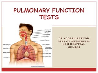
PULMONARY FUNCTION
TESTS
PULMONARY FUNCTION TESTS
 Includes a wide variety of Objective tests to assess
lung function.
 Provides Standardized measurements for assessing
the presence and severity of Respiratory
dysfunction.
GOALS
 To predict the presence of pulmonary dysfunction
 To know the functional nature of disease
(Obstructive or Restrictive)
 To assess the severity and progression of disease
 To identify patients at Perioperative risk of
Pulmonary complications
INDICATIONS OF PFT IN PAC
TISI GUIDELINES FOR PREOPERATIVE SPIROMETRY
 Age > 70 yrs.
 Morbid obesity
 Thoracic surgery
 Upper abdominal surgery
 Smoking history and cough
 Any pulmonary disease
INDICATIONS FOR PREOPERATIVE
SPIROMETRY
 ACP GUIDELINES FOR PREOPERATIVE
SPIROMETRY
 Lung resection
 H/o smoking, dyspnoea
 Cardiac surgery
 Upper abdominal surgery
 Lower abdominal surgery
 Uncharacterized pulmonary disease
(defined as history of pulmonary Disease or symptoms
and no PFT in last 60 days)
Lung Volumes and Capacities
 Four Lung volumes:
Tidal volume
Inspiratory reserve volume
Expiratory reserve volume
Residual volume
 Five capacities:
Inspiratory capacity
Expiratory capacity
Vital capacity
Functional residual capacity
Total lung capacity
Addition of 2 or more Volumes comprises a Capacity.
TIDAL VOLUME (TV)
 Volume of air inhaled/
exhaled in each breath
during quite respiration.
 N – ~6-8 ml/kg.
 TV FALLS WITH-
1. Decrease in compliance
2. Decreased ventilatory
muscle strength
INSPIRATORY RESERVE VOLUME (IRV)
 Maximum volume of air
which can be Inspired
after a Normal Tidal
inspiration i.e. from end
inspiration point.
 N- 1900 ml- 3300 ml.
EXPIRATORY RESERVE VOLUME (ERV)
 Maximum Volume of air
which can be expired
after a normal tidal
expiration i.e.
from end expiration
point
 N- 700 ml – 1000 ml
RESIDUAL VOLUME (RV)
 Volume of air remaining in
the lungs after a maximum
expiration.
 N- 20-25ml/kg
(1700- 2100 ml)
 Indirectly measured-
FRC-ERV
 Cannot be measured by
Spirometry
INSPIRATORY CAPACITY (IC)
 Maximum volume of air
which can be inspired
after a normal tidal
expiration.
 IC = TV + IRV
 N-2400 ml – 3800 ml
 Detects extrathoracic
airway obstruction
 Changes parallel changes in
VC
VITAL CAPACITY (VC)
 Maximum volume of air
expired after a maximum
Inspiration
 VC= TV+ERV+IRV
 N- 3100ml-4800ml
(60-70 ml/kg)
VITAL CAPACITY- CONTD
 Coined by John Hutchinson
 VC is considered abnormal if ≤ 80% of predicted value
 Factors Influencing VC
 PHYSIOLOGICAL :
 Physical dimensions- directly proportional to height
 SEX – More in males : large chest size, more muscle
power.
 AGE – decreases with increasing age
 STRENGTH OF RESPIRATORY MUSCLES
 POSTURE – decreases in supine position
 PREGNANCY- unchanged or increases by 10%
( increase in AP diameter in pregnancy)
FACTORS DECREASING VC
1. Alteration in muscle power- d/t drugs, n-m
diseases.
2. Pulmonary diseases – pneumonia, chronic
bronchitis, asthma, fibrosis, emphysema,
pulmonary edema,.
3. Space occupying lesions in chest- tumours,
pleural/pericardial effusion, kyphoscoliosis
4. Abdominal tumours, ascites.
5. Depression of respiration : Opioids/ Volatile
agents
6. Abdominal Splinting – Abdominal binders,
tight bandages, hip spica.
7.Abdominal pain – decreases by 50% & 75% in
lower & upper abdominal Surgeries respectively.
8. Posture
DIFFERENT POSTURES AFFECTING VC
POSITION DECREASE IN VC
TREDELENBERG 14.5%
LITHOTOMY 18%
PRONE 10%
RT LATERAL 12%
LT LATERAL 10%
CLINICAL SIGNIFICANCE OF VC
VC correlates with capability for deep
breathing and effective cough.
So in Post Operative period if VC falls below 3
times TV– Artificial Respiration is needed to
maintain airway clear of secretions.
FUNCTIONAL RESIDUAL CAPACITY (FRC)
 Volume of air remaining in the lungs after normal
tidal expiration.
 N- 2300ml -3300ml or 30-35 ml/kg
 FRC = RV + ERV
 Decreases under anaesthesia-
-With Spontaneous Respiration – decreases by 20%
-With paralysis – decreases by 16%
FACTORS AFFECTING FRC
 FRC increases with-
Increased height
Erect position (30% more than in supine)
Decreased lung recoil (e.G. Emphysema)- Gas Trapping
 FRC decreases with-
Obesity
Muscle paralysis (especially in supine)
Supine position
Pleural Effusion
Restrictive lung disease (e.G. Fibrosis, pregnancy)
Anaesthesia
FRC does NOT change with age.
FUNCTIONS OF FRC
Oxygen store
Buffer for maintaining a steady arterial po2
Partial inflation helps prevent atelectasis
Minimise the work of breathing
Minimise pulmonary vascular resistance
Minimised V/Q mismatch
Keep airway resistance low
TOTAL LUNG CAPACITY (TLC)
Maximum volume of air attained in lungs after
maximal inspiration.
N- 4000ml-6000ml or 80-100 ml/kg
TLC= VC + RV
DEFINITIONS
1. Forced Vital Capacity(FVC)-
Max vol. of air which can be expired out as forcefully and rapidly
as possible, following a maximal inspiration.
 Normal healthy subjects have VC = FVC.
2. FORCED VITAL CAPACITY IN 1 SEC. (FEV1)-
Forced expired volume in 1 sec during FVC maneuver.
 Expressed as an absolute value or % of FVC
 N- FEV1 (1 SEC)- 75-85% OF FVC
 FEV2 (2 SEC)- 94% OF FVC
 FEV3 (3 SEC)- 97% OF FVC
CLINICAL RANGE(FEV1) PATIENT GROUP
 3 - 4.5 L
 1.5 – 2.5 L
 <1 L
 0.8 L
 0.5 L
 NORMAL ADULT
 MILD.OBSTRUCTION
 MOD.OBSTRUCTION
 HANDICAPPED
 DISABILITY
 SEVERE EMPHYSEMA
 FEV1 – Decreased in both obstructive & restrictive lung
disorders.
 FEV1/FVC – Reduced in obstructive disorders.
 NORMAL VALUE (FEV1/FVC) 75 – 85 %
 < 70% of predicted value – Mild obstruction
 < 60% of predicted value – Moderate obstruction
 < 50% of predicted value – Severe obstruction
DISEASE
STATES
FVC FEV1 FEV1/FVC
1) OBSTRUCTIVE NORMAL ↓ ↓
2) STIFF LUNGS ↓ ↓ NORMAL
3 ) RESP.
MUSCLE
WEAKNESS
↓ ↓ NORMAL
PEAK EXPIRATORY FLOW RATE (PEFR)
 It is the maximum flow rate during FVC maneuver in
the initial 0.1 sec.
 Normal value in young adults (<40 yrs)= 500l/min
 Clinical significance - values of <200l/min- impaired
coughing & hence likelihood of post-op complication
FORCED MID-EXPIRATORY FLOW RATE
(FEF25%-75%):
 Maximum flow rate during the mid-expiratory part of
FVC maneuver.
 value – 4.5-5 l/sec. Or 300 l/min.
 CLINICAL SIGNIFICANCE: SENSITIVE & IST
INDICATOR OF OBSTRUCTION OF SMALL
DISTAL AIRWAYS
MAXIMUM BREATHING CAPACITY:
(MBC/MVV)
 Largest volume that can be breathed per minute by
voluntary effort , as hard & as fast as possible.
 N – 150-175 l/min.
 Measured for 12 secs – extrapolated for 1 min.
MVV(max voluntary ventilation) = FEV1 X 35
 Discrepancy b/w FEV1 and MVV means inconsistent /
submaximal inspiratory effort
 MBC/MVV altered by- airway resistance
- Elastic property
-Muscle strength
- Learning
- Coordination
- Motivation
BED SIDE PFTS
1) Sabrasez breath holding test:
• Ask the patient to take a full but not too deep breath & hold it as
long as possible.
- >25 SEC.- NORMAL Cardiopulmonary Reserve (CPR)
- 15-25 SEC- LIMITED CPR
- <15 SEC- VERY POOR CPR (Contraindication for elective surgery)
25- 30 SEC - 3500 ml VC (normal-3100-4800ml)
20 – 25 SEC - 3000 ml VC
15 - 20 SEC - 2500 ml VC
10 - 15 SEC - 2000 ml VC
5 - 10 SEC - 1500 ml VC
2) Single breath count:
After deep breath, hold it and start counting
till the next breath.
Indicates vital capacity
N- 30-40 COUNT
BED SIDE PFTS
3) SCHNEIDER’S MATCH BLOWING TEST:
(MEASURES Maximum Breathing Capacity)
Ask the patient to blow a match stick from a
distance of 6” (15 cms) with-
 Mouth wide open
 Chin rested/supported
 No purse lipping
 No head movement
 No air movement in the room
 Mouth and match at the same level
BED SIDE PFTS
 Can not blow out a match
 MBC < 60 L/min
 FEV1 < 1.6L
 Able to blow out a match
 MBC > 60 L/min
 FEV1 > 1.6L
 MODIFIED MATCH TEST:
DISTANCE MBC (N-150-175 L/min)
9” >150 L/MIN.
6” >60 L/MIN.
3” > 40 L/MIN.
BED SIDE PFTS
4) COUGH TEST: DEEP BREATH F/BY COUGH
 ABILITY TO COUGH
 STRENGTH
 EFFECTIVENESS
-VC ~ 3 TIMES TV FOR EFFECTIVE COUGH.
A wet productive cough / self propagated paroxysms of coughing –
patient susceptible for pulmonary Complication.
5) WHEEZE TEST :
 Patient asked to take 5 deep breaths, then auscultated between
shoulder blades to check presence or absence of wheeze.
BED SIDE PFTS
6) FORCED EXPIRATORY TIME:
After deep breath, exhale maximally and forcefully & keep
stethoscope over trachea & listen.
N FET – 3-5 SECS.
OBS.LUNG DIS. - > 6 SEC
RES. LUNG DIS.- < 3 SEC
7) DEBONOs WHISTLE BLOWING TEST: MEASURES PEFR.
Patient blows down a wide bore tube at the end of which is a
whistle, on the side is a hole with adjustable knob.
As subject blows → whistle blows, leak hole is gradually increased
till the intensity of whistle disappears.
At the last position at which the whistle can be blown , the PEFR
can be read off the scale.
BED SIDE PFTS
DEBONO’S WHISTLE
8) Wright Respirometer : measures TV, MV
 Simple and rapid
 Can be connected to endotracheal tube
or face mask
 Prior explanation to patients needed.
 Ideally done in sitting position
 MV- instrument records for 1 min and reads directly.
 TV-calculated by dividing MV by counting Respiratory Rate.
9) BED SIDE PULSE OXIMETRY
10) ABG.
CATEGORIZATION OF PFTs
1. MECHANICAL VENTILATORY FUNCTIONS OF LUNG /
CHEST WALL:
A) STATIC LUNG VOLUMES & CAPACITIES – VC, IC,
IRV, ERV, RV, FRC.
B) DYNAMIC LUNG VOLUMES –FVC, FEV1, FEF 25-75%, PEFR,
MVV, RESP. MUSCLE STRENGTH
C) VENTILATION TESTS – TV, MV, RR.
2) GAS- EXCHANGE TESTS:
A) Alveolar-arterial pO2 gradient
B) Diffusion capacity
C) Gas distribution tests -Single breath N2 test.
- Multiple Breath N2 test
- Helium dilution method.
D) Ventilation – Perfusion tests
A) ABG
B) single breath CO elimination test
CATEGORIZATION OF PFTs
3) CARDIOPULMONARY INTERACTION:
A) Qualitative tests:
- History , Examination
- ABG
- Stair Climbing Test
B) Quantitative tests
- 6 min Walk test (Gold standard)
CATEGORIZATION OF PFTs
SPIROMETRY
 CORNERSTONE OF ALL PFTs.
 John hutchinson – invented spirometer
 “Spirometry is a medical test that measures the
volume of air an individual inhales or exhales as a
function of time.”
 MEASURES - VC, FVC, FEV1, PEFR.
 CAN’T MEASURE – FRC, RV, TLC
Flow-Volume Curves and Spirograms
 Two ways to record results of FVC maneuver:
 Flow-volume curve--- Flow meter measures flow rate in
L/s upon exhalation; Flow plotted as Function of Volume
 Classic Spirogram---Volume as a Function of Time
Measurements Obtained from the FVC Curve
 FEV1---the volume exhaled during the first second of the FVC
maneuver
 FEF 25-75%---the mean expiratory flow during the middle half
of the FVC maneuver; reflects flow through the small (<2 mm in
diameter) airways
 FEV1/FVC---the ratio of FEV1 to FVC X 100 (expressed as a
percent); an important value because a reduction of this ratio
from expected values is specific for obstructive rather than
restrictive diseases
OBSTRUCTIVE DISORDERS RESTRICTIVE DISORDERS
 Limitation of expiratory
airflow as airways cannot
empty as rapidly compared
to normal (e.g. narrowed
airways from bronchospasm,
inflammation, etc.)
Examples:
 Asthma
 Emphysema
 Cystic Fibrosis
 Characterized by reduced
lung volumes/decreased lung
compliance
Examples:
 Interstitial Fibrosis
 Scoliosis
 Obesity
 Lung Resection
 Neuromuscular diseases
 Cystic Fibrosis
Spirometry Interpretation:
Obstructive vs. Restrictive Defect
 Obstructive Disorders
 FVC normal or ↓
 FEV1 ↓
 FEF25-75% ↓
 FEV1/FVC ↓
 TLC normal or ↑
 Restrictive Disorders
 FVC ↓
 FEV1 ↓
 FEF 25-75% normal to ↓
 FEV1/FVC normal to ↑
 TLC ↓
Normal vs. Obstructive vs. Restrictive
Spirometry Interpretation:
What do the numbers mean?
FVC
 Interpretation of %
predicted:
 80-120% Normal
 70-79% Mild reduction
 50%-69% Moderate
reduction
 <50% Severe reduction
FEV1
 Interpretation of %
predicted:
 >75% Normal
 60%-75% Mild obstruction
 50-59% Moderate
obstruction
 <49% Severe obstruction
FEF 25-75%
 Interpretation of %
predicted:
 >79% Normal
 60-79% Mild obstruction
 40-59% Moderate
obstruction
 <40% Severe obstruction
FEV1/FVC
 Interpretation of absolute
value:
 80 or Higher
Normal
 79 or Lower
Abnormal
Spirometry Interpretation:
What do the numbers mean?
Lung Volumes and Obstructive and Restrictive Disease?
MEASUREMENTS OF VOLUMES
 TLC, RV, FRC – MEASURED USING
Nitrogen washout method
Inert gas (helium) dilution method
Total body plethysmography
1) N2 WASH OUT METHOD
 Patient breathes in 100% oxygen and on expiration all nitrogen
is washed out.
 The exhaled volume and nitrogen conc. in it is measured.
 The difference in nitrogen volume at the initial concentration
and at the final exhaled concentration allows a calcul;ation of
the intrathoracic volume, usually the FRC
2) HELIUM DILUTION METHOD:
Patient breathes in and out of a spirometer filled with 10% helium
and 90% O2, till conc. in spirometer and lung becomes same
(equilibirium) as no helium is lost; (as He is insoluble in blood)
C1 X V1 = C2 ( V1 + V2)
V2 = V1 ( C1 – C2)
C2
V1= VOL. OF SPIROMETER
V2= FRC
C1= Conc.of He in the spirometer before equilibrium
C2 = Conc, of He in the spirometer after equilibrium
3) TOTAL BODY PLETHYSMOGRAPHY
Subject sits in an air tight box. At the end of normal exhalation –
shuttle of mouthpiece closed and pt. is asked to make resp.
efforts. As subject inhales – expands gas volume in the lung so
lung vol. increases and box pressure rises and box vol.
decreases.
BOYLE’S LAW:
PV = CONSTANT (at constant temp.)
For Box – p1v1 = p2 (v1- ∆v)
For Subject – p3 x v2 =p4 (v2 - ∆v)
P1- initial box pr. P2- final box pr.
V1- initial box vol. ∆ v- change in box vol.
P3- initial mouth pr., p4- final mouth pr.
V2- FRC
MEASUREMENT OF AIRWAY RESISTANCE
1) Body Plethysmography
2) Forced expiratory maneuvers:
 Peak expiratory flow (PEFR)
FEV1
3) Response to bronchodilators (FEV1)
 Patients with small airway obstruction tested twice-
before and after administration of bronchodilators to
evaluate responsiveness.
 If 2 out of 3 measurements improve, patient has a
reversible airway obstruction that is responsive to
medication.
1) FVC- increase of 10% or more
2) FEV1- increase of 200ml or 15% of baseline
3) FEF25%-75%- increase of 20% or more
Spirometry Pre and Post Bronchodilator
FLOW VOLUME LOOPS
 Do FVC maneuver and then inhale as rapidly and as
much as possible
 This makes an Inspiratory curve.
 The Expiratory and Inspiratory Flow Volume Curves
put together make a Flow Volume Loop.
TESTS FOR GAS EXCHANGE FUNCTION
1) ALVEOLAR-ARTERIAL O2 TENSION GRADIENT:
 Sensitive indicator of detecting regional V/Q inequality
 Normal value in young adult at room air = 8-25 mm Hg.
 Abnormal high values at room air is seen in
asymptomatic smokers & chr. Bronchitis.
2)DIFFUSING CAPACITY OF LUNG:
- defined as the rate at which gas enters into blood.
 DL IS MEASURED BY USING CO:
A) High affinity for Hb which is approx. 200 times
that of O2 , so does not rapidly build up in plasma
B) Under N condition it has low blood conc ≈ 0
C) Therefore, pulm conc.≈0
 Pt inspires a dilute mixture of CO and hold the
breath for 10 secs.
 CO taken up is determined by infrared analysis:
DLCO = CO ml/min/mmHg
PACO – PaCO
 N range 20- 30 ml/min./mmhg
SINGLE BREATH TEST USING CO
DLCO decreases in-
 Emphysema, lung resection, Pul. Embolism, Anaemia
 Pulmonary fibrosis, sarcoidosis- increased thickness
DLCO increases in:
(Cond. Which increase pulmonary blood flow)
 Supine position
 Exercise
 Obesity
 L-R shunt
TESTS FOR CARDIOPLULMONARY INTERACTIONS
 Reflect gas exchange, ventilation, tissue O2, CO2.
 QUALITATIVE-
History, examination, ABG, Stair climbing test
 QUANTITATIVE- 6 minute walk test
 1) STAIR CLIMBING TEST:
 If able to climb 3 flights of stairs without stopping/dyspnoea - ↓ed
morbidity & mortality
 If not able to climb 2 flights – high risk
 2) 6 MINUTE WALK TEST:
- Gold standard
- C.P. reserve is measured by estimating max. O2 uptake during
exercise
- Modified if pt. can’t walk – bicycle/ arm exercises
- If pt. is able to walk for >2000 feet during 6 min
- VO2 max > 15 ml/kg/min
- If 1080 feet in 6 mins : VO2 of 12ml/kg/min
- Simultaneously oximetry is done & if Spo2 falls >4%- high risk
PREDICTION OF POSTOPERATIVE PULMONARY
COMPLICATIONS
 1) Nunn and Miledge criteria:
a. FEV1<1L, N PaO2, PaCO2- Low risk of POPC
b. FEV1<1L, Low PaO2, N PaCO2- patient will need prolonged
O2 supplementation
c. FEV1<1L, Low PaO2, High PaCO2- patient may need postop
ventilation
 2) Based on Spirometry:
a. Predicted FVC< 50%
b. Predicted FEV1 < 50% or <2 L
c. Predicted MVV <50% or < 50L/min
PATIENT WITHOUT CHEST OPTIMIZATION FOR
GENERAL ANESTHESIA IS AN EXTRA BURDON
ON ANESTHETIST…….!
Thanks! Any Questions?

Más contenido relacionado

La actualidad más candente (20)

Copd and anaesthetic management
Copd and anaesthetic managementCopd and anaesthetic management
Copd and anaesthetic management
 
One lung ventilation
One lung ventilationOne lung ventilation
One lung ventilation
 
Capnography
CapnographyCapnography
Capnography
 
One lung ventilation
One lung ventilationOne lung ventilation
One lung ventilation
 
Et co2
Et co2Et co2
Et co2
 
Principles of mechanical ventilation part 1
Principles of mechanical ventilation part 1Principles of mechanical ventilation part 1
Principles of mechanical ventilation part 1
 
Inhalational Agents
Inhalational AgentsInhalational Agents
Inhalational Agents
 
Respiratory function and importance to anesthesia final
Respiratory function and importance to anesthesia  finalRespiratory function and importance to anesthesia  final
Respiratory function and importance to anesthesia final
 
Breathing circuits
Breathing circuitsBreathing circuits
Breathing circuits
 
Cardiopulmonary exercise testing
Cardiopulmonary exercise testingCardiopulmonary exercise testing
Cardiopulmonary exercise testing
 
Pre-oxygenation
Pre-oxygenationPre-oxygenation
Pre-oxygenation
 
Double Lumen Endobronchial Tubes ppt
Double Lumen Endobronchial Tubes pptDouble Lumen Endobronchial Tubes ppt
Double Lumen Endobronchial Tubes ppt
 
Artificial airways
Artificial airwaysArtificial airways
Artificial airways
 
Approach to hypoxemia
Approach to hypoxemiaApproach to hypoxemia
Approach to hypoxemia
 
Difficult airway
Difficult airwayDifficult airway
Difficult airway
 
High flow nasal cannula (hfnc) linkden
High flow nasal cannula (hfnc) linkdenHigh flow nasal cannula (hfnc) linkden
High flow nasal cannula (hfnc) linkden
 
Airway anatomy
Airway anatomy Airway anatomy
Airway anatomy
 
Context-Sensitive Half-Time in Anaesthetic Practice
Context-Sensitive Half-Time in Anaesthetic PracticeContext-Sensitive Half-Time in Anaesthetic Practice
Context-Sensitive Half-Time in Anaesthetic Practice
 
hypercarbia
 hypercarbia hypercarbia
hypercarbia
 
Weaning from mechanical ventilation
Weaning from mechanical ventilationWeaning from mechanical ventilation
Weaning from mechanical ventilation
 

Similar a Pulmonary Function Tests Guide

Pulmonary function test
Pulmonary function testPulmonary function test
Pulmonary function testravindrajha10
 
Pulmonary Function Tests
Pulmonary Function TestsPulmonary Function Tests
Pulmonary Function TestsAntara Banerji
 
Bedside respiratory assessment & spirometry
Bedside respiratory assessment & spirometryBedside respiratory assessment & spirometry
Bedside respiratory assessment & spirometryArjun Chhetri
 
14. pulmonary-function-tests
14. pulmonary-function-tests14. pulmonary-function-tests
14. pulmonary-function-testsSuhail Khan
 
Anaecon India - Spirometery
Anaecon India - SpirometeryAnaecon India - Spirometery
Anaecon India - SpirometerySarthak Jain
 
PULMONARY FUNCT TEST.pptx
PULMONARY FUNCT TEST.pptxPULMONARY FUNCT TEST.pptx
PULMONARY FUNCT TEST.pptxSaumya Singh
 
Pulmonary-Function-Tests and it's importance ppt
Pulmonary-Function-Tests and it's importance pptPulmonary-Function-Tests and it's importance ppt
Pulmonary-Function-Tests and it's importance pptpriadharshini31
 
lung volumes new v.pptx
lung volumes new v.pptxlung volumes new v.pptx
lung volumes new v.pptxudayasree30
 
Pulmonary fuction test seminar
Pulmonary fuction test seminar Pulmonary fuction test seminar
Pulmonary fuction test seminar Abhishek Verma
 
Pulmonary Function Tests Nonotes
Pulmonary Function Tests NonotesPulmonary Function Tests Nonotes
Pulmonary Function Tests Nonotesguest2379201
 
Lung volumes and capacities.pptx
Lung volumes and capacities.pptxLung volumes and capacities.pptx
Lung volumes and capacities.pptxManoj Aryal
 
Assessment of lung function before surgery.dr quiyum
Assessment of lung function before surgery.dr quiyumAssessment of lung function before surgery.dr quiyum
Assessment of lung function before surgery.dr quiyumMD Quiyumm
 
Pulmonary function test Dr Kavita.pptx
Pulmonary function test Dr Kavita.pptxPulmonary function test Dr Kavita.pptx
Pulmonary function test Dr Kavita.pptxKavitaKadyan1
 
SPIROMETRY.pptx
SPIROMETRY.pptxSPIROMETRY.pptx
SPIROMETRY.pptxDoraTimis1
 

Similar a Pulmonary Function Tests Guide (20)

Pulmonary function test
Pulmonary function testPulmonary function test
Pulmonary function test
 
Pft 10.12.14
Pft 10.12.14Pft 10.12.14
Pft 10.12.14
 
Pulmonary Function Tests
Pulmonary Function TestsPulmonary Function Tests
Pulmonary Function Tests
 
Bedside respiratory assessment & spirometry
Bedside respiratory assessment & spirometryBedside respiratory assessment & spirometry
Bedside respiratory assessment & spirometry
 
14. pulmonary-function-tests
14. pulmonary-function-tests14. pulmonary-function-tests
14. pulmonary-function-tests
 
Anaecon India - Spirometery
Anaecon India - SpirometeryAnaecon India - Spirometery
Anaecon India - Spirometery
 
pulmo test.pptx
pulmo test.pptxpulmo test.pptx
pulmo test.pptx
 
PULMONARY FUNCT TEST.pptx
PULMONARY FUNCT TEST.pptxPULMONARY FUNCT TEST.pptx
PULMONARY FUNCT TEST.pptx
 
Pulmonary-Function-Tests and it's importance ppt
Pulmonary-Function-Tests and it's importance pptPulmonary-Function-Tests and it's importance ppt
Pulmonary-Function-Tests and it's importance ppt
 
lung volumes new v.pptx
lung volumes new v.pptxlung volumes new v.pptx
lung volumes new v.pptx
 
Pulmonary fuction test seminar
Pulmonary fuction test seminar Pulmonary fuction test seminar
Pulmonary fuction test seminar
 
Lvcp
LvcpLvcp
Lvcp
 
Pulmonary function testing
Pulmonary function testingPulmonary function testing
Pulmonary function testing
 
Pulmonary Function Tests Nonotes
Pulmonary Function Tests NonotesPulmonary Function Tests Nonotes
Pulmonary Function Tests Nonotes
 
Spirometry
SpirometrySpirometry
Spirometry
 
Basic Concepts -Neo Ventilation.pptx
Basic Concepts -Neo Ventilation.pptxBasic Concepts -Neo Ventilation.pptx
Basic Concepts -Neo Ventilation.pptx
 
Lung volumes and capacities.pptx
Lung volumes and capacities.pptxLung volumes and capacities.pptx
Lung volumes and capacities.pptx
 
Assessment of lung function before surgery.dr quiyum
Assessment of lung function before surgery.dr quiyumAssessment of lung function before surgery.dr quiyum
Assessment of lung function before surgery.dr quiyum
 
Pulmonary function test Dr Kavita.pptx
Pulmonary function test Dr Kavita.pptxPulmonary function test Dr Kavita.pptx
Pulmonary function test Dr Kavita.pptx
 
SPIROMETRY.pptx
SPIROMETRY.pptxSPIROMETRY.pptx
SPIROMETRY.pptx
 

Más de logon2kingofkings

Más de logon2kingofkings (11)

Journal club
Journal clubJournal club
Journal club
 
Hm
HmHm
Hm
 
Atc &amp; hm
Atc &amp; hmAtc &amp; hm
Atc &amp; hm
 
Traumatic brain injury
Traumatic brain injuryTraumatic brain injury
Traumatic brain injury
 
Current role of hyperbaric therapy (hbot)
Current role of hyperbaric therapy (hbot)Current role of hyperbaric therapy (hbot)
Current role of hyperbaric therapy (hbot)
 
TRAUMATIC BRAIN INJURY
TRAUMATIC BRAIN INJURYTRAUMATIC BRAIN INJURY
TRAUMATIC BRAIN INJURY
 
Renal replacement therapy
Renal replacement therapyRenal replacement therapy
Renal replacement therapy
 
Prone cpcr
Prone cpcrProne cpcr
Prone cpcr
 
Acute fatty liver of pregnency
Acute fatty liver of pregnencyAcute fatty liver of pregnency
Acute fatty liver of pregnency
 
Perioperative fluid therapy
Perioperative fluid therapyPerioperative fluid therapy
Perioperative fluid therapy
 
Anticoagulant and regional anaesthesia
Anticoagulant and regional anaesthesiaAnticoagulant and regional anaesthesia
Anticoagulant and regional anaesthesia
 

Último

L1.INTRODUCTION to ENDOCRINOLOGY MEDICINE.pptx
L1.INTRODUCTION to ENDOCRINOLOGY MEDICINE.pptxL1.INTRODUCTION to ENDOCRINOLOGY MEDICINE.pptx
L1.INTRODUCTION to ENDOCRINOLOGY MEDICINE.pptxDr Bilal Natiq
 
Nutrition of OCD for my Nutritional Neuroscience Class
Nutrition of OCD for my Nutritional Neuroscience ClassNutrition of OCD for my Nutritional Neuroscience Class
Nutrition of OCD for my Nutritional Neuroscience Classmanuelazg2001
 
Wessex Health Partners Wessex Integrated Care, Population Health, Research & ...
Wessex Health Partners Wessex Integrated Care, Population Health, Research & ...Wessex Health Partners Wessex Integrated Care, Population Health, Research & ...
Wessex Health Partners Wessex Integrated Care, Population Health, Research & ...Wessex Health Partners
 
medico legal aspects of wound - forensic medicine
medico legal aspects of wound - forensic medicinemedico legal aspects of wound - forensic medicine
medico legal aspects of wound - forensic medicinethanaram patel
 
PULMONARY EDEMA AND ITS MANAGEMENT.pdf
PULMONARY EDEMA AND  ITS  MANAGEMENT.pdfPULMONARY EDEMA AND  ITS  MANAGEMENT.pdf
PULMONARY EDEMA AND ITS MANAGEMENT.pdfDolisha Warbi
 
SGK HÓA SINH NĂNG LƯỢNG SINH HỌC 2006.pdf
SGK HÓA SINH NĂNG LƯỢNG SINH HỌC 2006.pdfSGK HÓA SINH NĂNG LƯỢNG SINH HỌC 2006.pdf
SGK HÓA SINH NĂNG LƯỢNG SINH HỌC 2006.pdfHongBiThi1
 
ANTI-DIABETICS DRUGS - PTEROCARPUS AND GYMNEMA
ANTI-DIABETICS DRUGS - PTEROCARPUS AND GYMNEMAANTI-DIABETICS DRUGS - PTEROCARPUS AND GYMNEMA
ANTI-DIABETICS DRUGS - PTEROCARPUS AND GYMNEMADivya Kanojiya
 
COVID-19 (NOVEL CORONA VIRUS DISEASE PANDEMIC ).pptx
COVID-19  (NOVEL CORONA  VIRUS DISEASE PANDEMIC ).pptxCOVID-19  (NOVEL CORONA  VIRUS DISEASE PANDEMIC ).pptx
COVID-19 (NOVEL CORONA VIRUS DISEASE PANDEMIC ).pptxBibekananda shah
 
Radiation Dosimetry Parameters and Isodose Curves.pptx
Radiation Dosimetry Parameters and Isodose Curves.pptxRadiation Dosimetry Parameters and Isodose Curves.pptx
Radiation Dosimetry Parameters and Isodose Curves.pptxDr. Dheeraj Kumar
 
Tans femoral Amputee : Prosthetics Knee Joints.pptx
Tans femoral Amputee : Prosthetics Knee Joints.pptxTans femoral Amputee : Prosthetics Knee Joints.pptx
Tans femoral Amputee : Prosthetics Knee Joints.pptxKezaiah S
 
Statistical modeling in pharmaceutical research and development.
Statistical modeling in pharmaceutical research and development.Statistical modeling in pharmaceutical research and development.
Statistical modeling in pharmaceutical research and development.ANJALI
 
SYNDESMOTIC INJURY- ANATOMICAL REPAIR.pptx
SYNDESMOTIC INJURY- ANATOMICAL REPAIR.pptxSYNDESMOTIC INJURY- ANATOMICAL REPAIR.pptx
SYNDESMOTIC INJURY- ANATOMICAL REPAIR.pptxdrashraf369
 
Study on the Impact of FOCUS-PDCA Management Model on the Disinfection Qualit...
Study on the Impact of FOCUS-PDCA Management Model on the Disinfection Qualit...Study on the Impact of FOCUS-PDCA Management Model on the Disinfection Qualit...
Study on the Impact of FOCUS-PDCA Management Model on the Disinfection Qualit...MehranMouzam
 
Presentació "Real-Life VR Integration for Mild Cognitive Impairment Rehabilit...
Presentació "Real-Life VR Integration for Mild Cognitive Impairment Rehabilit...Presentació "Real-Life VR Integration for Mild Cognitive Impairment Rehabilit...
Presentació "Real-Life VR Integration for Mild Cognitive Impairment Rehabilit...Badalona Serveis Assistencials
 
Music Therapy's Impact in Palliative Care| IAPCON2024| Dr. Tara Rajendran
Music Therapy's Impact in Palliative Care| IAPCON2024| Dr. Tara RajendranMusic Therapy's Impact in Palliative Care| IAPCON2024| Dr. Tara Rajendran
Music Therapy's Impact in Palliative Care| IAPCON2024| Dr. Tara RajendranTara Rajendran
 
Presentation on General Anesthetics pdf.
Presentation on General Anesthetics pdf.Presentation on General Anesthetics pdf.
Presentation on General Anesthetics pdf.Prerana Jadhav
 
Big Data Analysis Suggests COVID Vaccination Increases Excess Mortality Of ...
Big Data Analysis Suggests COVID  Vaccination Increases Excess Mortality Of  ...Big Data Analysis Suggests COVID  Vaccination Increases Excess Mortality Of  ...
Big Data Analysis Suggests COVID Vaccination Increases Excess Mortality Of ...sdateam0
 
History and Development of Pharmacovigilence.pdf
History and Development of Pharmacovigilence.pdfHistory and Development of Pharmacovigilence.pdf
History and Development of Pharmacovigilence.pdfSasikiranMarri
 
Culture and Health Disorders Social change.pptx
Culture and Health Disorders Social change.pptxCulture and Health Disorders Social change.pptx
Culture and Health Disorders Social change.pptxDr. Dheeraj Kumar
 
VarSeq 2.6.0: Advancing Pharmacogenomics and Genomic Analysis
VarSeq 2.6.0: Advancing Pharmacogenomics and Genomic AnalysisVarSeq 2.6.0: Advancing Pharmacogenomics and Genomic Analysis
VarSeq 2.6.0: Advancing Pharmacogenomics and Genomic AnalysisGolden Helix
 

Último (20)

L1.INTRODUCTION to ENDOCRINOLOGY MEDICINE.pptx
L1.INTRODUCTION to ENDOCRINOLOGY MEDICINE.pptxL1.INTRODUCTION to ENDOCRINOLOGY MEDICINE.pptx
L1.INTRODUCTION to ENDOCRINOLOGY MEDICINE.pptx
 
Nutrition of OCD for my Nutritional Neuroscience Class
Nutrition of OCD for my Nutritional Neuroscience ClassNutrition of OCD for my Nutritional Neuroscience Class
Nutrition of OCD for my Nutritional Neuroscience Class
 
Wessex Health Partners Wessex Integrated Care, Population Health, Research & ...
Wessex Health Partners Wessex Integrated Care, Population Health, Research & ...Wessex Health Partners Wessex Integrated Care, Population Health, Research & ...
Wessex Health Partners Wessex Integrated Care, Population Health, Research & ...
 
medico legal aspects of wound - forensic medicine
medico legal aspects of wound - forensic medicinemedico legal aspects of wound - forensic medicine
medico legal aspects of wound - forensic medicine
 
PULMONARY EDEMA AND ITS MANAGEMENT.pdf
PULMONARY EDEMA AND  ITS  MANAGEMENT.pdfPULMONARY EDEMA AND  ITS  MANAGEMENT.pdf
PULMONARY EDEMA AND ITS MANAGEMENT.pdf
 
SGK HÓA SINH NĂNG LƯỢNG SINH HỌC 2006.pdf
SGK HÓA SINH NĂNG LƯỢNG SINH HỌC 2006.pdfSGK HÓA SINH NĂNG LƯỢNG SINH HỌC 2006.pdf
SGK HÓA SINH NĂNG LƯỢNG SINH HỌC 2006.pdf
 
ANTI-DIABETICS DRUGS - PTEROCARPUS AND GYMNEMA
ANTI-DIABETICS DRUGS - PTEROCARPUS AND GYMNEMAANTI-DIABETICS DRUGS - PTEROCARPUS AND GYMNEMA
ANTI-DIABETICS DRUGS - PTEROCARPUS AND GYMNEMA
 
COVID-19 (NOVEL CORONA VIRUS DISEASE PANDEMIC ).pptx
COVID-19  (NOVEL CORONA  VIRUS DISEASE PANDEMIC ).pptxCOVID-19  (NOVEL CORONA  VIRUS DISEASE PANDEMIC ).pptx
COVID-19 (NOVEL CORONA VIRUS DISEASE PANDEMIC ).pptx
 
Radiation Dosimetry Parameters and Isodose Curves.pptx
Radiation Dosimetry Parameters and Isodose Curves.pptxRadiation Dosimetry Parameters and Isodose Curves.pptx
Radiation Dosimetry Parameters and Isodose Curves.pptx
 
Tans femoral Amputee : Prosthetics Knee Joints.pptx
Tans femoral Amputee : Prosthetics Knee Joints.pptxTans femoral Amputee : Prosthetics Knee Joints.pptx
Tans femoral Amputee : Prosthetics Knee Joints.pptx
 
Statistical modeling in pharmaceutical research and development.
Statistical modeling in pharmaceutical research and development.Statistical modeling in pharmaceutical research and development.
Statistical modeling in pharmaceutical research and development.
 
SYNDESMOTIC INJURY- ANATOMICAL REPAIR.pptx
SYNDESMOTIC INJURY- ANATOMICAL REPAIR.pptxSYNDESMOTIC INJURY- ANATOMICAL REPAIR.pptx
SYNDESMOTIC INJURY- ANATOMICAL REPAIR.pptx
 
Study on the Impact of FOCUS-PDCA Management Model on the Disinfection Qualit...
Study on the Impact of FOCUS-PDCA Management Model on the Disinfection Qualit...Study on the Impact of FOCUS-PDCA Management Model on the Disinfection Qualit...
Study on the Impact of FOCUS-PDCA Management Model on the Disinfection Qualit...
 
Presentació "Real-Life VR Integration for Mild Cognitive Impairment Rehabilit...
Presentació "Real-Life VR Integration for Mild Cognitive Impairment Rehabilit...Presentació "Real-Life VR Integration for Mild Cognitive Impairment Rehabilit...
Presentació "Real-Life VR Integration for Mild Cognitive Impairment Rehabilit...
 
Music Therapy's Impact in Palliative Care| IAPCON2024| Dr. Tara Rajendran
Music Therapy's Impact in Palliative Care| IAPCON2024| Dr. Tara RajendranMusic Therapy's Impact in Palliative Care| IAPCON2024| Dr. Tara Rajendran
Music Therapy's Impact in Palliative Care| IAPCON2024| Dr. Tara Rajendran
 
Presentation on General Anesthetics pdf.
Presentation on General Anesthetics pdf.Presentation on General Anesthetics pdf.
Presentation on General Anesthetics pdf.
 
Big Data Analysis Suggests COVID Vaccination Increases Excess Mortality Of ...
Big Data Analysis Suggests COVID  Vaccination Increases Excess Mortality Of  ...Big Data Analysis Suggests COVID  Vaccination Increases Excess Mortality Of  ...
Big Data Analysis Suggests COVID Vaccination Increases Excess Mortality Of ...
 
History and Development of Pharmacovigilence.pdf
History and Development of Pharmacovigilence.pdfHistory and Development of Pharmacovigilence.pdf
History and Development of Pharmacovigilence.pdf
 
Culture and Health Disorders Social change.pptx
Culture and Health Disorders Social change.pptxCulture and Health Disorders Social change.pptx
Culture and Health Disorders Social change.pptx
 
VarSeq 2.6.0: Advancing Pharmacogenomics and Genomic Analysis
VarSeq 2.6.0: Advancing Pharmacogenomics and Genomic AnalysisVarSeq 2.6.0: Advancing Pharmacogenomics and Genomic Analysis
VarSeq 2.6.0: Advancing Pharmacogenomics and Genomic Analysis
 

Pulmonary Function Tests Guide

  • 1. D R Y O G E S H R A T H O D D E P T O F A N E S T H E S I A K E M H O S P I T A L M U M B A I PULMONARY FUNCTION TESTS
  • 2. PULMONARY FUNCTION TESTS  Includes a wide variety of Objective tests to assess lung function.  Provides Standardized measurements for assessing the presence and severity of Respiratory dysfunction.
  • 3. GOALS  To predict the presence of pulmonary dysfunction  To know the functional nature of disease (Obstructive or Restrictive)  To assess the severity and progression of disease  To identify patients at Perioperative risk of Pulmonary complications
  • 4. INDICATIONS OF PFT IN PAC TISI GUIDELINES FOR PREOPERATIVE SPIROMETRY  Age > 70 yrs.  Morbid obesity  Thoracic surgery  Upper abdominal surgery  Smoking history and cough  Any pulmonary disease
  • 5. INDICATIONS FOR PREOPERATIVE SPIROMETRY  ACP GUIDELINES FOR PREOPERATIVE SPIROMETRY  Lung resection  H/o smoking, dyspnoea  Cardiac surgery  Upper abdominal surgery  Lower abdominal surgery  Uncharacterized pulmonary disease (defined as history of pulmonary Disease or symptoms and no PFT in last 60 days)
  • 6. Lung Volumes and Capacities  Four Lung volumes: Tidal volume Inspiratory reserve volume Expiratory reserve volume Residual volume  Five capacities: Inspiratory capacity Expiratory capacity Vital capacity Functional residual capacity Total lung capacity Addition of 2 or more Volumes comprises a Capacity.
  • 7.
  • 8. TIDAL VOLUME (TV)  Volume of air inhaled/ exhaled in each breath during quite respiration.  N – ~6-8 ml/kg.  TV FALLS WITH- 1. Decrease in compliance 2. Decreased ventilatory muscle strength
  • 9. INSPIRATORY RESERVE VOLUME (IRV)  Maximum volume of air which can be Inspired after a Normal Tidal inspiration i.e. from end inspiration point.  N- 1900 ml- 3300 ml.
  • 10. EXPIRATORY RESERVE VOLUME (ERV)  Maximum Volume of air which can be expired after a normal tidal expiration i.e. from end expiration point  N- 700 ml – 1000 ml
  • 11. RESIDUAL VOLUME (RV)  Volume of air remaining in the lungs after a maximum expiration.  N- 20-25ml/kg (1700- 2100 ml)  Indirectly measured- FRC-ERV  Cannot be measured by Spirometry
  • 12. INSPIRATORY CAPACITY (IC)  Maximum volume of air which can be inspired after a normal tidal expiration.  IC = TV + IRV  N-2400 ml – 3800 ml  Detects extrathoracic airway obstruction  Changes parallel changes in VC
  • 13. VITAL CAPACITY (VC)  Maximum volume of air expired after a maximum Inspiration  VC= TV+ERV+IRV  N- 3100ml-4800ml (60-70 ml/kg)
  • 14. VITAL CAPACITY- CONTD  Coined by John Hutchinson  VC is considered abnormal if ≤ 80% of predicted value  Factors Influencing VC  PHYSIOLOGICAL :  Physical dimensions- directly proportional to height  SEX – More in males : large chest size, more muscle power.  AGE – decreases with increasing age  STRENGTH OF RESPIRATORY MUSCLES  POSTURE – decreases in supine position  PREGNANCY- unchanged or increases by 10% ( increase in AP diameter in pregnancy)
  • 15. FACTORS DECREASING VC 1. Alteration in muscle power- d/t drugs, n-m diseases. 2. Pulmonary diseases – pneumonia, chronic bronchitis, asthma, fibrosis, emphysema, pulmonary edema,. 3. Space occupying lesions in chest- tumours, pleural/pericardial effusion, kyphoscoliosis 4. Abdominal tumours, ascites.
  • 16. 5. Depression of respiration : Opioids/ Volatile agents 6. Abdominal Splinting – Abdominal binders, tight bandages, hip spica. 7.Abdominal pain – decreases by 50% & 75% in lower & upper abdominal Surgeries respectively. 8. Posture
  • 17. DIFFERENT POSTURES AFFECTING VC POSITION DECREASE IN VC TREDELENBERG 14.5% LITHOTOMY 18% PRONE 10% RT LATERAL 12% LT LATERAL 10%
  • 18. CLINICAL SIGNIFICANCE OF VC VC correlates with capability for deep breathing and effective cough. So in Post Operative period if VC falls below 3 times TV– Artificial Respiration is needed to maintain airway clear of secretions.
  • 19. FUNCTIONAL RESIDUAL CAPACITY (FRC)  Volume of air remaining in the lungs after normal tidal expiration.  N- 2300ml -3300ml or 30-35 ml/kg  FRC = RV + ERV  Decreases under anaesthesia- -With Spontaneous Respiration – decreases by 20% -With paralysis – decreases by 16%
  • 20. FACTORS AFFECTING FRC  FRC increases with- Increased height Erect position (30% more than in supine) Decreased lung recoil (e.G. Emphysema)- Gas Trapping  FRC decreases with- Obesity Muscle paralysis (especially in supine) Supine position Pleural Effusion Restrictive lung disease (e.G. Fibrosis, pregnancy) Anaesthesia FRC does NOT change with age.
  • 21. FUNCTIONS OF FRC Oxygen store Buffer for maintaining a steady arterial po2 Partial inflation helps prevent atelectasis Minimise the work of breathing Minimise pulmonary vascular resistance Minimised V/Q mismatch Keep airway resistance low
  • 22. TOTAL LUNG CAPACITY (TLC) Maximum volume of air attained in lungs after maximal inspiration. N- 4000ml-6000ml or 80-100 ml/kg TLC= VC + RV
  • 23. DEFINITIONS 1. Forced Vital Capacity(FVC)- Max vol. of air which can be expired out as forcefully and rapidly as possible, following a maximal inspiration.  Normal healthy subjects have VC = FVC. 2. FORCED VITAL CAPACITY IN 1 SEC. (FEV1)- Forced expired volume in 1 sec during FVC maneuver.  Expressed as an absolute value or % of FVC  N- FEV1 (1 SEC)- 75-85% OF FVC  FEV2 (2 SEC)- 94% OF FVC  FEV3 (3 SEC)- 97% OF FVC
  • 24. CLINICAL RANGE(FEV1) PATIENT GROUP  3 - 4.5 L  1.5 – 2.5 L  <1 L  0.8 L  0.5 L  NORMAL ADULT  MILD.OBSTRUCTION  MOD.OBSTRUCTION  HANDICAPPED  DISABILITY  SEVERE EMPHYSEMA
  • 25.  FEV1 – Decreased in both obstructive & restrictive lung disorders.  FEV1/FVC – Reduced in obstructive disorders.  NORMAL VALUE (FEV1/FVC) 75 – 85 %  < 70% of predicted value – Mild obstruction  < 60% of predicted value – Moderate obstruction  < 50% of predicted value – Severe obstruction
  • 26. DISEASE STATES FVC FEV1 FEV1/FVC 1) OBSTRUCTIVE NORMAL ↓ ↓ 2) STIFF LUNGS ↓ ↓ NORMAL 3 ) RESP. MUSCLE WEAKNESS ↓ ↓ NORMAL
  • 27. PEAK EXPIRATORY FLOW RATE (PEFR)  It is the maximum flow rate during FVC maneuver in the initial 0.1 sec.  Normal value in young adults (<40 yrs)= 500l/min  Clinical significance - values of <200l/min- impaired coughing & hence likelihood of post-op complication
  • 28. FORCED MID-EXPIRATORY FLOW RATE (FEF25%-75%):  Maximum flow rate during the mid-expiratory part of FVC maneuver.  value – 4.5-5 l/sec. Or 300 l/min.  CLINICAL SIGNIFICANCE: SENSITIVE & IST INDICATOR OF OBSTRUCTION OF SMALL DISTAL AIRWAYS
  • 29. MAXIMUM BREATHING CAPACITY: (MBC/MVV)  Largest volume that can be breathed per minute by voluntary effort , as hard & as fast as possible.  N – 150-175 l/min.  Measured for 12 secs – extrapolated for 1 min. MVV(max voluntary ventilation) = FEV1 X 35  Discrepancy b/w FEV1 and MVV means inconsistent / submaximal inspiratory effort  MBC/MVV altered by- airway resistance - Elastic property -Muscle strength - Learning - Coordination - Motivation
  • 30. BED SIDE PFTS 1) Sabrasez breath holding test: • Ask the patient to take a full but not too deep breath & hold it as long as possible. - >25 SEC.- NORMAL Cardiopulmonary Reserve (CPR) - 15-25 SEC- LIMITED CPR - <15 SEC- VERY POOR CPR (Contraindication for elective surgery) 25- 30 SEC - 3500 ml VC (normal-3100-4800ml) 20 – 25 SEC - 3000 ml VC 15 - 20 SEC - 2500 ml VC 10 - 15 SEC - 2000 ml VC 5 - 10 SEC - 1500 ml VC
  • 31. 2) Single breath count: After deep breath, hold it and start counting till the next breath. Indicates vital capacity N- 30-40 COUNT BED SIDE PFTS
  • 32. 3) SCHNEIDER’S MATCH BLOWING TEST: (MEASURES Maximum Breathing Capacity) Ask the patient to blow a match stick from a distance of 6” (15 cms) with-  Mouth wide open  Chin rested/supported  No purse lipping  No head movement  No air movement in the room  Mouth and match at the same level BED SIDE PFTS
  • 33.  Can not blow out a match  MBC < 60 L/min  FEV1 < 1.6L  Able to blow out a match  MBC > 60 L/min  FEV1 > 1.6L  MODIFIED MATCH TEST: DISTANCE MBC (N-150-175 L/min) 9” >150 L/MIN. 6” >60 L/MIN. 3” > 40 L/MIN. BED SIDE PFTS
  • 34. 4) COUGH TEST: DEEP BREATH F/BY COUGH  ABILITY TO COUGH  STRENGTH  EFFECTIVENESS -VC ~ 3 TIMES TV FOR EFFECTIVE COUGH. A wet productive cough / self propagated paroxysms of coughing – patient susceptible for pulmonary Complication. 5) WHEEZE TEST :  Patient asked to take 5 deep breaths, then auscultated between shoulder blades to check presence or absence of wheeze. BED SIDE PFTS
  • 35. 6) FORCED EXPIRATORY TIME: After deep breath, exhale maximally and forcefully & keep stethoscope over trachea & listen. N FET – 3-5 SECS. OBS.LUNG DIS. - > 6 SEC RES. LUNG DIS.- < 3 SEC 7) DEBONOs WHISTLE BLOWING TEST: MEASURES PEFR. Patient blows down a wide bore tube at the end of which is a whistle, on the side is a hole with adjustable knob. As subject blows → whistle blows, leak hole is gradually increased till the intensity of whistle disappears. At the last position at which the whistle can be blown , the PEFR can be read off the scale. BED SIDE PFTS
  • 37. 8) Wright Respirometer : measures TV, MV  Simple and rapid  Can be connected to endotracheal tube or face mask  Prior explanation to patients needed.  Ideally done in sitting position  MV- instrument records for 1 min and reads directly.  TV-calculated by dividing MV by counting Respiratory Rate. 9) BED SIDE PULSE OXIMETRY 10) ABG.
  • 38. CATEGORIZATION OF PFTs 1. MECHANICAL VENTILATORY FUNCTIONS OF LUNG / CHEST WALL: A) STATIC LUNG VOLUMES & CAPACITIES – VC, IC, IRV, ERV, RV, FRC. B) DYNAMIC LUNG VOLUMES –FVC, FEV1, FEF 25-75%, PEFR, MVV, RESP. MUSCLE STRENGTH C) VENTILATION TESTS – TV, MV, RR.
  • 39. 2) GAS- EXCHANGE TESTS: A) Alveolar-arterial pO2 gradient B) Diffusion capacity C) Gas distribution tests -Single breath N2 test. - Multiple Breath N2 test - Helium dilution method. D) Ventilation – Perfusion tests A) ABG B) single breath CO elimination test CATEGORIZATION OF PFTs
  • 40. 3) CARDIOPULMONARY INTERACTION: A) Qualitative tests: - History , Examination - ABG - Stair Climbing Test B) Quantitative tests - 6 min Walk test (Gold standard) CATEGORIZATION OF PFTs
  • 41. SPIROMETRY  CORNERSTONE OF ALL PFTs.  John hutchinson – invented spirometer  “Spirometry is a medical test that measures the volume of air an individual inhales or exhales as a function of time.”  MEASURES - VC, FVC, FEV1, PEFR.  CAN’T MEASURE – FRC, RV, TLC
  • 42. Flow-Volume Curves and Spirograms  Two ways to record results of FVC maneuver:  Flow-volume curve--- Flow meter measures flow rate in L/s upon exhalation; Flow plotted as Function of Volume  Classic Spirogram---Volume as a Function of Time
  • 43.
  • 44. Measurements Obtained from the FVC Curve  FEV1---the volume exhaled during the first second of the FVC maneuver  FEF 25-75%---the mean expiratory flow during the middle half of the FVC maneuver; reflects flow through the small (<2 mm in diameter) airways  FEV1/FVC---the ratio of FEV1 to FVC X 100 (expressed as a percent); an important value because a reduction of this ratio from expected values is specific for obstructive rather than restrictive diseases
  • 45. OBSTRUCTIVE DISORDERS RESTRICTIVE DISORDERS  Limitation of expiratory airflow as airways cannot empty as rapidly compared to normal (e.g. narrowed airways from bronchospasm, inflammation, etc.) Examples:  Asthma  Emphysema  Cystic Fibrosis  Characterized by reduced lung volumes/decreased lung compliance Examples:  Interstitial Fibrosis  Scoliosis  Obesity  Lung Resection  Neuromuscular diseases  Cystic Fibrosis Spirometry Interpretation: Obstructive vs. Restrictive Defect
  • 46.  Obstructive Disorders  FVC normal or ↓  FEV1 ↓  FEF25-75% ↓  FEV1/FVC ↓  TLC normal or ↑  Restrictive Disorders  FVC ↓  FEV1 ↓  FEF 25-75% normal to ↓  FEV1/FVC normal to ↑  TLC ↓
  • 47. Normal vs. Obstructive vs. Restrictive
  • 48. Spirometry Interpretation: What do the numbers mean? FVC  Interpretation of % predicted:  80-120% Normal  70-79% Mild reduction  50%-69% Moderate reduction  <50% Severe reduction FEV1  Interpretation of % predicted:  >75% Normal  60%-75% Mild obstruction  50-59% Moderate obstruction  <49% Severe obstruction
  • 49. FEF 25-75%  Interpretation of % predicted:  >79% Normal  60-79% Mild obstruction  40-59% Moderate obstruction  <40% Severe obstruction FEV1/FVC  Interpretation of absolute value:  80 or Higher Normal  79 or Lower Abnormal Spirometry Interpretation: What do the numbers mean?
  • 50. Lung Volumes and Obstructive and Restrictive Disease?
  • 51. MEASUREMENTS OF VOLUMES  TLC, RV, FRC – MEASURED USING Nitrogen washout method Inert gas (helium) dilution method Total body plethysmography
  • 52. 1) N2 WASH OUT METHOD  Patient breathes in 100% oxygen and on expiration all nitrogen is washed out.  The exhaled volume and nitrogen conc. in it is measured.  The difference in nitrogen volume at the initial concentration and at the final exhaled concentration allows a calcul;ation of the intrathoracic volume, usually the FRC
  • 53. 2) HELIUM DILUTION METHOD: Patient breathes in and out of a spirometer filled with 10% helium and 90% O2, till conc. in spirometer and lung becomes same (equilibirium) as no helium is lost; (as He is insoluble in blood) C1 X V1 = C2 ( V1 + V2) V2 = V1 ( C1 – C2) C2 V1= VOL. OF SPIROMETER V2= FRC C1= Conc.of He in the spirometer before equilibrium C2 = Conc, of He in the spirometer after equilibrium
  • 54. 3) TOTAL BODY PLETHYSMOGRAPHY Subject sits in an air tight box. At the end of normal exhalation – shuttle of mouthpiece closed and pt. is asked to make resp. efforts. As subject inhales – expands gas volume in the lung so lung vol. increases and box pressure rises and box vol. decreases. BOYLE’S LAW: PV = CONSTANT (at constant temp.) For Box – p1v1 = p2 (v1- ∆v) For Subject – p3 x v2 =p4 (v2 - ∆v) P1- initial box pr. P2- final box pr. V1- initial box vol. ∆ v- change in box vol. P3- initial mouth pr., p4- final mouth pr. V2- FRC
  • 55. MEASUREMENT OF AIRWAY RESISTANCE 1) Body Plethysmography 2) Forced expiratory maneuvers:  Peak expiratory flow (PEFR) FEV1 3) Response to bronchodilators (FEV1)
  • 56.  Patients with small airway obstruction tested twice- before and after administration of bronchodilators to evaluate responsiveness.  If 2 out of 3 measurements improve, patient has a reversible airway obstruction that is responsive to medication. 1) FVC- increase of 10% or more 2) FEV1- increase of 200ml or 15% of baseline 3) FEF25%-75%- increase of 20% or more Spirometry Pre and Post Bronchodilator
  • 57. FLOW VOLUME LOOPS  Do FVC maneuver and then inhale as rapidly and as much as possible  This makes an Inspiratory curve.  The Expiratory and Inspiratory Flow Volume Curves put together make a Flow Volume Loop.
  • 58.
  • 59. TESTS FOR GAS EXCHANGE FUNCTION 1) ALVEOLAR-ARTERIAL O2 TENSION GRADIENT:  Sensitive indicator of detecting regional V/Q inequality  Normal value in young adult at room air = 8-25 mm Hg.  Abnormal high values at room air is seen in asymptomatic smokers & chr. Bronchitis.
  • 60. 2)DIFFUSING CAPACITY OF LUNG: - defined as the rate at which gas enters into blood.  DL IS MEASURED BY USING CO: A) High affinity for Hb which is approx. 200 times that of O2 , so does not rapidly build up in plasma B) Under N condition it has low blood conc ≈ 0 C) Therefore, pulm conc.≈0
  • 61.  Pt inspires a dilute mixture of CO and hold the breath for 10 secs.  CO taken up is determined by infrared analysis: DLCO = CO ml/min/mmHg PACO – PaCO  N range 20- 30 ml/min./mmhg SINGLE BREATH TEST USING CO
  • 62. DLCO decreases in-  Emphysema, lung resection, Pul. Embolism, Anaemia  Pulmonary fibrosis, sarcoidosis- increased thickness DLCO increases in: (Cond. Which increase pulmonary blood flow)  Supine position  Exercise  Obesity  L-R shunt
  • 63. TESTS FOR CARDIOPLULMONARY INTERACTIONS  Reflect gas exchange, ventilation, tissue O2, CO2.  QUALITATIVE- History, examination, ABG, Stair climbing test  QUANTITATIVE- 6 minute walk test
  • 64.  1) STAIR CLIMBING TEST:  If able to climb 3 flights of stairs without stopping/dyspnoea - ↓ed morbidity & mortality  If not able to climb 2 flights – high risk  2) 6 MINUTE WALK TEST: - Gold standard - C.P. reserve is measured by estimating max. O2 uptake during exercise - Modified if pt. can’t walk – bicycle/ arm exercises - If pt. is able to walk for >2000 feet during 6 min - VO2 max > 15 ml/kg/min - If 1080 feet in 6 mins : VO2 of 12ml/kg/min - Simultaneously oximetry is done & if Spo2 falls >4%- high risk
  • 65. PREDICTION OF POSTOPERATIVE PULMONARY COMPLICATIONS  1) Nunn and Miledge criteria: a. FEV1<1L, N PaO2, PaCO2- Low risk of POPC b. FEV1<1L, Low PaO2, N PaCO2- patient will need prolonged O2 supplementation c. FEV1<1L, Low PaO2, High PaCO2- patient may need postop ventilation  2) Based on Spirometry: a. Predicted FVC< 50% b. Predicted FEV1 < 50% or <2 L c. Predicted MVV <50% or < 50L/min
  • 66. PATIENT WITHOUT CHEST OPTIMIZATION FOR GENERAL ANESTHESIA IS AN EXTRA BURDON ON ANESTHETIST…….!