Cluster classification of the mycobacteriophage bruce
1. Cluster classification of the mycobacteriophage Bruce
Lysander Borrero-Romero and Alberto Citrón-Colón
MBRS-RISE Program
University of Puerto Rico at Cayey
Abstract (The abstract has too many run-on sentences. It is an indication of not
enough revision of the text by the group.)
Mycobacteriophages are viruses that infect a certain type of bacteria called
mycobacteria. The objective of this experiment was to classify and identify the cluster of
mycobacteriophages. These viruses can have either one of two cycles. One of the cycles
is called the lytic cycle which occurs when the virus injects its DNA into the bacteria and
more viruses form resulting in the destruction of the host. The other cycle is called the
lysogenic cycle . It can have two phases. One occurs when the virus injects its DNA into
the bacteria host, but this time the DNA is incorporated into the bacteria’s chromosome
and divides along with the bacteria without destroying it immediately. The other phase
of the lysogenic cycle is that it can transform into a lytic cycle. Furthermore, we used
different techniques like PCR and electrophoresis for this experiment. We added different
primers containing the clusters to our mycobacteriophage so we could determine, if they
appeared, to what clusters they belonged . In the electrophoresis technique we saw a
band which meant that our mycobacteriophage, called Bruce, had regions complementary
to those of the cluster. As a result, Bruce was complementary to only one of the clusters.
We compared our results with the mycobacteriophages of our other classmates, and it
seemed that Bruce, along with other mycobacteriophages, were from the same cluster.
Introduction
The viruses called bacteriophages are
responsible for infecting other kinds of
bacteria such as E. Coli (Simmons and
Snustad, 2012). Mycobacteriophages,
just like bacteriophages, are viruses that
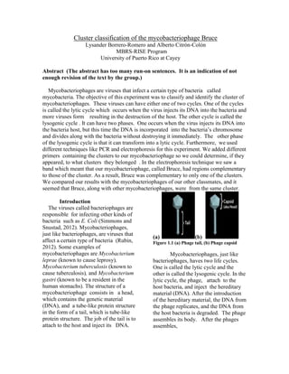
(a) (b)
affect a certain type of bacteria (Rubin, Figure 1.1 (a) Phage tail, (b) Phage capsid
2012). Some examples of
mycobacteriophages are Mycobacterium Mycobacteriophages, just like
leprae (known to cause leprosy), bacteriophages, haves two life cycles.
Mycobacterium tuberculosis (known to One is called the lytic cycle and the
cause tuberculosis), and Mycobacterium other is called the lysogenic cycle. In the
gastri (known to be a resident in the lytic cycle, the phage, attach to the
human stomachs). The structure of a host bacteria, and inject the hereditary
mycobacteriophage consists in a head, material (DNA). After the introduction
which contains the genetic material of the hereditary material, the DNA from
(DNA), and a tube-like protein structure the phage replicates, and the DNA from
in the form of a tail, which is tube-like the host bacteria is degraded. The phage
protein structure. The job of the tail is to assembles its body. After the phages
attach to the host and inject its DNA. assembles,
2. it is ready, and the host bacteria is lysed.
Next, the progeny is released, and it is
able to infect other bacteria. The
characteristic step in this life cycle is the
lysis, and that is why it is called lytic
cycle.
Figure 1.3 Lysogenic Cycle
The purpose of studying
mycobacteriophages is to learn and
understand the life cycle. They can also
Figure 1.2 Lytic Cycle be used in the area of medicine to treat
diseases against mycobacterias.
In contrast, the lysogenic cycle
involves both the lytic and the lysogenic Materials and methods
cycle. In the lysogenic life cycle, the In order to do the cluster
phage gets attached to the bacteria and classification, first the
injects its genome. The phage’s genome mycobacteriophage was prepared. The
is integrated into the host bacteria mycobacteriophage was prepared by
chromosome, and it replicates along with transferring 1ml of mycobacteriophage
it when the cell divides. The phage stays HTPL into a sterile microtube. Then, it
in the lysogenic cycle as long as the is centrifuged 10,000x for 1 hour, and
conditions are favorable, if not they go this way the particles from the
through the lytic life cycle, and it starts mycobacteriophage get to be more
all over again. When the phage genome concentrated. A micropipette is used to
is going to leave the host chromosome remove 950 micro liters of the
the process is called excision, and it is supernatant, which is the material
considered a recombination mechanism. floating on the surface. The preparation
The lysogenic life cycle is better, of the primers is done by re-suspending
because you have more options. You primers in PCR grade water to a
can either stay in the chromosome or concentration of 10 micrograms (ug) /
undergo lysis When the genome is micro liters (ul).
attached to the chromosome, it is better The Polymerase Chain Reactions
protected. If the phage stays with the are going to be set up for the assigned
chromosome of the host and keeps mycobacteriophage genome using each
dividing when it enters the lytic cycle, it of the cluster specific primer
will have a lot of progeny. combination by adding the indicated
volume for each reagent. ???. The
reagent, in accordance with their
respective volumes, are: nano pure PCR,
grade water (5ul), assigned
mycobacteriophage genomic DNA from
step #3 (5ul), cluster specific forward
3. primer (1ul), cluster specific reverse do not have any bands, therefore they are
primer (1ul), PCR master mix (which is not complementary.
composed of Taq polymerase, buffer,
nucleotides, Mg+) (12ul) ?? I don’t
understand the use of so many
parentheses.. The total volume is going
to be 24 micro liters (ul). After adding
the PCR reaction components, the PCR
tubes are added in the thermocycler, and
are amplified using the cycling condition
methods.
The 2% agarose gel is prepared
by adding 2 grams of agarose, 10ml of
Figure 1.4 Bruce Cluster
10x TAE gel running buffer, and 90 ml Electrophoresis
of distilled water. Afterwards, you
microwave the gel on medium power Discussion
and let it gently boil for about 1 minute. According with the electrophoresis
Use gloves to add 4 ul of Ethidium technique, it suggests that Bruce has
Bromidium (EtBr). After adding EtBr, regions complementary to only one
let it cool and pour it into a clean gel particular primer. The primer was the
apparatus. Next, mold it with the cluster B2 primer and we can see this
comb??? in order to make the wells. Let result because it is the only band in the
the gel solidify for around 1 hour. Later, gel. This means that Bruce may belong
2 micro liters of loading dye is added to to that specific cluster. Determining the
10 micro liters of the PCR reaction after cluster in which Bruce belongs to, is a
thermocycling, and load the 2% great advantage, because we can have an
agarose gel. After loading the agarose idea of the genome of the phage and the
gel, the gel will be run at 80 volts for 1 target DNA of the mycobacteriophage.
hour and 45 minutes. Finally, the gel We can also assume that Bruce, Cemi,
will be photographed using a gel Lorenzoveg, and NovaAndreas are in the
documentation system. Every group same cluster because they all have bands
had different mycobacteriophages to test on the well that had the cluster B2
with different clusters in the experiment. primer.
Our group did electrophoresis to
separate molecules based on their mass
and charge. We added the cluster Acknowledgements
specific primers so we could see a band Dr. Michael Rubin, for providing
in the gel which meant that the us with the essential information
mycobacteriophage contained regions and materials to do the both the
complementary to the designed primers. paper and the lab.
Results Dr. Eneida Díaz and Dr. Elena
Our result with the mycobacteriophage Gonzalez, for allowing Dr. Rubin
called Bruce was that apparently it had be our profesor for the day.
regions which were complementary to
the cluster B2 primer as Figure 1.4
below demonstrates. The other regions
4. References Rico. Howard Hughes and RISE
Programs. Puerto Rico pp. 4, 5,
Ross, Robert. 2012. General 6, 13, 14, 15, 17.
Botany Study Guide. Department Simmons, Michael J., Snustad,
of Biology UPR Cayey. Puerto D. Peter. 2012. Principles of
Rico pp xxvii, xxviii, xxix. Genetics. John Wiley & Sons,
Rubin, Michael. 2012. Cluster Inc. New Jersey pp. 165, 167,
Classification of 168.
Mycobacteriophages Isolated
from Tropical Soils of Puerto