SlideShare una empresa de Scribd logo
1 de 101
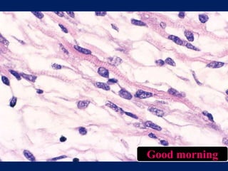
Good morning
Seminar on
Spindle cell tumors of the oral cavity
Part - II
By: Dr. Madhusudhanreddy
III year PG
VDC
• Part I seminar
I. Tumors of fibrous origin
II. Tumors of Fibro histiocytic origin
III.Tumors of adipose tissue origin
IV.Tumors of Smooth muscle origin
7. Neural tumors
Benign Malignant
Traumatic
neuroma
Neurofibrosarcoma
Neurofibroma
Schwannoma
Benign Malignant
Cellular
rhabdomyoma
Spindle cell
rhabdomyosarcoma
Benign Malignant
Spindle cell
hemangioendothelioma
Angiosarcoma
Kaposi
sarcoma
5. Skeletal muscle tumors
6. Vascular tumors
Skeletal muscle tumors
• Benign
• Rabdomyoma
• Malignant
• Rabdomyosarcoma
Rabdomyoma
• Definition: Benign mesenchymal tumor with skeletal
muscle differentiation divided into the adult, fetal, and
genital types, according to degree of differentiation and
location
• Named by Zenker in 1864
• Rare benign tumor that exhibits mature skeletal muscle
differentiation.
• Demographics:
• Age: 4 years (age range - 3 days to 58 years)
• Sex: M:F ratio is 2.4:1
• Site: Histologic types
– Myxoid type – preauricular and post auricular
– Intermediate type – head and neck
• Clinical features:
• Kapadia et al
– 42% - younger than 1 year old
– 25% - congenital
– 50% - older than 15 years of age
• Symptoms: Solitary mass involving soft tissue or mucosa
• Some times fetal rhabdomyoma may be associated with
nevoid basal cell carcinoma syndrome.
• Adult Rhabdomyoma
• Fetal Rhabdomyoma
Douglas R.Gnepp; diagnostic surgical pathology of head and neck; second edition
• Clinical differential diagnosis
• Fibrosarcoma
• Leiomyosarcoma
• Rhabdomyosarcoma
• Kaposi sarcoma
• Lymphoma
• Neuroblastoma
Histopathology:
Varying size and shape of muscle cells
with bipolar nucleus and eosinophillic
cytoplasm in myxoid background
Mixture of undifferentiated and
Differentiated skeletal muscle cells
Cross striation in the in the myofibrils
• Cross striation – H&E stain, PTAH, Masson trichrome stain
• IHC markers – Desmin, Muscle specific actin (MSA)
• Histological differential diagnosis
• Spindle cell Rhabdomyosarcoma
• Infantile fibromatosis
• Treatment
• Total surgical excision is the treatment of choice
• Recurrence rate – up to 40% - incomplete excision
Rabdomyosarcoma
• Initially described by Weber in 1854.
• First published was probably a tongue lesion
• Accounts for 6% of all malignancies in children under 15
years of age
• RMS of the oral cavity accounts for 10 –12% of all the head
and neck RMS cases
• Definition: A primitive soft tissue sarcoma, showing a
variable degree of embryonic skeletal muscle differentiation
Ankita Tandon et al; Oral Rhabdomyosarcoma: A review; J Clin Exp Dent. 2012;4(5):e302-8.
• Demographics:
• Age: First and second decades
• Sex: Males have slight high predelection
• Site: Head and neck > Orbit (35%) > trunk and extremities
> intra-abdominal organs and genitourinary tract (23%)
• Oral cavity – Tongue > soft palate > hard palate > baccal
mucosa
• Signs and symptoms: pain, parasthesia, loss of teeth and
trismus
Ankita Tandon et al; Oral Rhabdomyosarcoma: A review; J Clin Exp Dent. 2012;4(5):e302-8.
Clinical features:
Smooth or lobulated surface, sometimes botryoid or grape cluster–like in
appearance and become fixed to surrounding tissues
• Clinical differential diagnosis
• Fibrosarcoma
• Leiomyosarcoma
• Rhabdomyosarcoma
• Alveolar soft-part sarcoma
• Kaposi sarcoma
• Lymphoma
Ovoid and spindled tumor cells with
dark nuclei in myxoid stroma
Cells are polygonal or elongated
with eosinophilic cytoplasm
Histopathology:
Moderate nuclear pleomorphism and
“strap” cell
More cellular area with mitotic
figures
Primitive and anaplastic muscle fibers
• Histological differential diagnosis
• Ewings sarcoma
• Neuroblastoma
• IHC markers:
– Desmin
– Myogenin/MyoD1
• Treatment
• Surgical excision
• Followed by chemotherapy and radiotherapy
• 66- 90% of 5 years survival rate
Vascular tumors
• Benign
• Spindle cell hemangioendothelioma
• Malignant
• Angiosarcoma
• Kaposi sarcoma
Spindle cell hemangioendothelioma
• Definition:
• Vascular tumor of intermediate malignancy
• Weiss and Enzinger described in 1986
• It was considered to be an intermediate or low-grade
malignancy, with a biologic behavior between a hemangioma
and an angiosarcoma
• Relatively uncommon lesion
• Fletcher et al, Imayama et al - nonneoplastic reactive
vascular proliferation
• Later Perkins and Weiss
– “spindle cell hemangioma” (SCH) - solitary lesions
– “spindle cell hemangiomatosis” - multifocal lesions
K.I. Tosios et al; Spindle cell hemangioma (hemangioendothelioma) of the head and neck: case report
of an unusual (or underdiagnosed) tumor; Oral Surg Oral Med Oral Pathol Oral Radiol Endod
2008;105:216-21
• Demographics:
• Age: Young Adults
• Sex: no sex predelection
• Site: dermis and subcutis of distal extremities, axilla,
trunk, vulva, penis, spleen, pancreas, bones, head and
neck
• Clinical features:
• Solitary tumor or multiple asymptomatic nodules
clustered
• Color - Normal or bluish
• Consistency - firm
• Size - few millimeters to a few centimeters - less than
2.0 cm.
Histopathology:
Spindle cell arranged in the form of
glomeruloid nests
Capillary hemangioma like areas
• Histological differential diagnosis
• Kaposi sarcoma
• Cavernous hemangioma
• Epithelioid hemangioma
• Intravascular papillary endothelial hyperplasia
• Kaposiform hemangioendothelioma
• Epithelioid and spindle cell hemangioma
• IHC markers:
• Vimentin
• CD31
• CD34
• SMA
• Factor VIII–related antigen,
• HAM-56
CD34
SMA
• Treatment
• surgical excision
• Recurrence may develop many years after the initial
excision
• No metastasis
Angiosarcoma
• Definition: Fully malignant sarcoma showing endothelial
differentiation.
• Rarely affecting the oral cavity
• Demographics:
• Age: 1 year to 20 years with an average of 7.3 years
• Sex: Equal sex predilection
• Site: Tongue > Parotid gland > Lip > Submandibular gland
> Palate
• Clinical features:
• Poorly demarcated
• Round or ovoid nodules
• Red-blue to purplish
• Painful
• Spontaneously bleeding
• Firm on palpation
• Clinical differential diagnosis
• Hemangioma
• Pyogenic granuloma
• Melanoma
• Erythroplakia
• Diffuse angiomatosis
• Atypical vascular proliferations after irradiation
Atypical polygonal or spindle cells
with hyperchromatic nuclei
Histopathology:
Clear-cut vasoformation and clearly
malignant endothelial cells
Sheet-like proliferation malignant
epithelioid cells, Mimicking Carcinoma
Pleomorphic tumor with rudimentary lumen
formation
• Histological differential diagnosis
• Haemangioma and its variants
• Pyogenic granuloma
• Spindle cell carcinoma
• Mucosal malignant melanoma
• Rhabdomyosarcoma
• Leiomyosarcoma
• Liposarcoma
• Synovial sarcoma
• Fibro-histiocytic sarcomas, MPNST and epithelioid
sarcoma
• Malignant myoepithelioma of salivary gland
• Anaplastic non-Hodgkin’s lymphoma with spindle cell
features
G. Favia et al; Angiosarcoma of the head and neck with intra-oral presentation. A clinico-pathological
study of four cases; Oral Oncology 38 (2002) 757–762
• IHC markers:
• Vimentin
• CD31
• CD34
• FLI-1 protein
• D2-40 and VEGFR-3
• Low-molecular-weight cytokeratin expression in 25%
of cases, particularly epithelioid tumors
G. Favia et al; Angiosarcoma of the head and neck with intra-oral presentation. A clinico-pathological
study of four cases; Oral Oncology 38 (2002) 757–762
Factor VIII-related antigen
Positive for CD34
MIB-1 labeling
CD31 immunostain for
angiosarcoma cells
• Treatment
• Radiotherapy of 4000–5000 rads,
• Followed by radical surgery - most effective procedure
• Prognosis
• Survival - longer in low grade angiosarcoma than high grade
angiosarcoma
• 50% - die within 15 months from the diagnosis
• 12% - survive 5 years or longer
• Death is usually due to uncontrolled local progression and/or
distant metastases to the lungs, liver and bone
G. Favia et al; Angiosarcoma of the head and neck with intra-oral presentation. A clinico-pathological
study of four cases; Oral Oncology 38 (2002) 757–762
Kaposi sarcoma
• Definition: Malignant vascular tumor caused by infection
with HHV-8, most often in immunocompromised patients
• Kaposi sarcoma (KS) - multifocal angioproliferative
disorder of vascular endothelium, primarily affecting
mucocutaneous tissues with the potential to involve viscera
Mahnaz Fatahzadeh; Kaposi sarcoma: review and medical management update; Oral Surg Oral Med
Oral Pathol Oral Radiol 2012;113:2-16
• Demographics:
Clinical
variants of KS
Etiology Age sex site
Classic KS - 5th decade older men
M:F, 17:1
Skin and extrimities
Endemic KS - children and
young
male
individuals
lymph node and soft
tissue
Iatrogenic KS - - - kidney transplant
patients
Epidemic KS Homosexual male
intravenous drug
Abusers
Adults Males skin,
mucosal
and visceral locations
Clinical features:
Solitary, mutifocal, or multicentric
macules, plaques, or nodules of different
sizes
Clinical features
• Clinical differential diagnosis
• Hemangioma
• Pyogenic granuloma
• Melanoma
• Erythroplakia
Histopathology:
Short fascicles of spindle cells
with abundant fresh hemorrhage
Spindle cells show mild nuclear
pleomorphism and mitotic activity
Proliferation of spindle cells with dilated vessels and
hemosiderin
Spindle cells with mild nuclear
atypia
Lumen formation and presence of
hyaline globules
• Histological differential diagnosis
• Angiosarcoma
• Sindle cell hemangioma
• Spindle cell sarcomas
• Hemangioma
• Pyogenic granuloma
• Angiopericytoma
• IHC markers
• CD31
• CD34
• FLI-1 protein
• D2-40 and VEGFR-3 expression in most cases
• HH V-8 LANA protein expression
D2-40
CD31 in majority of the cells
LANA1 of HHV8
• Treatment
• Treatment is divided into
– Local/regional therapy
– Systemic therapy
• Local/regional therapy
– Surgical excision
– Cryotherapy
– Sclerotherapy
– Intralesional vinca-alkaloids
– Radiotherapy
– Laser therapy
Mahnaz Fatahzadeh; Kaposi sarcoma: review and medical management update; Oral Surg Oral Med
Oral Pathol Oral Radiol 2012;113:2-16
• HAART
– NNRT-based therapy
(non-nucleoside reverse
transcriptase)
– PI-based therapy
(protease inhibitor)
• Chemotherapy
– Combination agent ABV
or ABVb
– Vincristine,
Vinblastine,Vinorebline
– Bleomycin
• Immune modulators
– Interferon-alpha
• Experimental & targeted
therapies
– Antiherpes therapy
– Angiogenic inhibitors (e.g.,
thalidomide)
– VEGF inhibitors
– Tyrosine kinase inhibitors
– Matrix metalloproteinases
Systemic therapy
Mahnaz Fatahzadeh; Kaposi sarcoma: review and medical management update; Oral Surg Oral Med
Oral Pathol Oral Radiol 2012;113:2-16
Neural tumors
• Benign
• Traumatic neuroma
• Neurofibroma
• Schwannoma
• Malignant
• Neurofibrosarcoma
Traumatic neuroma
• Definition: Disorganized proliferation of nerve in response
to injury or disruption.
• Occurs in the area of any nerve that has been surgically or
auto-amputated
• Demographics:
• Age: Middle-aged adults
• Sex: Slightly more common in females than in males
• Site: Mental foramen area
lateral tongue
lower lip
• Clinical features
• Patient has firm, painful nodule
• Constant or intermittent aching, a burning sensation,
or severe radiating pain
• Clinical differential diagnosis
• Traumatic fibroma
• Mucocele
• Solitary neurofibroma
Histopathology:
Nerve fibers of varying sizes with
fibrosis
Prominent fibrosis
Nerve fibers with thickened
endoneurium
• Histological differential diagnosis
• PEN
• Mucosal neuroma
• Morton’s neuroma
• Neurofibroma
• IHC markers
• S-100
• CD-57
• Treatment
• Excision
Neurofilament protein positivity
CD57 (Leu-7) in traumatic neuroma
Neurofibroma
• Definition:
• Neurofibromas are the most frequent benign neoplasms
originating from the peripheral nerve sheath and occur as
solitary or multiple tumours when associated to the von
Recklinghausen disease, better known as neurofibromatosis
• Demographics:
• Age: Most patients are younger than 45 years of age
• Sex: Females are more frequently affected,
• F:M ratio being 2:1.
• Site:
• Buccal mucosa, palate, alveolar ridge, vestibule and
tongue, lips and gingival
• Intraosseous lesions are quite rare.
• Most common site being the posterior mandible
• Clinical features
• ORAL NEUROFIBROMA - uncommon benign tumor
of the oral cavity
• Neurofibroma occurs as a
– Solitary lesion – unknown cause
– As part of the generalized syndrome of
neurofibromatosis (von Recklinghausen disease of
the skin) - inherited as an autosomal dominant trait
• 50% of cases are reported to be the result of
spontaneous mutation in
– Neurofibromatosis type 1 (NF1) gene
– Neurofibromatosis type 2 (NF2) gene
• Solitary oral lesions - non-ulcerated nodules, which
tend to be of same color of normal mucosa.
• Lesions associated with neurofibromatosis – solitary
lesions, iris Lisch nodules, cafe-au-lait spots and
freckling in the axillary region.
Intraoral involvement
Multiple tumors of the trunk
and cafe-au-lait spots
• Central neurofibromas
– Early stages – Asymptomatic
– Grow in size - Cause expansion of the cortical plates
of the mandible, with or without destruction,
– Pain and anesthesia or paresthesia of the lower lip
• Radiographically - Well-circumscribed or poorly
demarcated radiolucent lesion
• Clinical differential diagnosis
• Mucocele
• Lipoma
• Pleomorphic adenoma
Histopathology
Unencapsulated lesion
Spindle cells, curvilinear nuclei,
delicate collagen, and mast cells
S-100 positivity
• Histological differential diagnosis
• Nodular fasciitis (absence of thin wavy nucleus)
• Focal mucinosis
• Palasaded encapsulated neuroma
• IHC markers
• S-100
• CD34
• Neurofilament
• Factor VIII
• Treatment
• Surgical excision
• Malignant transformation - is reported to occur in 5%
to 16% of patients
Schwannoma
• Once termed “neurilemoma,” - Stout, 1935
• Benign tumor, apparently derived from the Schwann cells
• Definition: A peripheral nerve sheath tumor arising
eccentric to nerve
• Demographics:
• Age: 20 to 50 years common in 2nd and 3rd decades
• Sex: Female Sex Predilection
• Site: Tongue > Palate > Buccal mucosa
• Some times intraosseous – mandible
• Bone expansion, pain, parasthesia
Clinical features
• Clinical differential diagnosis
• Lipoma
• Fibroma
• Solitary neurofibroma
• Hemangioma
• Eosinophillic granuloma
• Epidermoid and dermoid cysts
• Epithelial hyperplasia
• Granular cell tumor
• Leiomyoma
• Lymphangioma
• Salivary gland tumors
Monoela D M et al; Intraoral schwannoma: case report and litrature review; indian journal of dental
res, 20(1), 2009.
Histological varients
Histopathology
Marked cystic degeneration of stroma
Encapsulation
• Verocay bodies – Antoni A pattern
Eosinophilic basement membrane
material between palisaded nuclei
Spindle cells – Antoni B pattern
Antoni A- nuclear palasading
Antoni A- without nuclear
palasading
Verocay bodies and adjacent Antoni
type B tissue
Delicately collagenized and myxoid
stroma
Spindle cell proliferation
perivascular hyalinization, mostly
Antoni type B tissue
Palisading of nuclei forming
unusual Verocay body
• Histological differential diagnosis
• Neurofibroma
• Melanoma
• Leiomyosarcoma
• MPNST
• Solitary fibrous tumor
• Solitary circumscribed neuroma
• IHC markers:
• S-100
S-100 positivity
• Treatment
• Conservative surgical excision
• No evidence of recurrence if completely excised
• Malignant transformation - controversial
Neurofibrosarcoma
• Definition: A malignant neoplasm with a poor
prognosis of perineural fibroblasts or schwann cells,
with propensity to rapidly extend along the associated
nerve trunk.
• WHO has grouped this lesion under the heading of
MPNSTs
– Mlignant schwannoma
– Malignant neurilemmoma
– Neurogenic sarcoma
– Neurofibrosarcoma
• Demographics
• Age: 20 – 50 yrs of age
• Sex: no sex predelection
• Site: any where in the body but most commonly in skin
• Clinical features
• Exact mechanism of occurance of the lesion is not yet
identified
• Occurs as a complication of neurofibromatosis I (15%)
• Or occurs denovo
• Mostly occurs in soft tissue
• Intraosseous neurofibrosarcoma
– Due to the nerve present in the neutrient canal
– Due invasion into the bone from soft tissue
– Symptoms include pain, parasthesia
Neurofibromatosis-I patient with
neurofibrosarcoma of the left cheek
Histopathology
Spindle cell proliferation with
numerous mitotic figures
Fascicles of spindle cells
resembling fibrosarcoma
Patchy positivity for S-100
Vimentin positivity
• Histological differential diagnosis
• Monophasic syanovial sarcoma
• Leiomyosarcoma
• Fibrosarcoma
• IHC markers
• S-100 (weak or negative)
• GFAP
• Neurofilament
• NSE
• Leu-7
• Myelin basic protein
• Ki67
• Treatment
• Highly aggressive tumor and difficult to treat
• Prognosis - Grade of tumor, tumor size
• Overall survival rate is 40-75%
• Early detection and appropriate treatment is crucial
for survival
Referrences
• Brad . W . Neville , Douglas D . Damm , Carl M . Allen Oral and
maxillofacial pathology 2nd edition 2004
• SHAFER; Text book of oral pathology 5th edition 2006
• Enzinger and weiss; Soft tissue tumors fifth edition
• Douglas .R. Gnepp; Diagnostic surgical pathology of head and neck; second
edition
• Andrew L.Folpe, Carrie Y. Inwards; Bone and soft tissue pathology
foundation in surgical pathology.
• Sook-Bin Woo; Oral pathology A comprehensive atlas and text
• Net source and articles

Más contenido relacionado

La actualidad más candente

Benign tumors of oral cavity sonal
Benign tumors of oral cavity  sonalBenign tumors of oral cavity  sonal
Benign tumors of oral cavity sonalSonal Aggarwal
 
Odontogenic and Non-odontogenic Tumors - Update from the 4th Edition of WHO 2...
Odontogenic and Non-odontogenic Tumors - Update from the 4th Edition of WHO 2...Odontogenic and Non-odontogenic Tumors - Update from the 4th Edition of WHO 2...
Odontogenic and Non-odontogenic Tumors - Update from the 4th Edition of WHO 2...Himanshu Soni
 
Clear cell lesions of head & neck.
Clear cell lesions of head & neck.Clear cell lesions of head & neck.
Clear cell lesions of head & neck.doctorpeace
 
NON ODONTOGENIC TUMORS OF ORAL CAVITY-I
NON ODONTOGENIC TUMORS OF ORAL CAVITY-I NON ODONTOGENIC TUMORS OF ORAL CAVITY-I
NON ODONTOGENIC TUMORS OF ORAL CAVITY-I Krupali Gandhi
 
Adenoid cystic carcinoma (Doctor Faris Alabeedi MSc, MMedSc, PgDip, BDS.)
Adenoid cystic carcinoma (Doctor Faris Alabeedi MSc, MMedSc, PgDip, BDS.)Adenoid cystic carcinoma (Doctor Faris Alabeedi MSc, MMedSc, PgDip, BDS.)
Adenoid cystic carcinoma (Doctor Faris Alabeedi MSc, MMedSc, PgDip, BDS.)Doctor Faris Alabeedi
 
Salivary gland pathology
Salivary gland pathologySalivary gland pathology
Salivary gland pathologyPrasad CSBR
 
Case presentation of Gorlin Goltz syndrome
Case presentation of Gorlin Goltz syndromeCase presentation of Gorlin Goltz syndrome
Case presentation of Gorlin Goltz syndromeAnushan Madushanka
 
Peripheral giant cell granuloma (giant cell epulis
Peripheral giant cell granuloma (giant cell epulisPeripheral giant cell granuloma (giant cell epulis
Peripheral giant cell granuloma (giant cell epulisKhin Soe
 
Pigmented lesion
Pigmented lesionPigmented lesion
Pigmented lesionIAU Dent
 
METASTATIC TUMORS OF THE JAW
METASTATIC  TUMORS OF THE JAWMETASTATIC  TUMORS OF THE JAW
METASTATIC TUMORS OF THE JAWUpama Sishan
 
Salivary gland tumors classification
Salivary gland tumors classificationSalivary gland tumors classification
Salivary gland tumors classificationDr Preeti Sharma
 
Epithelial dysplasia
Epithelial dysplasiaEpithelial dysplasia
Epithelial dysplasiaasmaa1996
 
malignant epithelial tumors of oral cavity
malignant epithelial tumors of oral cavitymalignant epithelial tumors of oral cavity
malignant epithelial tumors of oral cavitymadhusudhan reddy
 
Mucoepidermoid carcinoma
Mucoepidermoid carcinomaMucoepidermoid carcinoma
Mucoepidermoid carcinomaNehal mohamed
 
Ameloblastoma (Odontogenic Tumor) Oral Pathology
Ameloblastoma (Odontogenic Tumor) Oral Pathology Ameloblastoma (Odontogenic Tumor) Oral Pathology
Ameloblastoma (Odontogenic Tumor) Oral Pathology Sarang Suresh Hotchandani
 
Spindle cell lesions of oral cavity part III
Spindle cell lesions of oral cavity part IIISpindle cell lesions of oral cavity part III
Spindle cell lesions of oral cavity part IIImadhusudhan reddy
 
oral field cancerization - Dr Sanjana Ravindra
oral field cancerization - Dr Sanjana Ravindraoral field cancerization - Dr Sanjana Ravindra
oral field cancerization - Dr Sanjana RavindraDr. Sanjana Ravindra
 

La actualidad más candente (20)

Basal Cell Adenoma
Basal Cell AdenomaBasal Cell Adenoma
Basal Cell Adenoma
 
Benign tumors of oral cavity sonal
Benign tumors of oral cavity  sonalBenign tumors of oral cavity  sonal
Benign tumors of oral cavity sonal
 
Odontogenic and Non-odontogenic Tumors - Update from the 4th Edition of WHO 2...
Odontogenic and Non-odontogenic Tumors - Update from the 4th Edition of WHO 2...Odontogenic and Non-odontogenic Tumors - Update from the 4th Edition of WHO 2...
Odontogenic and Non-odontogenic Tumors - Update from the 4th Edition of WHO 2...
 
Clear cell lesions of head & neck.
Clear cell lesions of head & neck.Clear cell lesions of head & neck.
Clear cell lesions of head & neck.
 
NON ODONTOGENIC TUMORS OF ORAL CAVITY-I
NON ODONTOGENIC TUMORS OF ORAL CAVITY-I NON ODONTOGENIC TUMORS OF ORAL CAVITY-I
NON ODONTOGENIC TUMORS OF ORAL CAVITY-I
 
Adenoid cystic carcinoma (Doctor Faris Alabeedi MSc, MMedSc, PgDip, BDS.)
Adenoid cystic carcinoma (Doctor Faris Alabeedi MSc, MMedSc, PgDip, BDS.)Adenoid cystic carcinoma (Doctor Faris Alabeedi MSc, MMedSc, PgDip, BDS.)
Adenoid cystic carcinoma (Doctor Faris Alabeedi MSc, MMedSc, PgDip, BDS.)
 
Salivary gland pathology
Salivary gland pathologySalivary gland pathology
Salivary gland pathology
 
Case presentation of Gorlin Goltz syndrome
Case presentation of Gorlin Goltz syndromeCase presentation of Gorlin Goltz syndrome
Case presentation of Gorlin Goltz syndrome
 
Ameloblastoma
AmeloblastomaAmeloblastoma
Ameloblastoma
 
Peripheral giant cell granuloma (giant cell epulis
Peripheral giant cell granuloma (giant cell epulisPeripheral giant cell granuloma (giant cell epulis
Peripheral giant cell granuloma (giant cell epulis
 
Pigmented lesion
Pigmented lesionPigmented lesion
Pigmented lesion
 
METASTATIC TUMORS OF THE JAW
METASTATIC  TUMORS OF THE JAWMETASTATIC  TUMORS OF THE JAW
METASTATIC TUMORS OF THE JAW
 
Salivary gland tumors classification
Salivary gland tumors classificationSalivary gland tumors classification
Salivary gland tumors classification
 
Aot,ceot
Aot,ceotAot,ceot
Aot,ceot
 
Epithelial dysplasia
Epithelial dysplasiaEpithelial dysplasia
Epithelial dysplasia
 
malignant epithelial tumors of oral cavity
malignant epithelial tumors of oral cavitymalignant epithelial tumors of oral cavity
malignant epithelial tumors of oral cavity
 
Mucoepidermoid carcinoma
Mucoepidermoid carcinomaMucoepidermoid carcinoma
Mucoepidermoid carcinoma
 
Ameloblastoma (Odontogenic Tumor) Oral Pathology
Ameloblastoma (Odontogenic Tumor) Oral Pathology Ameloblastoma (Odontogenic Tumor) Oral Pathology
Ameloblastoma (Odontogenic Tumor) Oral Pathology
 
Spindle cell lesions of oral cavity part III
Spindle cell lesions of oral cavity part IIISpindle cell lesions of oral cavity part III
Spindle cell lesions of oral cavity part III
 
oral field cancerization - Dr Sanjana Ravindra
oral field cancerization - Dr Sanjana Ravindraoral field cancerization - Dr Sanjana Ravindra
oral field cancerization - Dr Sanjana Ravindra
 

Similar a Spindle cell lesions of oral cavity part II

softtissuesarcomafinal-220603060631-961c47c7.pptx
softtissuesarcomafinal-220603060631-961c47c7.pptxsofttissuesarcomafinal-220603060631-961c47c7.pptx
softtissuesarcomafinal-220603060631-961c47c7.pptxssuserc0817d
 
Malignant bone tumors
Malignant bone tumorsMalignant bone tumors
Malignant bone tumorsSaachiGupta4
 
SALIVORY GLAND FINAL YR.pptx
SALIVORY GLAND FINAL YR.pptxSALIVORY GLAND FINAL YR.pptx
SALIVORY GLAND FINAL YR.pptxafzal mohd
 
topic seminar sudha-1...............pptx
topic  seminar sudha-1...............pptxtopic  seminar sudha-1...............pptx
topic seminar sudha-1...............pptxHarishankarSharma27
 
Salivary gland tumors.pptx
Salivary gland tumors.pptxSalivary gland tumors.pptx
Salivary gland tumors.pptxSatishray9
 
Tumors-of-nerves-and-muscles(Part-2)-20208191427460.pptx
Tumors-of-nerves-and-muscles(Part-2)-20208191427460.pptxTumors-of-nerves-and-muscles(Part-2)-20208191427460.pptx
Tumors-of-nerves-and-muscles(Part-2)-20208191427460.pptxPRAGYARATHORE24
 
Pathology of oral cancer
Pathology of oral cancerPathology of oral cancer
Pathology of oral cancerSanika Kulkarni
 
Ocular and Orbital Tumours.pptx
Ocular and Orbital Tumours.pptxOcular and Orbital Tumours.pptx
Ocular and Orbital Tumours.pptxMedinfopedia Blog
 

Similar a Spindle cell lesions of oral cavity part II (20)

Bone and soft tissue pathology
Bone and soft tissue pathology  Bone and soft tissue pathology
Bone and soft tissue pathology
 
Adrenal
AdrenalAdrenal
Adrenal
 
Salivary gland tumors
Salivary gland tumorsSalivary gland tumors
Salivary gland tumors
 
SOFT TISSUE SARCOMA
SOFT TISSUE SARCOMASOFT TISSUE SARCOMA
SOFT TISSUE SARCOMA
 
softtissuesarcomafinal-220603060631-961c47c7.pptx
softtissuesarcomafinal-220603060631-961c47c7.pptxsofttissuesarcomafinal-220603060631-961c47c7.pptx
softtissuesarcomafinal-220603060631-961c47c7.pptx
 
Git 7-csbrp
Git 7-csbrpGit 7-csbrp
Git 7-csbrp
 
Malignant bone tumors
Malignant bone tumorsMalignant bone tumors
Malignant bone tumors
 
Neoplasma 1
Neoplasma 1Neoplasma 1
Neoplasma 1
 
Osteosarcoma
OsteosarcomaOsteosarcoma
Osteosarcoma
 
SALIVORY GLAND FINAL YR.pptx
SALIVORY GLAND FINAL YR.pptxSALIVORY GLAND FINAL YR.pptx
SALIVORY GLAND FINAL YR.pptx
 
topic seminar sudha-1...............pptx
topic  seminar sudha-1...............pptxtopic  seminar sudha-1...............pptx
topic seminar sudha-1...............pptx
 
Salivary gland tumors.pptx
Salivary gland tumors.pptxSalivary gland tumors.pptx
Salivary gland tumors.pptx
 
MENINGIOMA-neurosurgery
MENINGIOMA-neurosurgeryMENINGIOMA-neurosurgery
MENINGIOMA-neurosurgery
 
Orbital tumors.pptx
Orbital tumors.pptxOrbital tumors.pptx
Orbital tumors.pptx
 
Tumors-of-nerves-and-muscles(Part-2)-20208191427460.pptx
Tumors-of-nerves-and-muscles(Part-2)-20208191427460.pptxTumors-of-nerves-and-muscles(Part-2)-20208191427460.pptx
Tumors-of-nerves-and-muscles(Part-2)-20208191427460.pptx
 
Testicular carcinoma
Testicular carcinomaTesticular carcinoma
Testicular carcinoma
 
Giant cell lesion.pptx
Giant cell lesion.pptxGiant cell lesion.pptx
Giant cell lesion.pptx
 
Pathology of oral cancer
Pathology of oral cancerPathology of oral cancer
Pathology of oral cancer
 
Round cell tumors
Round cell tumorsRound cell tumors
Round cell tumors
 
Ocular and Orbital Tumours.pptx
Ocular and Orbital Tumours.pptxOcular and Orbital Tumours.pptx
Ocular and Orbital Tumours.pptx
 

Más de madhusudhan reddy

benign tumors of epithelial origin of oral cavity
benign tumors of epithelial origin of oral cavitybenign tumors of epithelial origin of oral cavity
benign tumors of epithelial origin of oral cavitymadhusudhan reddy
 
maxillary sinus anatomy histology.pptx
maxillary sinus anatomy histology.pptxmaxillary sinus anatomy histology.pptx
maxillary sinus anatomy histology.pptxmadhusudhan reddy
 
class traits of premolars and Maxillary 1st premolar.pptx
class traits of  premolars and Maxillary 1st premolar.pptxclass traits of  premolars and Maxillary 1st premolar.pptx
class traits of premolars and Maxillary 1st premolar.pptxmadhusudhan reddy
 
non neoplastic disorders of salivary glands
non neoplastic disorders of salivary glands non neoplastic disorders of salivary glands
non neoplastic disorders of salivary glands madhusudhan reddy
 
permanent maxillary lateral incisor.pptx
permanent maxillary lateral incisor.pptxpermanent maxillary lateral incisor.pptx
permanent maxillary lateral incisor.pptxmadhusudhan reddy
 
EPEDERMOLYSIS BULLOSA SLE SCLERODERMA .ppt
EPEDERMOLYSIS BULLOSA SLE SCLERODERMA .pptEPEDERMOLYSIS BULLOSA SLE SCLERODERMA .ppt
EPEDERMOLYSIS BULLOSA SLE SCLERODERMA .pptmadhusudhan reddy
 
vesiculobullous lesions, pempigus ppt
vesiculobullous lesions, pempigus  pptvesiculobullous lesions, pempigus  ppt
vesiculobullous lesions, pempigus pptmadhusudhan reddy
 
lichen planus and lichenoid reaction 4 .ppt
lichen planus and lichenoid reaction 4 .pptlichen planus and lichenoid reaction 4 .ppt
lichen planus and lichenoid reaction 4 .pptmadhusudhan reddy
 
psoriasis and erythema multiformae 3 .ppt
psoriasis and erythema multiformae 3 .pptpsoriasis and erythema multiformae 3 .ppt
psoriasis and erythema multiformae 3 .pptmadhusudhan reddy
 
ectodermal dysplasia and white spongy nevus.ppt
ectodermal dysplasia and white spongy nevus.pptectodermal dysplasia and white spongy nevus.ppt
ectodermal dysplasia and white spongy nevus.pptmadhusudhan reddy
 
Diseases of pulp and periapical tissues
Diseases of pulp and periapical tissues Diseases of pulp and periapical tissues
Diseases of pulp and periapical tissues madhusudhan reddy
 
Non odontogenic cysts or fissural cysts
Non odontogenic cysts or fissural cystsNon odontogenic cysts or fissural cysts
Non odontogenic cysts or fissural cystsmadhusudhan reddy
 
cysts of the oral and maxillofacial region
cysts of the oral and maxillofacial regioncysts of the oral and maxillofacial region
cysts of the oral and maxillofacial regionmadhusudhan reddy
 

Más de madhusudhan reddy (20)

benign tumors of epithelial origin of oral cavity
benign tumors of epithelial origin of oral cavitybenign tumors of epithelial origin of oral cavity
benign tumors of epithelial origin of oral cavity
 
mandibular premolars.pptx
mandibular premolars.pptxmandibular premolars.pptx
mandibular premolars.pptx
 
Periodontal Ligament.ppt
Periodontal Ligament.pptPeriodontal Ligament.ppt
Periodontal Ligament.ppt
 
maxillary sinus anatomy histology.pptx
maxillary sinus anatomy histology.pptxmaxillary sinus anatomy histology.pptx
maxillary sinus anatomy histology.pptx
 
class traits of premolars and Maxillary 1st premolar.pptx
class traits of  premolars and Maxillary 1st premolar.pptxclass traits of  premolars and Maxillary 1st premolar.pptx
class traits of premolars and Maxillary 1st premolar.pptx
 
Enamel
EnamelEnamel
Enamel
 
non neoplastic disorders of salivary glands
non neoplastic disorders of salivary glands non neoplastic disorders of salivary glands
non neoplastic disorders of salivary glands
 
permanent maxillary lateral incisor.pptx
permanent maxillary lateral incisor.pptxpermanent maxillary lateral incisor.pptx
permanent maxillary lateral incisor.pptx
 
EPEDERMOLYSIS BULLOSA SLE SCLERODERMA .ppt
EPEDERMOLYSIS BULLOSA SLE SCLERODERMA .pptEPEDERMOLYSIS BULLOSA SLE SCLERODERMA .ppt
EPEDERMOLYSIS BULLOSA SLE SCLERODERMA .ppt
 
vesiculobullous lesions, pempigus ppt
vesiculobullous lesions, pempigus  pptvesiculobullous lesions, pempigus  ppt
vesiculobullous lesions, pempigus ppt
 
lichen planus and lichenoid reaction 4 .ppt
lichen planus and lichenoid reaction 4 .pptlichen planus and lichenoid reaction 4 .ppt
lichen planus and lichenoid reaction 4 .ppt
 
psoriasis and erythema multiformae 3 .ppt
psoriasis and erythema multiformae 3 .pptpsoriasis and erythema multiformae 3 .ppt
psoriasis and erythema multiformae 3 .ppt
 
ectodermal dysplasia and white spongy nevus.ppt
ectodermal dysplasia and white spongy nevus.pptectodermal dysplasia and white spongy nevus.ppt
ectodermal dysplasia and white spongy nevus.ppt
 
Skin terminologies.ppt
Skin terminologies.pptSkin terminologies.ppt
Skin terminologies.ppt
 
Diseases of bones and joint
Diseases of bones and jointDiseases of bones and joint
Diseases of bones and joint
 
Diseases of pulp and periapical tissues
Diseases of pulp and periapical tissues Diseases of pulp and periapical tissues
Diseases of pulp and periapical tissues
 
healing of oral wounds
healing of oral woundshealing of oral wounds
healing of oral wounds
 
Spread of oral infections
Spread of oral infectionsSpread of oral infections
Spread of oral infections
 
Non odontogenic cysts or fissural cysts
Non odontogenic cysts or fissural cystsNon odontogenic cysts or fissural cysts
Non odontogenic cysts or fissural cysts
 
cysts of the oral and maxillofacial region
cysts of the oral and maxillofacial regioncysts of the oral and maxillofacial region
cysts of the oral and maxillofacial region
 

Último

Measurement of Radiation and Dosimetric Procedure.pptx
Measurement of Radiation and Dosimetric Procedure.pptxMeasurement of Radiation and Dosimetric Procedure.pptx
Measurement of Radiation and Dosimetric Procedure.pptxDr. Dheeraj Kumar
 
April 2024 ONCOLOGY CARTOON by DR KANHU CHARAN PATRO
April 2024 ONCOLOGY CARTOON by  DR KANHU CHARAN PATROApril 2024 ONCOLOGY CARTOON by  DR KANHU CHARAN PATRO
April 2024 ONCOLOGY CARTOON by DR KANHU CHARAN PATROKanhu Charan
 
Nutrition of OCD for my Nutritional Neuroscience Class
Nutrition of OCD for my Nutritional Neuroscience ClassNutrition of OCD for my Nutritional Neuroscience Class
Nutrition of OCD for my Nutritional Neuroscience Classmanuelazg2001
 
Study on the Impact of FOCUS-PDCA Management Model on the Disinfection Qualit...
Study on the Impact of FOCUS-PDCA Management Model on the Disinfection Qualit...Study on the Impact of FOCUS-PDCA Management Model on the Disinfection Qualit...
Study on the Impact of FOCUS-PDCA Management Model on the Disinfection Qualit...MehranMouzam
 
Biomechanics- Shoulder Joint!!!!!!!!!!!!
Biomechanics- Shoulder Joint!!!!!!!!!!!!Biomechanics- Shoulder Joint!!!!!!!!!!!!
Biomechanics- Shoulder Joint!!!!!!!!!!!!ibtesaam huma
 
PNEUMOTHORAX AND ITS MANAGEMENTS.pdf
PNEUMOTHORAX   AND  ITS  MANAGEMENTS.pdfPNEUMOTHORAX   AND  ITS  MANAGEMENTS.pdf
PNEUMOTHORAX AND ITS MANAGEMENTS.pdfDolisha Warbi
 
Culture and Health Disorders Social change.pptx
Culture and Health Disorders Social change.pptxCulture and Health Disorders Social change.pptx
Culture and Health Disorders Social change.pptxDr. Dheeraj Kumar
 
Presentation on Parasympathetic Nervous System
Presentation on Parasympathetic Nervous SystemPresentation on Parasympathetic Nervous System
Presentation on Parasympathetic Nervous SystemPrerana Jadhav
 
Wessex Health Partners Wessex Integrated Care, Population Health, Research & ...
Wessex Health Partners Wessex Integrated Care, Population Health, Research & ...Wessex Health Partners Wessex Integrated Care, Population Health, Research & ...
Wessex Health Partners Wessex Integrated Care, Population Health, Research & ...Wessex Health Partners
 
Presentation on General Anesthetics pdf.
Presentation on General Anesthetics pdf.Presentation on General Anesthetics pdf.
Presentation on General Anesthetics pdf.Prerana Jadhav
 
The next social challenge to public health: the information environment.pptx
The next social challenge to public health:  the information environment.pptxThe next social challenge to public health:  the information environment.pptx
The next social challenge to public health: the information environment.pptxTina Purnat
 
Giftedness: Understanding Everyday Neurobiology for Self-Knowledge
Giftedness: Understanding Everyday Neurobiology for Self-KnowledgeGiftedness: Understanding Everyday Neurobiology for Self-Knowledge
Giftedness: Understanding Everyday Neurobiology for Self-Knowledgeassessoriafabianodea
 
epilepsy and status epilepticus for undergraduate.pptx
epilepsy and status epilepticus  for undergraduate.pptxepilepsy and status epilepticus  for undergraduate.pptx
epilepsy and status epilepticus for undergraduate.pptxMohamed Rizk Khodair
 
Statistical modeling in pharmaceutical research and development.
Statistical modeling in pharmaceutical research and development.Statistical modeling in pharmaceutical research and development.
Statistical modeling in pharmaceutical research and development.ANJALI
 
VarSeq 2.6.0: Advancing Pharmacogenomics and Genomic Analysis
VarSeq 2.6.0: Advancing Pharmacogenomics and Genomic AnalysisVarSeq 2.6.0: Advancing Pharmacogenomics and Genomic Analysis
VarSeq 2.6.0: Advancing Pharmacogenomics and Genomic AnalysisGolden Helix
 
SWD (Short wave diathermy)- Physiotherapy.ppt
SWD (Short wave diathermy)- Physiotherapy.pptSWD (Short wave diathermy)- Physiotherapy.ppt
SWD (Short wave diathermy)- Physiotherapy.pptMumux Mirani
 
MedDRA-A-Comprehensive-Guide-to-Standardized-Medical-Terminology.pdf
MedDRA-A-Comprehensive-Guide-to-Standardized-Medical-Terminology.pdfMedDRA-A-Comprehensive-Guide-to-Standardized-Medical-Terminology.pdf
MedDRA-A-Comprehensive-Guide-to-Standardized-Medical-Terminology.pdfSasikiranMarri
 
low cost antibiotic cement nail for infected non union.pptx
low cost antibiotic cement nail for infected non union.pptxlow cost antibiotic cement nail for infected non union.pptx
low cost antibiotic cement nail for infected non union.pptxdrashraf369
 
Presentation for Bella Mahl 2024-03-28-24-MW-Overview-Bella.pptx
Presentation for Bella Mahl 2024-03-28-24-MW-Overview-Bella.pptxPresentation for Bella Mahl 2024-03-28-24-MW-Overview-Bella.pptx
Presentation for Bella Mahl 2024-03-28-24-MW-Overview-Bella.pptxpdamico1
 
SGK HÓA SINH NĂNG LƯỢNG SINH HỌC 2006.pdf
SGK HÓA SINH NĂNG LƯỢNG SINH HỌC 2006.pdfSGK HÓA SINH NĂNG LƯỢNG SINH HỌC 2006.pdf
SGK HÓA SINH NĂNG LƯỢNG SINH HỌC 2006.pdfHongBiThi1
 

Último (20)

Measurement of Radiation and Dosimetric Procedure.pptx
Measurement of Radiation and Dosimetric Procedure.pptxMeasurement of Radiation and Dosimetric Procedure.pptx
Measurement of Radiation and Dosimetric Procedure.pptx
 
April 2024 ONCOLOGY CARTOON by DR KANHU CHARAN PATRO
April 2024 ONCOLOGY CARTOON by  DR KANHU CHARAN PATROApril 2024 ONCOLOGY CARTOON by  DR KANHU CHARAN PATRO
April 2024 ONCOLOGY CARTOON by DR KANHU CHARAN PATRO
 
Nutrition of OCD for my Nutritional Neuroscience Class
Nutrition of OCD for my Nutritional Neuroscience ClassNutrition of OCD for my Nutritional Neuroscience Class
Nutrition of OCD for my Nutritional Neuroscience Class
 
Study on the Impact of FOCUS-PDCA Management Model on the Disinfection Qualit...
Study on the Impact of FOCUS-PDCA Management Model on the Disinfection Qualit...Study on the Impact of FOCUS-PDCA Management Model on the Disinfection Qualit...
Study on the Impact of FOCUS-PDCA Management Model on the Disinfection Qualit...
 
Biomechanics- Shoulder Joint!!!!!!!!!!!!
Biomechanics- Shoulder Joint!!!!!!!!!!!!Biomechanics- Shoulder Joint!!!!!!!!!!!!
Biomechanics- Shoulder Joint!!!!!!!!!!!!
 
PNEUMOTHORAX AND ITS MANAGEMENTS.pdf
PNEUMOTHORAX   AND  ITS  MANAGEMENTS.pdfPNEUMOTHORAX   AND  ITS  MANAGEMENTS.pdf
PNEUMOTHORAX AND ITS MANAGEMENTS.pdf
 
Culture and Health Disorders Social change.pptx
Culture and Health Disorders Social change.pptxCulture and Health Disorders Social change.pptx
Culture and Health Disorders Social change.pptx
 
Presentation on Parasympathetic Nervous System
Presentation on Parasympathetic Nervous SystemPresentation on Parasympathetic Nervous System
Presentation on Parasympathetic Nervous System
 
Wessex Health Partners Wessex Integrated Care, Population Health, Research & ...
Wessex Health Partners Wessex Integrated Care, Population Health, Research & ...Wessex Health Partners Wessex Integrated Care, Population Health, Research & ...
Wessex Health Partners Wessex Integrated Care, Population Health, Research & ...
 
Presentation on General Anesthetics pdf.
Presentation on General Anesthetics pdf.Presentation on General Anesthetics pdf.
Presentation on General Anesthetics pdf.
 
The next social challenge to public health: the information environment.pptx
The next social challenge to public health:  the information environment.pptxThe next social challenge to public health:  the information environment.pptx
The next social challenge to public health: the information environment.pptx
 
Giftedness: Understanding Everyday Neurobiology for Self-Knowledge
Giftedness: Understanding Everyday Neurobiology for Self-KnowledgeGiftedness: Understanding Everyday Neurobiology for Self-Knowledge
Giftedness: Understanding Everyday Neurobiology for Self-Knowledge
 
epilepsy and status epilepticus for undergraduate.pptx
epilepsy and status epilepticus  for undergraduate.pptxepilepsy and status epilepticus  for undergraduate.pptx
epilepsy and status epilepticus for undergraduate.pptx
 
Statistical modeling in pharmaceutical research and development.
Statistical modeling in pharmaceutical research and development.Statistical modeling in pharmaceutical research and development.
Statistical modeling in pharmaceutical research and development.
 
VarSeq 2.6.0: Advancing Pharmacogenomics and Genomic Analysis
VarSeq 2.6.0: Advancing Pharmacogenomics and Genomic AnalysisVarSeq 2.6.0: Advancing Pharmacogenomics and Genomic Analysis
VarSeq 2.6.0: Advancing Pharmacogenomics and Genomic Analysis
 
SWD (Short wave diathermy)- Physiotherapy.ppt
SWD (Short wave diathermy)- Physiotherapy.pptSWD (Short wave diathermy)- Physiotherapy.ppt
SWD (Short wave diathermy)- Physiotherapy.ppt
 
MedDRA-A-Comprehensive-Guide-to-Standardized-Medical-Terminology.pdf
MedDRA-A-Comprehensive-Guide-to-Standardized-Medical-Terminology.pdfMedDRA-A-Comprehensive-Guide-to-Standardized-Medical-Terminology.pdf
MedDRA-A-Comprehensive-Guide-to-Standardized-Medical-Terminology.pdf
 
low cost antibiotic cement nail for infected non union.pptx
low cost antibiotic cement nail for infected non union.pptxlow cost antibiotic cement nail for infected non union.pptx
low cost antibiotic cement nail for infected non union.pptx
 
Presentation for Bella Mahl 2024-03-28-24-MW-Overview-Bella.pptx
Presentation for Bella Mahl 2024-03-28-24-MW-Overview-Bella.pptxPresentation for Bella Mahl 2024-03-28-24-MW-Overview-Bella.pptx
Presentation for Bella Mahl 2024-03-28-24-MW-Overview-Bella.pptx
 
SGK HÓA SINH NĂNG LƯỢNG SINH HỌC 2006.pdf
SGK HÓA SINH NĂNG LƯỢNG SINH HỌC 2006.pdfSGK HÓA SINH NĂNG LƯỢNG SINH HỌC 2006.pdf
SGK HÓA SINH NĂNG LƯỢNG SINH HỌC 2006.pdf
 

Spindle cell lesions of oral cavity part II

  • 2. Seminar on Spindle cell tumors of the oral cavity Part - II By: Dr. Madhusudhanreddy III year PG VDC
  • 3. • Part I seminar I. Tumors of fibrous origin II. Tumors of Fibro histiocytic origin III.Tumors of adipose tissue origin IV.Tumors of Smooth muscle origin
  • 4. 7. Neural tumors Benign Malignant Traumatic neuroma Neurofibrosarcoma Neurofibroma Schwannoma Benign Malignant Cellular rhabdomyoma Spindle cell rhabdomyosarcoma Benign Malignant Spindle cell hemangioendothelioma Angiosarcoma Kaposi sarcoma 5. Skeletal muscle tumors 6. Vascular tumors
  • 5. Skeletal muscle tumors • Benign • Rabdomyoma • Malignant • Rabdomyosarcoma
  • 7. • Definition: Benign mesenchymal tumor with skeletal muscle differentiation divided into the adult, fetal, and genital types, according to degree of differentiation and location • Named by Zenker in 1864 • Rare benign tumor that exhibits mature skeletal muscle differentiation.
  • 8. • Demographics: • Age: 4 years (age range - 3 days to 58 years) • Sex: M:F ratio is 2.4:1 • Site: Histologic types – Myxoid type – preauricular and post auricular – Intermediate type – head and neck
  • 9. • Clinical features: • Kapadia et al – 42% - younger than 1 year old – 25% - congenital – 50% - older than 15 years of age • Symptoms: Solitary mass involving soft tissue or mucosa • Some times fetal rhabdomyoma may be associated with nevoid basal cell carcinoma syndrome. • Adult Rhabdomyoma • Fetal Rhabdomyoma Douglas R.Gnepp; diagnostic surgical pathology of head and neck; second edition
  • 10. • Clinical differential diagnosis • Fibrosarcoma • Leiomyosarcoma • Rhabdomyosarcoma • Kaposi sarcoma • Lymphoma • Neuroblastoma
  • 11. Histopathology: Varying size and shape of muscle cells with bipolar nucleus and eosinophillic cytoplasm in myxoid background Mixture of undifferentiated and Differentiated skeletal muscle cells
  • 12. Cross striation in the in the myofibrils
  • 13. • Cross striation – H&E stain, PTAH, Masson trichrome stain • IHC markers – Desmin, Muscle specific actin (MSA) • Histological differential diagnosis • Spindle cell Rhabdomyosarcoma • Infantile fibromatosis • Treatment • Total surgical excision is the treatment of choice • Recurrence rate – up to 40% - incomplete excision
  • 15. • Initially described by Weber in 1854. • First published was probably a tongue lesion • Accounts for 6% of all malignancies in children under 15 years of age • RMS of the oral cavity accounts for 10 –12% of all the head and neck RMS cases • Definition: A primitive soft tissue sarcoma, showing a variable degree of embryonic skeletal muscle differentiation Ankita Tandon et al; Oral Rhabdomyosarcoma: A review; J Clin Exp Dent. 2012;4(5):e302-8.
  • 16. • Demographics: • Age: First and second decades • Sex: Males have slight high predelection • Site: Head and neck > Orbit (35%) > trunk and extremities > intra-abdominal organs and genitourinary tract (23%) • Oral cavity – Tongue > soft palate > hard palate > baccal mucosa • Signs and symptoms: pain, parasthesia, loss of teeth and trismus Ankita Tandon et al; Oral Rhabdomyosarcoma: A review; J Clin Exp Dent. 2012;4(5):e302-8.
  • 17. Clinical features: Smooth or lobulated surface, sometimes botryoid or grape cluster–like in appearance and become fixed to surrounding tissues
  • 18. • Clinical differential diagnosis • Fibrosarcoma • Leiomyosarcoma • Rhabdomyosarcoma • Alveolar soft-part sarcoma • Kaposi sarcoma • Lymphoma
  • 19. Ovoid and spindled tumor cells with dark nuclei in myxoid stroma Cells are polygonal or elongated with eosinophilic cytoplasm Histopathology:
  • 20. Moderate nuclear pleomorphism and “strap” cell More cellular area with mitotic figures
  • 21. Primitive and anaplastic muscle fibers
  • 22. • Histological differential diagnosis • Ewings sarcoma • Neuroblastoma • IHC markers: – Desmin – Myogenin/MyoD1 • Treatment • Surgical excision • Followed by chemotherapy and radiotherapy • 66- 90% of 5 years survival rate
  • 23. Vascular tumors • Benign • Spindle cell hemangioendothelioma • Malignant • Angiosarcoma • Kaposi sarcoma
  • 25. • Definition: • Vascular tumor of intermediate malignancy • Weiss and Enzinger described in 1986 • It was considered to be an intermediate or low-grade malignancy, with a biologic behavior between a hemangioma and an angiosarcoma • Relatively uncommon lesion
  • 26. • Fletcher et al, Imayama et al - nonneoplastic reactive vascular proliferation • Later Perkins and Weiss – “spindle cell hemangioma” (SCH) - solitary lesions – “spindle cell hemangiomatosis” - multifocal lesions K.I. Tosios et al; Spindle cell hemangioma (hemangioendothelioma) of the head and neck: case report of an unusual (or underdiagnosed) tumor; Oral Surg Oral Med Oral Pathol Oral Radiol Endod 2008;105:216-21
  • 27. • Demographics: • Age: Young Adults • Sex: no sex predelection • Site: dermis and subcutis of distal extremities, axilla, trunk, vulva, penis, spleen, pancreas, bones, head and neck
  • 28. • Clinical features: • Solitary tumor or multiple asymptomatic nodules clustered • Color - Normal or bluish • Consistency - firm • Size - few millimeters to a few centimeters - less than 2.0 cm.
  • 29. Histopathology: Spindle cell arranged in the form of glomeruloid nests Capillary hemangioma like areas
  • 30. • Histological differential diagnosis • Kaposi sarcoma • Cavernous hemangioma • Epithelioid hemangioma • Intravascular papillary endothelial hyperplasia • Kaposiform hemangioendothelioma • Epithelioid and spindle cell hemangioma
  • 31. • IHC markers: • Vimentin • CD31 • CD34 • SMA • Factor VIII–related antigen, • HAM-56
  • 33. • Treatment • surgical excision • Recurrence may develop many years after the initial excision • No metastasis
  • 35. • Definition: Fully malignant sarcoma showing endothelial differentiation. • Rarely affecting the oral cavity • Demographics: • Age: 1 year to 20 years with an average of 7.3 years • Sex: Equal sex predilection • Site: Tongue > Parotid gland > Lip > Submandibular gland > Palate
  • 36. • Clinical features: • Poorly demarcated • Round or ovoid nodules • Red-blue to purplish • Painful • Spontaneously bleeding • Firm on palpation
  • 37. • Clinical differential diagnosis • Hemangioma • Pyogenic granuloma • Melanoma • Erythroplakia • Diffuse angiomatosis • Atypical vascular proliferations after irradiation
  • 38. Atypical polygonal or spindle cells with hyperchromatic nuclei
  • 39. Histopathology: Clear-cut vasoformation and clearly malignant endothelial cells Sheet-like proliferation malignant epithelioid cells, Mimicking Carcinoma
  • 40. Pleomorphic tumor with rudimentary lumen formation
  • 41. • Histological differential diagnosis • Haemangioma and its variants • Pyogenic granuloma • Spindle cell carcinoma • Mucosal malignant melanoma • Rhabdomyosarcoma • Leiomyosarcoma • Liposarcoma • Synovial sarcoma • Fibro-histiocytic sarcomas, MPNST and epithelioid sarcoma • Malignant myoepithelioma of salivary gland • Anaplastic non-Hodgkin’s lymphoma with spindle cell features G. Favia et al; Angiosarcoma of the head and neck with intra-oral presentation. A clinico-pathological study of four cases; Oral Oncology 38 (2002) 757–762
  • 42. • IHC markers: • Vimentin • CD31 • CD34 • FLI-1 protein • D2-40 and VEGFR-3 • Low-molecular-weight cytokeratin expression in 25% of cases, particularly epithelioid tumors G. Favia et al; Angiosarcoma of the head and neck with intra-oral presentation. A clinico-pathological study of four cases; Oral Oncology 38 (2002) 757–762
  • 44. MIB-1 labeling CD31 immunostain for angiosarcoma cells
  • 45. • Treatment • Radiotherapy of 4000–5000 rads, • Followed by radical surgery - most effective procedure • Prognosis • Survival - longer in low grade angiosarcoma than high grade angiosarcoma • 50% - die within 15 months from the diagnosis • 12% - survive 5 years or longer • Death is usually due to uncontrolled local progression and/or distant metastases to the lungs, liver and bone G. Favia et al; Angiosarcoma of the head and neck with intra-oral presentation. A clinico-pathological study of four cases; Oral Oncology 38 (2002) 757–762
  • 47. • Definition: Malignant vascular tumor caused by infection with HHV-8, most often in immunocompromised patients • Kaposi sarcoma (KS) - multifocal angioproliferative disorder of vascular endothelium, primarily affecting mucocutaneous tissues with the potential to involve viscera
  • 48. Mahnaz Fatahzadeh; Kaposi sarcoma: review and medical management update; Oral Surg Oral Med Oral Pathol Oral Radiol 2012;113:2-16 • Demographics: Clinical variants of KS Etiology Age sex site Classic KS - 5th decade older men M:F, 17:1 Skin and extrimities Endemic KS - children and young male individuals lymph node and soft tissue Iatrogenic KS - - - kidney transplant patients Epidemic KS Homosexual male intravenous drug Abusers Adults Males skin, mucosal and visceral locations
  • 49. Clinical features: Solitary, mutifocal, or multicentric macules, plaques, or nodules of different sizes
  • 51. • Clinical differential diagnosis • Hemangioma • Pyogenic granuloma • Melanoma • Erythroplakia
  • 52. Histopathology: Short fascicles of spindle cells with abundant fresh hemorrhage Spindle cells show mild nuclear pleomorphism and mitotic activity
  • 53. Proliferation of spindle cells with dilated vessels and hemosiderin
  • 54. Spindle cells with mild nuclear atypia Lumen formation and presence of hyaline globules
  • 55. • Histological differential diagnosis • Angiosarcoma • Sindle cell hemangioma • Spindle cell sarcomas • Hemangioma • Pyogenic granuloma • Angiopericytoma
  • 56. • IHC markers • CD31 • CD34 • FLI-1 protein • D2-40 and VEGFR-3 expression in most cases • HH V-8 LANA protein expression D2-40
  • 57. CD31 in majority of the cells LANA1 of HHV8
  • 58. • Treatment • Treatment is divided into – Local/regional therapy – Systemic therapy • Local/regional therapy – Surgical excision – Cryotherapy – Sclerotherapy – Intralesional vinca-alkaloids – Radiotherapy – Laser therapy Mahnaz Fatahzadeh; Kaposi sarcoma: review and medical management update; Oral Surg Oral Med Oral Pathol Oral Radiol 2012;113:2-16
  • 59. • HAART – NNRT-based therapy (non-nucleoside reverse transcriptase) – PI-based therapy (protease inhibitor) • Chemotherapy – Combination agent ABV or ABVb – Vincristine, Vinblastine,Vinorebline – Bleomycin • Immune modulators – Interferon-alpha • Experimental & targeted therapies – Antiherpes therapy – Angiogenic inhibitors (e.g., thalidomide) – VEGF inhibitors – Tyrosine kinase inhibitors – Matrix metalloproteinases Systemic therapy Mahnaz Fatahzadeh; Kaposi sarcoma: review and medical management update; Oral Surg Oral Med Oral Pathol Oral Radiol 2012;113:2-16
  • 61. • Benign • Traumatic neuroma • Neurofibroma • Schwannoma • Malignant • Neurofibrosarcoma
  • 63. • Definition: Disorganized proliferation of nerve in response to injury or disruption. • Occurs in the area of any nerve that has been surgically or auto-amputated • Demographics: • Age: Middle-aged adults • Sex: Slightly more common in females than in males • Site: Mental foramen area lateral tongue lower lip
  • 64. • Clinical features • Patient has firm, painful nodule • Constant or intermittent aching, a burning sensation, or severe radiating pain • Clinical differential diagnosis • Traumatic fibroma • Mucocele • Solitary neurofibroma
  • 65. Histopathology: Nerve fibers of varying sizes with fibrosis
  • 66. Prominent fibrosis Nerve fibers with thickened endoneurium
  • 67. • Histological differential diagnosis • PEN • Mucosal neuroma • Morton’s neuroma • Neurofibroma • IHC markers • S-100 • CD-57 • Treatment • Excision
  • 68. Neurofilament protein positivity CD57 (Leu-7) in traumatic neuroma
  • 70. • Definition: • Neurofibromas are the most frequent benign neoplasms originating from the peripheral nerve sheath and occur as solitary or multiple tumours when associated to the von Recklinghausen disease, better known as neurofibromatosis
  • 71. • Demographics: • Age: Most patients are younger than 45 years of age • Sex: Females are more frequently affected, • F:M ratio being 2:1. • Site: • Buccal mucosa, palate, alveolar ridge, vestibule and tongue, lips and gingival • Intraosseous lesions are quite rare. • Most common site being the posterior mandible
  • 72. • Clinical features • ORAL NEUROFIBROMA - uncommon benign tumor of the oral cavity • Neurofibroma occurs as a – Solitary lesion – unknown cause – As part of the generalized syndrome of neurofibromatosis (von Recklinghausen disease of the skin) - inherited as an autosomal dominant trait • 50% of cases are reported to be the result of spontaneous mutation in – Neurofibromatosis type 1 (NF1) gene – Neurofibromatosis type 2 (NF2) gene
  • 73. • Solitary oral lesions - non-ulcerated nodules, which tend to be of same color of normal mucosa. • Lesions associated with neurofibromatosis – solitary lesions, iris Lisch nodules, cafe-au-lait spots and freckling in the axillary region.
  • 74. Intraoral involvement Multiple tumors of the trunk and cafe-au-lait spots
  • 75. • Central neurofibromas – Early stages – Asymptomatic – Grow in size - Cause expansion of the cortical plates of the mandible, with or without destruction, – Pain and anesthesia or paresthesia of the lower lip • Radiographically - Well-circumscribed or poorly demarcated radiolucent lesion
  • 76. • Clinical differential diagnosis • Mucocele • Lipoma • Pleomorphic adenoma
  • 77. Histopathology Unencapsulated lesion Spindle cells, curvilinear nuclei, delicate collagen, and mast cells
  • 79. • Histological differential diagnosis • Nodular fasciitis (absence of thin wavy nucleus) • Focal mucinosis • Palasaded encapsulated neuroma • IHC markers • S-100 • CD34 • Neurofilament • Factor VIII • Treatment • Surgical excision • Malignant transformation - is reported to occur in 5% to 16% of patients
  • 81. • Once termed “neurilemoma,” - Stout, 1935 • Benign tumor, apparently derived from the Schwann cells • Definition: A peripheral nerve sheath tumor arising eccentric to nerve • Demographics: • Age: 20 to 50 years common in 2nd and 3rd decades • Sex: Female Sex Predilection • Site: Tongue > Palate > Buccal mucosa • Some times intraosseous – mandible • Bone expansion, pain, parasthesia
  • 83. • Clinical differential diagnosis • Lipoma • Fibroma • Solitary neurofibroma • Hemangioma • Eosinophillic granuloma • Epidermoid and dermoid cysts • Epithelial hyperplasia • Granular cell tumor • Leiomyoma • Lymphangioma • Salivary gland tumors Monoela D M et al; Intraoral schwannoma: case report and litrature review; indian journal of dental res, 20(1), 2009.
  • 85. Histopathology Marked cystic degeneration of stroma Encapsulation
  • 86. • Verocay bodies – Antoni A pattern Eosinophilic basement membrane material between palisaded nuclei Spindle cells – Antoni B pattern
  • 87. Antoni A- nuclear palasading Antoni A- without nuclear palasading
  • 88. Verocay bodies and adjacent Antoni type B tissue Delicately collagenized and myxoid stroma
  • 89. Spindle cell proliferation perivascular hyalinization, mostly Antoni type B tissue Palisading of nuclei forming unusual Verocay body
  • 90. • Histological differential diagnosis • Neurofibroma • Melanoma • Leiomyosarcoma • MPNST • Solitary fibrous tumor • Solitary circumscribed neuroma • IHC markers: • S-100
  • 92. • Treatment • Conservative surgical excision • No evidence of recurrence if completely excised • Malignant transformation - controversial
  • 94. • Definition: A malignant neoplasm with a poor prognosis of perineural fibroblasts or schwann cells, with propensity to rapidly extend along the associated nerve trunk. • WHO has grouped this lesion under the heading of MPNSTs – Mlignant schwannoma – Malignant neurilemmoma – Neurogenic sarcoma – Neurofibrosarcoma
  • 95. • Demographics • Age: 20 – 50 yrs of age • Sex: no sex predelection • Site: any where in the body but most commonly in skin • Clinical features • Exact mechanism of occurance of the lesion is not yet identified • Occurs as a complication of neurofibromatosis I (15%) • Or occurs denovo
  • 96. • Mostly occurs in soft tissue • Intraosseous neurofibrosarcoma – Due to the nerve present in the neutrient canal – Due invasion into the bone from soft tissue – Symptoms include pain, parasthesia Neurofibromatosis-I patient with neurofibrosarcoma of the left cheek
  • 97. Histopathology Spindle cell proliferation with numerous mitotic figures Fascicles of spindle cells resembling fibrosarcoma
  • 98. Patchy positivity for S-100 Vimentin positivity
  • 99. • Histological differential diagnosis • Monophasic syanovial sarcoma • Leiomyosarcoma • Fibrosarcoma • IHC markers • S-100 (weak or negative) • GFAP • Neurofilament • NSE • Leu-7 • Myelin basic protein • Ki67
  • 100. • Treatment • Highly aggressive tumor and difficult to treat • Prognosis - Grade of tumor, tumor size • Overall survival rate is 40-75% • Early detection and appropriate treatment is crucial for survival
  • 101. Referrences • Brad . W . Neville , Douglas D . Damm , Carl M . Allen Oral and maxillofacial pathology 2nd edition 2004 • SHAFER; Text book of oral pathology 5th edition 2006 • Enzinger and weiss; Soft tissue tumors fifth edition • Douglas .R. Gnepp; Diagnostic surgical pathology of head and neck; second edition • Andrew L.Folpe, Carrie Y. Inwards; Bone and soft tissue pathology foundation in surgical pathology. • Sook-Bin Woo; Oral pathology A comprehensive atlas and text • Net source and articles