

Recomendados
Recomendados
Más contenido relacionado
La actualidad más candente
La actualidad más candente (20)
Destacado
Destacado (20)
Similar a Ge
Similar a Ge (20)
Más de cardilogy
Más de cardilogy (20)
Último
Último (20)
Ge
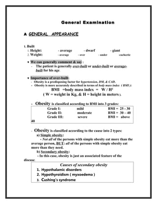
- 1. General Examination A GENERAL APPEARANCE 1. Built 1. Height: - average - dwarf - giant 2. Weight: - average - over - under -cachectic ♥ We can generally comment & say : - The patient is generally over-built or under-built or average- built for his age. ♥ Importance of over-built: - Obesity is a predisposing factor for hypertension, DM, & CAD. - Obesity is more accurately described in terms of body mass index ( BMI ): BMI =body mass index = W / H² ( W = weight in Kg, & H = height in meters ). - Obesity is classified according to BMI into 3 grades: Grade I: mild BMI = 25 - 30 Grade II: moderate BMI = 30 – 40 Grade III: severe BMI = above 40 - Obesity is classified according to the cause into 2 types: a) Simple obesity: - Not all of the persons with simple obesity eat more than the average person, BUT: all of the persons with simple obesity eat more than they need. b) Secondary obesity: - In this case, obesity is just an associated feature of the disease: Causes of secondary obesity 1. Hypothalamic disorders 2. Hypothyroidism ( myxoedema ) 3. Cushing’s syndrome
- 2. 4. Corticosteroids intake ♥ Importance of under-built: “ loss of weight ” - Congenital heart diseases with chronic infections & chronic hypoxia. - Cardiac cachexia in severe chronic heart failure. - Diabetes mellitus. - Thyrotoxicosis - Malignancy - Malabsorption & malnutrition 2. Decubitus “ position in bed ” 1. Semi sitting: is encountered in left-sided heart failure ( orthopnea ). 2. Sitting and leaning forward: is encountered in pericardial effusion ( prayer’s position ). 3. Squatting is encountered in the Tetralogy of Fallot. 3. Consciousness & mentality disturbed ( drowsy, confused, or disoriented ) in: ▪ COPD & respiratory failure: CO2 narcosis. ▪ Liver cell failure: Hepatic encephalopathy. ▪ Renal failure: Uremic encephalopathy. B FACE EXAMINATION 1. Pallor - We search for pallor in: - face , conjunctiva , lips, tongue , hands, palmer creases - In cardiac disease, pallor may be due to: 1. Anemia 2. Rheumatic activity 3. Infective endocarditis
- 3. 4. Low cardiac output 5. Shock Types of anemia on the clinical back ground: i. IRON DEFICIENCY ANEMIA ii. HAEMOLYTIC ANEMIA iii. MEGALOBLASTIC ANEMIA IRON DEFICIENCY ANEMIA: This is the commonest type of anemia. There is usually a clue for blood lose in the history. You have to look for the evidence of chronicity which are : Angular stomatitis ”soreness at mouth angles” Atrophic glossitis:” smooth tongue” Kilonychia ”spoon shaped nails” HAEMOLYTIC ANEMIA: This will result from the reduction of life span of red blood cells to 100 days as this will present with pallor and jaundice . Causes of haemolysis: A. Congenital 1.Abnomality of the shape-spheroctosisi, elliptocytosis 2.Abnormality of haemoglobin – thalasemia alpha and beta 3.Abnormality of RBC enzymes – glucose 6 phosphate dehydrogenase DEF. B. Acquired 1. Atypical pneumonia – mycoplasma 2. Virus – HIV , HAV 3. parasite –malaria 4. Connective tissues diseases= SLE 5. Lymphoma and leukemia 6. prosthetic valve 7. drug induced –methyl dopa 8. Autoimmune heamolysis
- 4. MEGLOBLASTIC ANEMIA: It is due to vitamin b12 or folic acid deficiency. It is commonly due to vit.b12 deficiency which is commonly caused by pernicious anemia “autoimmune” In pernicious anemia , The patient is asthenic , anemic , albinism with atrophic gastritis , amnesia. The presence of GIT symptoms , CNS symptoms ,LOWER LIMB weakness gives a clue toward the diagnosis.
- 5. 2. Cyanosis ♥ Definition: - Bluish discoloration of skin & mucous membranes due to the presence of excessive blue blood ( unoxygenated ) in the circulation ( more than 5 gm % reduced Hb ) or decrease blood flow peripherally. It could be Central cyanosis, or Peripheral cyanosis. The bluish color is more readily apparent in those with high hemoglobin counts than it is with those with anemia. Also the bluer color is more difficult to detect on deeply pigmented skin. When signs of cyanosis first appear, such as on the lips or
- 6. fingers, intervention should be made within 3–5 minutes because a severe hypoxia or severe circulatory failure may have induced the cyanosis. ♥ Examination: Where do we search for cyanosis?? - Look at the tongue, lips & nails for the blue colour of cyanosis: ▪ Central cyanosis: blue colour in all “ tongue” ▪ Peripheral cyanosis: blue colour in nails only “hands and feet”. If the hands are cold ,it is likely to be peripheral cyanosis. The central cyanosis will result in warm hands with blue colour. ♥ Common causes: Cyanosis Central Peripheral Definition - Blood pumped in the aorta contains more than 5 gm % reduced Hb. - Blood pumped in the aorta is quit normal (oxygenated), BUT: there is stagnation of blood in the peripheral circulation, leading to extraction of more oxygen & development of cyanosis. Causes - Cyanotic congenital heart disease - Lung fibrosis , bronchiectasis & Pulmonary embolism, emphysema hypoxic corpulmonale. - Venous stagnation, e.g. CHF - Venous obstruction. Effect of warming No effect Improves Effect of oxygen inhalation May improve central cyanosis which is due to chronic lung disease only. No effect
- 7. ♥ Why do we particularly look for central cyanosis in the tongue?? - Normally the colour of a tissue depends on: 1 Sympathetic supply: state of blood vessels ( vasoconstriction or vasodilatation ). 2 Pigment granules. 3 Colour of the Hb pigment in the circulating blood. - The tongue is devoid of: 1 sympathetic supply, & is devoid of: 2 pigment granules, & so the tongue colour depends only on 3 colour of the Hb pigment in the circulating blood, & so the tongue colour is a good index of the blood colour. Causes of peripheral cyanosis: • All common causes of central cyanosis • Arterial obstruction • Cold exposure (due to vasoconstriction) • Raynaud phenomenon (vasoconstriction) • Reduced cardiac output (e.g. heart failure, hypovolaemia) • Vasoconstriction • Venous obstruction (e.g. deep vein thrombosis) Causes of central cyanosis: 1. Respiratory System: • Bronchospasm (e.g. Asthma) • brochiectasis • Pulmonary Hypertension • Pulmonary embolism • Hypoventilation • COPD (emphysema and chronic bronchitis) • Lung fibrosis 2. Cardiac Disorders: • Congenital heart disease (e.g. Tetralogy of Fallot, ) • Pulmonary hypertension • Heart valve disease • Myocardial infarction “shock” 3.blood • Methemoglobinemia
- 8. Differential cyanosis Differential cyanosis is the bluish coloration of the lower but not the upper extremity and the head. This is seen in patients with a patent ductus arteriosus. Patients with a large ductus develop progressive pulmonary vascular disease, and pressure overload of the right ventricle occurs. As soon as pulmonary pressure exceeds aortic pressure, shunt reversal (right-to-left shunt) occurs. The upper extremity remains pink because the brachiocephalic trunk, left common carotid trunk and the left subclavian trunk is given off proximal to the PDA. 4. Abnormal pigmentation - Malar flush: erythematous (red) rash in the butterfly area of the face in: ▪ Mitral stenosis ▪ Systemic lupus erythematosus 5. Eye examination a) Loss of hair in the outer 1/3 of the eye brows: - Myxoedema - Leprosy b) Puffiness of lower eye lids: - Diabetic nephropathy -Hypertensive patients - Graves disease - Chronic cough - Constrictive pericarditis & pericardial effusion - SVC obstruction - Severe hypoproteinemia & renal failure - Myxoedema c) Peticheal hemorrhage in the conjunctiva: - Malignant hypertension - Infective endocarditis - Purpura
- 9. Splinter hemorrhages(Panel A) are normally seen under the fingernails. They are usually linear and red for the firstr two to three days and brownish thereafter. Panel B shows conjunctival petechiae. Osler's nodes (Panel C)are tender, subcutaneous nodules, often in the pulp of the digits or the thenar eminence. Janeway's lesions (Panel D) are nontender, erythematous, hemorrhagic, or pustular lesions, often on the palms or soles. D) Exophthalmos: - Thyrotoxicosis E) Pallor: “ in the conjunctiva ” F) Jaundice: “ in the sclera ” ASK THE PATIENT TO LOOK DOWN AND PULL THE UPPER EYE LID GENTLY WITH YOUR THUMBS NEAR THE LINE OF EYE LASHES TO LOOK FOR SCLERA AND TO LOOK UPWARD TO PULL THE LOWER EYELID DOWNWARD WITH THE THUMB YO LOOK FOR CONJUNCTIVA FOR PALLOR.
- 10. You have to check the elastic tissues which has a high affinity for bilirubin. They are : the sclera and under the tongue. Three types of jaundice: 1.Prehepatic jaundice 2.Hepatic jaundice 3.Posthepatic jaundice In prehepatic jaundice= pallor and jaundice In hepatic jaundice, the commonest cause is chronic liver diseases “cirrhosis” The stigma of chronic liver disease should be detected once the patient has jaundice which are: • spider nevi • Palmar erythema= reddening of the palms at the thenar and hypothenar eminences • Clubbing • Ecchymosis • Kilonychia • leukonychia • Scratch marks • Gynaecomastia • Feminising hair distribution • Testicular atrophy • Wasting of small muscles of hand and muscle emaciation • Anaemia • Caput medusae (recanalisation of the umbilical vein) (Distended abdominal veins) Signs associated with decompensation • Drowsiness (encephalopathy) • Metabolic Flap/Asterixis (encephalopathy) • Jaundice (excretory dysfunction) • Ascites (portal hypertension and hypoalbuminaemia) • Peripheral oedema (hypoalbuminaemia) • Bruising (coagulopathy)
- 11. Signs associated with the aetiology • Dupuytren's contracture, Parotidomegally (Alcohol) • Peripheral neuropathy (Alcohol and some drugs) • Cerebellar signs (alcohol and Wilson's disease) • Hepatomegaly (alcohol, NAFLD, Haemochromatosis) • Kayser-Fleisher Rings (Wilson's) • Increased pigmentation of the skin (Haemochromatosis) • Signs of Right Heart Failure • Tattoos (Hepatitis C) Spider nevi A spider angioma ( spider nevus, vascular spider, and spider telangiectasia) is a type of telangiectasis found slightly beneath the skin surface, often containing a central red spot and reddish extensions which radiate outwards like a spider's web. They are common and may be benign, presenting in around 10-15% of healthy adults and young children. However, having more than five spider naevi may be a sign of liver disease.
- 12. Spider angiomas are found only in the distribution of the superior vena cava, and are thus commonly found on the face, neck, upper part of the trunk and arms. They may also be present on the backs of the hands and fingers in young children. Spider angiomas are due to failure of the sphincteric muscle surrounding a cutaneous arteriole. The central red dot is the dilated arteriole and the red "spider legs" are small veins carrying away the freely-flowing blood. If momentary pressure is applied, it is possible to see the emptied veins refilling from the centre. No other angiomas show this phenomenon.
- 13. The hepatic jaundice can be caused by any pathological process that involves the liver like hepatitis , hepatic carcinoma , congested liver , liver cirrhosis etc. Those patients commonly present with bleeding tendency with ecchymosis , Leg edema and jaundice. In posthepatic jaundice, there is obstruction in the common bile duct which shift the bilirubin to be excreted in the urine “increased urobilinogen, stercobilirubin” .This will result in change the colour of urine and stool. There will be 3 signs : 1.White colored stool –steatorrhea =clay colored stool 2.Tea colored urine –dark brown =haematuria 3.Olive green eyes as the jaundice is progressive It is a surgical jaundice that need surgical intervention. Causes of obstructive jaundice: 1. obstruction of common bile duct by Stone, tumor , stricture , blood clot , porta hepatis 2. carcinoma head of pancrease 3. carcinoma of ampulla of vater
- 14. 6. Tongue examination a) Cyanosis b) Coated tongue: - Fevers especially Typhoid. c) Dry tongue: - Dehydration & renal failure. d) Red glazed tongue: - Riboflavin deficiency ( Vitamin B2 deficiency ). e) Tremors: - Fine: Thyrotoxicosis - Flapping: Heart failure, renal or respiratory failure.
- 15. SUMMARY; 1. look for the patient and comment on the conscious level, position on bed , general looking , colour of skin. 2. Exam the hair for alopecia , upper eye lids , ask the patient to look down and pull the upper eye lids of the patient with your thumbs to look for jaundice. 3. Ask the patient to look up and pull the lower eye lids for conjunctiva for pallor , bleeding. 4. Exam the mouth- tongue and mucous membrane for cyanosis , pigmentation , pallor , atrophy , lose of papilla. 5.Ask the patient to set to look for the neck and exam for swellings if present and ask him to swallow for thyroid swelling. 5. Sit behind the patient while he is sitting and exame for lymph nodes. 6. Ask the patient to lie on the bed elevated 45 for neck veins. 7. Exam hands for pallor& cyanosis ,nails for clubbing , pulse. 8. Exam lower limbs for calf swelling, pedal edema, ischemic signs. C NECK EXAMINATION 1. Neck veins 2. Carotid arteries 3. Thyroid gland 4. Trachea 5. Lymph nodes 1. Neck Veins Jugular Venous Pulse ( JVP ) a) Identification: ▪ There are 2 jugular veins on each side of the neck: External Internal - Easy to see, but gives false impression of raised pressure. - More difficult to see, but is accurate for measurement of pressure & pulsations. - Runs from the midpoint of the clavicle upwards to cross the sternomastoid muscle obliquely. Runs deeply from the sternoclavicular joint upwards & laterally to the angle of the jaw. ▪ Both can be identified by asking the patient to perform a Valsalva manoeuvre.
- 16. b) Comment: Neck veins should be examined for: I Pressure: ( congested or not?? ) ♥ Positioning the patient: - No special position to measure the JV pressure. -Just make the patient relaxed & comfortable with his head slightly elevated on a pillow & his sternomastoid muscles relaxed. -Adjust the head of the bed so as to maximize the JV pulsations & make them visible in the lower half of the neck ( above the clavicle, but below the jaw.( -Usually, the head of the bed needs slight elevation (45º -However, when the patient’s venous pressure is increased, the head of the bed may need more elevation up to 60º or even 90º.
- 17. - In all these positions , the sternal angle remains about 5 cm above the right atrium. ♥ Measuring the pressure: - Identify the external jugular vein. - .Identify the pulsations of the internal jugular vein Because ,this vein lies deep to the sternomastoid you may not see the . ,vein itself Instead watch for the pulsations transmitted through the surrounding soft tissues. - Identify the highest point of pulsation in the internal jugular vein: ▫ With a centimeter ruler measure the vertical distance between this point & the sternal angle, as in the Fig. below. ▫ Venous pressure greater than 3 cm above the sternal angle is considered elevated ( with the patient in any position ). II Pulsations: ( pulsating or not?? ) ♥ JV pulsations reflect pressure changes in the right atrium.
- 18. ♥ Normally, the JV pulsations are wavy & consist of: a-wave : right atrial contraction. x-descent : right atrial relaxation. c-wave : - transmitted from adjacent carotids. - upward bulge of tricuspid valve into right atrium. v-wave : right atrial filling during ventricular systole. y-descent: right atrial empty & opening of tricuspid v. ♥ Normal systolic collapse: ▪ Normally, the x-descent coincides with the arterial pulsations. Important points in comment on the neck veins * Unilateral distension of the ext. jug. vein is usually due to local kinking or obstruction. * Empty neck veins when the patient is horizontal may indicate hypovolemia. * Full neck veins up to the angle of the jaw when the patient is upright may indicate severe CHF. * Hepato jugular reflux: - In CHF, exert firm & sustained pressure with your hand over the patient’s right hypochondrium for 1 minute. - Watch for an increase in the JV pressure. A rise of more than 1 cm is abnormal & confirms the presence of CHF ( positive hepato jugular reflux ). c) Clinical significance of neck veins: 1) Abnormal pressure: Congested neck veins Congested pulsating: - Right sided HF - Pericardial effusion & constrictive pericarditis - Hypervolemia Congested non-pulsating: - SVC obstruction - Mediastinal syndrome 2) Abnormal Pulsations: Abnormal waves a- wave : - GAINT: complete heart block,pulmonary hypertension, PS, TS - Absent : AF
- 19. x-descent: - Obliterated ( systolic expansion of neck veins ): * TR * AF * Cannon waves: . regular : nodal rhythm . occasional : A-V dissociation - Prominent : * Constrictive pericarditis * Pericardial effusion y-descent : - Prominent : * Constrictive pericarditis 2. Carotid Arteries a) Prominent pulsations: volume load e.g. Aortic regurgetation. b) Systolic thrill: 1- Propagated from the base of the heart: - Aortic stenosis: there will be thrill over the base of the heart. 2- Initiated in the vessel itself: - In cases with big pulse volume, e.g. Aortic regurge: there will be no thrill over the base of the heart. Jugular venous pulsations Carotid artery pulsations Impulse Wavy impulse One impulse Palpation Rarely palpable ( better seen ) Palpable Location Lateral to the sternomastoid Medial to the sternomastoid Upper level Has an upper level No upper level Congestion - Increases with gentle pressure - Increases with hepato jugular reflux - No effect - No effect - No effect - No effect
- 20. - Decreases with inspiration - Varies with position Head/Neck Lymph Nodes The greatest supply of lymph nodes are located in the head and neck. Sources tend to differ on the name of these lymph nodes. These particular names of the lymph nodes used correspond to adjacent structures. • Preauricular, in front of the ear • Posterior auricular (mastoid), superficial to the mastoid process • Occipital, at the base of the skull • Submental, midline, behind the tip of the mandible • Submandibular, halfway between the angle and the tip of the mandible • Jugulodigastric, under the angle of the mandible • Superficial cervical, overlying the sternomastoid muscle • Deep cervical, deep under the sternomastoid muscle • Posterior cervical, in the posterior triangle , the edge of the trapezius muscle • Supraclavicular, just above & behind the clavicle. Head & neck lymph nodes 1. SUBMENTAL 2. SUBMANDIBULAR
- 21. 3. PAROTID 4. PREAURICULAR 5. POSTAURICULAR 6. OCCIPITAL 7. ANTERIOR CERVICAL 8. SUPRACLAVICULAR 9. POSTERIOR CERVICAL When examining for lymph nodes you want to note: • Site: localised to one region or generalised? • Size: Large nodes are usually abnormal (greater than 1 cm) • Consistency: Hard nodes suggest carcinoma, soft may be normal and rubbery nodes may be due to lymphoma • Tenderness: Usually implies acute inflammation or infection • Fixation: Nodes that are fixed to underlying structures are more likely to be due to carcinoma • When examining one area, always compare to the other side • When examining for lymph nodes, usually an abdominal examination should take place. Particularly examining for splenomegaly, hepatomegaly, para-aortic nodes and any potential masses. HOW TO EXAM THE LYMPH NODES? Using a gentle circular motion with your finger pads palpate each lymph node in the order previously stated. It is a good idea to palpate both sides at the same time, comparing the two sides symmetrically. Normal cervical nodes should be less than one centimeter, movable, discrete, soft, and nontender. AXILLARY LYMPH NODES Patient position: Usually the patient is seated or reclined on a couch for this examination. The examiner tends to raise the patient’s arm, and using the left hand for the patients right axilla, (and vice versa) the examiner passes their extended fingers high into the patients axilla.
- 22. The patients arm is now brought to rest on the examiners forearm. Now the examiner should palpate for the following groups of nodes: 1) CENTRAL/APEX 2) LATERAL 3) PECTORAL (medial) 4)INFRACLAVICULAR 5) SUBSCAPULAR . Every effort should be made to feel for nodes in each of these areas. Axillary or armpit lymph nodes drain lymph from the arms, breast, chest wall and upper abdomen INGUINAL LYMPH NODES For clinical purposes, an oblique set of nodes along the inguinal ligament area and a longitudinal set overlying the femoral vessels are usually palpated for. Have the patient reclined on the couch for this assessment. You will need to expose the groin area. Note that small nodes can be commonly detected in otherwise normal patients. Causes of L.N. enlargement: 1. INFECTION 2. LEUKEMIA
- 23. 3. LYMPHOMA 4. HIV 5. METASTASES 6. CONNECTIVE TISSUE DISEASES D UPPER LIMBS 1. Clubbing. 2. Spooning: iron deficiency anemia. 3. Splinter hemorrhages: infective endocarditis. 4. Subcutaneous nodules: rheumatic fever. 5. Pallor or peripheral cyanosis. Clubbing ▪ Definition: - Tissue proliferation of the nail bed due to chronic toxemia or hypoxia . Clubbing develops in five steps: 1. Fluctuation and softening of the nail bed (increased ballotability) 2. Loss of the normal <165° angle (Lovibond angle) between the nailbed and the fold (cuticula) 3. Increased convexity of the nail fold 4. Thickening of the whole distal (end part of the) finger (resembling a drumstick) 5. Shiny aspect and striation of the nail and skin Schamroth's test or Schamroth's window test (originally demonstrated by South African cardiologist Leo Schamroth on himself) [5] is a popular test for clubbing. When the distal phalanges (bones nearest the fingertips) of corresponding fingers of opposite hands are directly opposed (place
- 24. fingernails of same finger on opposite hands against each other, nail to nail), a small diamond-shaped "window" is normally apparent between the nailbeds. If this window is obliterated, the test is positive and clubbing is present. ▪ Degrees: 1- First: - Obliteration of the angle of the nail bed. - Increased nail curvature in longitudinal &lateral axes. - When palpating the nail base, it gives a spongy or floating sensation. 2. Second: Parrot beak appearance. 3. Third: Drum stick appearance. ▪ Causes of clubbing: NO ACUTE CAUSE , IF PRESENT IT IS A CHRONIC CAUSE. A) Cardiac: Cyanotic congenital heart disease (cyanotic clubbing ) - Infective endocarditis ( pale clubbing ). - Atrial myxoma (benign tumor) B) Chest:
- 25. • Fibrosing alveolitis. • Lung cancer, mainly non-small-cell (54% of all cases), not seen frequently in small-cell lung cancer (< 5% of cases)[6] • Interstitial lung disease • Complicated tuberculosis • Suppurative lung disease: • lung abscess, empyema, bronchiectasis, cystic fibrosis • Mesothelioma of the pleura • Arteriovenous fistula or malformation` C) Hepatic & GIT: - Liver cirrhosis, especially biliary. - Ulcerative colitis & Crohn’s disease. - Bilharzial polyposis coli. D) Congenital Others: • Hyperthyroidism (thyroid acropachy)[9] • Familial and racial clubbing and "pseudoclubbing" (people of African descent often have what appears to be clubbing) • Vascular anomalies of the affected arm such as an axillary artery aneurysm (in unilateral clubbing) A special form of clubbing is hypertrophic pulmonary osteoarthropathy, known in continental Europe as Pierre Marie- Bamberger syndrome. This is the combination of clubbing and thickening of periosteum (connective tissue lining of the bones) and synovium (lining of joints), and is often initially diagnosed as arthritis. It is commonly associated with lung cancer. E LOWER LIMBS
- 26. 1. Oedema. 2. Dorsalis pedis pulsations. 3. Clubbing & peripheral cyanosis. 4. Chronic leg ulcers. Oedema 1. Examination: -Press firmly with your thumb for at least 10 seconds behind each medial malleolus, over the dorsum of each foot & over the shins - Look for pitting: a depression in the skin caused by pressure. - Check for sacral oedema in bed ridden patients. - Palpate the calf muscles for signs of DVT: calf muscles are firm, tense & tender. - Look for signs of inflammation: redness, hotness or discoloration. 2. Characteristics of cardiac oedema: 1. Occurs in the dependant parts of the body: Ankle oedema : in ambulant patients. Sacral oedema : in bed ridden patients. 2. Bilateral, but one side may be affected more due to: Deep venous thrombosis. Postural, in patients sleeping on one side. 3. Always pitting 4. Oedema of lower limbs always precedes appearance of ascites, except in two conditions in which ascites occurs first “Ascites precox”: a) Percardial effusion & constrictive pericardit - Kinking of hepatic veins causes early liver congestion & ascites. - Obstruction of lymphatics passing through the central tendon of the diaphragm causes accumulation of lymph in the peritoneum. b) Tricuspid incompetence: - Regurgitation of blood causes liver congestion & ascites. 4. Differential diagnosis of cardiac oedema:
- 27. 1. Renal Oedema: This occurs in nephritis or nephritic syndromes. Oedema occurs first in the eye lids and is associated with features of renal disease. Nephrotic syndrome: 1.protenuria 2.hypoalbuminemia 3.generalized anasarca 4.hyperlipidemia Nephritic syndrome: 1.Haematuria 2.Hypertension 3.Urine casts 2. Hepatic Oedema: Oedema of lower limbs occurs after ascites and is associated with features of liver disease. 3. Nutritional Oedema: This occurs with severe nutritional deficiency. 4. Angioneurotic Oedema: -This allergic oedema occurs with constant relation to certain factors, as eating certain food or inhaling certain substances. - Oedema occurs acutely especially in the lips, eye lids & larynx, but may be generalized . There is usually positive family history of oedema or other allergies, and the patient himself may have other forms of allergy. - Rapid response of oedema to antiallergic measures is characteristic. 5. Local Oedema
- 28. G VITAL SIGNS 1. Temperature, BP & respiratory rate 2. Pulse one's pulse represents the tactile arterial palpation of the heartbeat by trained fingertips. The pulse may be palpated in any place that allows an artery to be compressed against a bone, such as at the neck (carotid artery), at the wrist (radial artery), behind the knee (popliteal artery), on the inside of the elbow (brachial artery), and near the ankle joint (posterior tibial artery). ► Rate: • Normally: 60-100 beat / min. • Abnormally: bradycardia, or tachycardia. • Can be counted in half a min. & multiplied by two. ► Rhythm: • Regular or irregular. • If irregular: you should:
- 29. 1. Count the rate in one whole min. 2. Count the apical heart rate. • Irregularity is either: 1. Occasional: extrasystoles. 2. Marked: AF • Pulsus deficit (difference between apical rate & radial pulse rate): 1. In extrasystoles: Pulsus deficit is less than 10. 2. In AF: Pulsus deficit is more than 10. ► Volume: • Average. • Big: - AR & hyperdynamic circulation. • Small: - Decreased filling: Pericardial effusion & constrictive pericarditis. - Decreased pumping: HF & myocardial disease. - Obstruction: MS, AS & pulmonary hypertension. ► Special character: Water hammer: AR & hyperdynamic circulation. Pulsus alternans: Alternating strong & weak regular beats in LVF. Pulsus paradoxus: Diminished pulse volume on inspiration in: Pericardial effusion, constrictive pericarditis & COPD. Pulsus bigeminus: Alternating normal beats & extrasystoles in: - Digitalis toxicity - Very frequent extrasystoles. Pulsus bisferiens: A double systolic peak in double aortic & IHSS. Plateau pulse: Delayed upstroke, in AS. ► Arterial wall:
- 30. &Thickened felt in .atherosclerosis ► Equality of pulse volume on both sides: Normally, pulse volume is equal on both sides. Abnormally, there is unequal pulse volume on both sides, in: 1. Pressure from outside: - Cervical rib, or pancoast tumour. 2.Disease of the arterial wall: - Aneurysm of aortic arch, or subclavian artery. - Arteritis. 3. Occlusion of the lumen: - Thrombosis or embolism.