Extra-Osseous TaloTarsal Stabilization Devices
•
0 recomendaciones•932 vistas

This report describes the difference between a type I subtalar arthroereisis device and a type II non-arthroereisis device used to stabilization a talotarsal joint dislocation. Learn more at www.GraMedica.com.
Recomendados

Recomendados
Más contenido relacionado
Más de GraMedica
Más de GraMedica (20)
Último
Último (20)
Extra-Osseous TaloTarsal Stabilization Devices
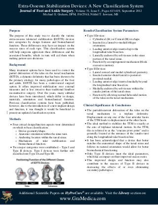
- 1. Extra-Osseous Stabilization Devices: A New Classification System Journal of Foot and Ankle Surgery, Volume 51, Issue 5 , Pages 613-619, September 2012 Michael E. Graham, DPM, FACFAS, Nikhil T. Jawrani, MS Purpose Results/Classification System Parameters The purpose of this study was to classify the various Type I Devices: extra-osseous talotarsal stabilization (EOTTS) devices o Cylindrical (IA) or Conical (IB) in shape. into categories by design features and biomechanical o Inserted in a lateral-to-medial/oblique function. These differences may have an impact on the orientation. success rates of each type. This classification system o Leading anterior edge inserted up to the will help surgeons appreciate these differences and the longitudinal talar bisection. associated benefits, which in turn will aid them when o Laterally anchored by soft tissues in the sinus making patient care decisions. portion of the tarsal sinus. o Function by an impingement mechanism (block Background excessive motion). Many treatment options have been used to correct the Type II Devices: partial dislocation of the talus on the tarsal mechanism o Lateral conical and medial cylindrical geometry. (RTTD), a dynamic deformity that has been shown to be o Inserted anterior-distal-lateral to posterior- the primary etiology for many pathologies of the foot proximal medial. and ankle. EOTTS has been a controversial surgical o Leading anterior edge inserted medially beyond option. It offers improved stabilization over external the longitudinal talar bisection. measures and is less invasive than traditional hindfoot o Medially anchored by soft tissues within the reconstructive surgery. Over the years, many subtalar canalis portion of the tarsal sinus. devices have been introduced, which differ in design, o Function by allowing normal helicoidal motion materials, orientation and biomechanical function. of the talus on the tarsal mechanism. Previous classification systems have been published, however, due to the introduction of a new implant design Clinical Significance & Conclusions and function, it was thought it would be beneficial to The partial/recurrent dislocation of the talus on the present an updated classification system. tarsal mechanism is a triplane deformity. Displacement on any one of the four articular facets Methods of the TTM leads to displacement at the other facets. Four critical design/function aspects were determined The ideal method to stabilize the TTM is exactly at on which to base classification: the axis of triplanar talotarsal motion. In the TTM, o Device geometry/shape. this is referred to as the “cruciate pivot point” and is o Anatomic orientation within the sinus tarsi. generally located at the entrance of the canalis tarsi o Anchoring location within the tarsal sinus. along the longitudinal talar bisection line. o Mechanism of talar stabilization and It has been acknowledged that a device that better biomechanical function. matches the anatomical shape of the tarsal sinus and Two major categories were established – Type I and follows its natural orientation would allow for better Type II devices. Type I devices were further sub- biomechanical functioning. classified into Type IA and Type IB. Only Type II devices meet the ideal parameters, which has an impact on their improved success rates. This improved design and function may also contribute to the success of Type II devices in decreasing the effects of or even eliminating secondary pathologies. Additional Scientific Papers on HyProCure® are available. Visit the Library section on: www.GraMedica.com