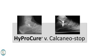
HyProCure Verse Calcaneo-Stop
•
5 likes•3,257 views

This document compares the HyProCure device to the Calcaneo-stop device for treating foot disorders. HyProCure is an extra-osseous device that stabilizes the talus bone at the subtalar joint axis without blocking motion, while Calcaneo-stop is an intra-osseous screw that blocks talus motion. HyProCure has advantages of allowing normal motion, lower removal rates, and no reports of bone fractures. In contrast, Calcaneo-stop blocks motion, has removal rates up to 100%, and complications of bone fractures. Studies show HyProCure decreases strain on soft tissues compared to no effect shown for Calcaneo-stop.

Recommended
Recommended
More Related Content
What's hot
What's hot (20)
Viewers also liked
Similar to HyProCure Verse Calcaneo-Stop
Similar to HyProCure Verse Calcaneo-Stop (20)
More from GraMedica
More from GraMedica (20)
Recently uploaded
Recently uploaded (20)
HyProCure Verse Calcaneo-Stop
- 2. One of these devices is far superior than the other…you be the judge.
- 3. What is Calcaneo-stop? • This is an Arthroereisis – joint blocking procedure. • Can also be called: Intra-osseous Talotarsal stabilization – a screw is partially drilled into talus or calcaneus. • The head of a screw blocks or restricts talar motion. • Functions against the normal TTJ range of motion. • Does not stabilize the talus at the axis of the subtalar joint.
- 4. What is HyProCure? • Non-arthroereisis/Non-joint blocking procedure. • An Extra-osseous talotarsal stabilization (EOTTS) device. • It is NOT inserted into bone. • Restores talotarsal joint motion at the axis point of the subtalar joint. • Does not block nor limit TTJ ROM. • Functions with TTJ motion, not against.
- 5. A Closer Side-by-Side Comparison HyProCure Extra-osseous HyProCure is not inserted into bone, rather is it positioned within the canalis and sinus portions of the sinus tarsi. There is no reported damage to the bone. Calcaneo-stop Intra-osseous Calcaneo-stop is partially drilled into either the talus or calcaneus. The integrity to the bone is weakened and could lead to a possible fracture of the talus or calcaneus. This is a reported complication.
- 6. Held in place by talocalcaneal interosseous ligament (TCIL) adherence along with the osseous chamber forming the sinus tarsi. Advantage/Disadvantage: There is no interruption to the integrity of bone. The TCIL is not functioning to stabilize the talus on the calcaneus. It is transected, the stent is placed, and the TCIL will heal back together to incorporate the stent within the fibers of the ligament. A potential problem occurs when the TCIL has atrophied due to chronic talar hypermobility. In that situation, the anchoring mechanism is compromised. Anchoring Method Partially drilled into the bone. Advantage/Disadvantage: There is perceived to be a less likelihood of device displacement, yet this is still a reported complication. A major disadvantage is that the integrity to the bone is compromised and that lead to a fracture of the bone. This is also a reported complication. A bone defect occurs upon removal of the device. HyProCure Calcaneo-stop
- 7. How is talar stabilization accomplished? • The smooth tapered section of HyProCure stabilizes the talus at the axis point of the subtalar joint. • The lateral process of the talus smashes into the head of the screw in the outer part of the sinus. HyProCure Calcaneo-stop
- 8. Location on the calcaneus where the talus is stabilized. CalcaneoStop device acts here: HyProCure internally stabilizes the talus at the axis point here. Anterior Posterior MedialLateral
- 9. Non-Arthroereisis vs. Arthroereisis Stabilization • Non-Arthroereisis • This means HyProCure maintains the alignment and stabilization of the talus while allowing a normal talotarsal joint range of motion. • HyProCure restores TTJ ROM. • Arthroereisis • This means that there is an abrupt blocking of talotarsal joint range of motion. • Calcaneo-stop blocks TTJ ROM. HyProCure Calcaneo-stop
- 10. Who is a candidate – Age Restrictions? HyProCure • Minimum age: 3 years • Maximum age: none Calcaneo-stop • Minimum age: 6 years • Maximum age: 14 years
- 11. Stand-alone or Combination of Procedures? HyProCure • Pediatric (3 and older) can be performed as a stand-alone, when indicated. • Adults – can be performed as a stand-alone or combined with other procedures, when indicated. Calcaneo-stop • Pediatric (8 – 14) can be a stand- alone, when indicated. • Adults – rarely recommended and only when combined with other surgical procedures.
- 12. Published Removal Rates? 6% or less HyProCure is designed to stay in place for the life of the patient. Up to 100% Calcaneo-stop screw is recommended to be removed within a few years. HyProCure Calcaneo-stop
- 13. What happens after device removal? Is there any supporting radiographic data? HyProCure • No scientific published paper that follows a patient after HyProCure has been removed. • The same statements can be made concerning HyProCure. Calcaneo-stop • No scientific published paper that follows a patient after the CalcaneoStop screw has been removed. • There is non-scientifically proven statements made in published literature that suggest that correction is maintain, yet there is no proof to substantiate this claim.
- 14. Reported Complications specific to the device: • Revision/Repositioning/Resizing • Permanent removal • Screw breakage • Resorption of cortical surfaces • Metatarsal stress fracture • Bone fracture • Bone destruction • Revision/Repositioning/Resizing • Permanent removal HyProCure Calcaneo-stop
- 15. Beliefs of the advocates of Calcaneo-stop? • The device is a rather inexpensive orthopedic screw. • The smooth head should limit damage to bone. • Because it is anchored into bone, there should be less likelihood for displacement. • It is extra-articular. • No sinus tarsi dissection is required. • Can be reversed. • Superior option over calcaneal osteotomy.
- 16. The price of Calcaneo-stop is less than HyProCure. • While it may appear that the actual Calcaneo-stop screw is less expensive, further considerations must be explored. • The removal rate of Calcaneo- stop is reported to be as high as 100% of the time. • How much does it cost to bring a patient back to the operating room for a second surgery? These devices cannot be removed in a private practice setting. • The hospital and surgeon may see this high removal rate as an advantage because they get paid to put the device into the patient’s foot and also get paid to take it out. • There is a financial incentive to use a device that is only slightly less expensive, that has a significantly higher removal rate.
- 17. The smooth head Calcaneo-stop should limit bone trauma. • Imagine that 5,000 to 10,000 times a day the ankle bone is smashing into the head of this screw. • Eventually, this will take a toll on the bone and could lead to serious issues. • That is why most surgeons want to remove the device after a few years. There are no long term studies to show that no bone damage occurs. • To the contrary, there are reports of complications including fracture of the talus and calcaneus.
- 18. Because Calcaneo-stop is anchored into bone, there is less likelihood of displacement. • Displacement still occurs, this is a reported complication. • There is no scientific data comparing displacement rates of HyProCure to Calcaneo-stop, however, there are more potential risks and complications associated with Calcaneo-stop.
- 19. Calcaneo-stop is Extra-Articular • Both HyProCure and Calcaneo-stop are placed outside of the articular facets of the talocalcaneal joints.
- 20. HyProCure and Calcaneo-stop are a superior option to calcaneal osteotomy. • Surgeon advocates of both HyProCure and Calcaneo-stop will agree that the insertion of these devices is a conservative minimally invasive, reversible option over a calcaneal osteotomy. A procedure that is aggressive, requires a longer recovery, and is not reversible.
- 21. Neuroproprioceptive theory as a benefit for the calcaneo-stop procedure. • There are opinions that the TCIL has “specialized” neurosensors that help to control foot motions. • There is no evidence to this –it still remains an hypothesis. • In fact, the TCIL is compromised in patients with a hypermobile talus. • There are no clinical supportive studies to confirm this only a neurohistologic cadaver analysis. • Patients have the contents of their sinus tarsi routinely removed without reported complication. The sinus tarsi is explored via arthroscopic surgery on a routine basis without reported complication. • The fact that there is no dissection of the sinus tarsi is pointless because HyProCure replaces the function of the TCIL. The TCIL is not removed, it is transected and will heal.
- 22. HyProCure Advocates: • Routinely used in pediatric and adult patients of all activity levels. • Simple, minimally invasive, conservative procedure to stabilize the talus on the tarsal mechanism without interrupting the integrity of the talus or calcaneus. • It is reversible, without bone- defect-formation. • Has a significantly less removal rate than Calcaneo-stop and other arthroereisis devices. • Extensive scientific basis. • Less associated complications than Calcaneo-stop. • No case reports of fracture to the talus or calcaneus with HyProCure.
- 23. Published Reports on the Calcaneo-stop Procedure • Most literature has less than 2 years of follow up • There are no long-term studies to support the fact that correction of the angular deformities is maintained after the screw is removed. • Many of the claims are unsubstantiated, rather they are opinion based. • It is not expected that the screw will remain in place for the life of the patient. Rather it is recommended to be removed in 100% of cases.
- 24. Average follow up 2.9 years 24% of screws already removed. Contra-indicated 13 years or older.
- 25. 100% removal rate of the calcaneo-stop screw.
- 26. Mean follow-up was less than 10 months.
- 28. Extra-osseous Talotarsal Stabilization Devices: A New Classification System J Foot & Ankle Surgery – (52), 613-619, 2012. • Stabilization of the talotarsal joint is a primary consideration to the treatment of many lower extremity pathologies. • There are 2 main types of extra-osseous (not inserted into bones): • Type I – Arthroereisis devices • Cylindrical or conically shaped • Inserted lateral to medial so that the medial tip is aligned with the horizontal bisection of the talus. • Laterally anchored by the soft tissues within the sinus portion of the sinus tarsi. • Functions via impingement of the lateral process of the talus to block talar motion. • Works against normal talotarsal joint motion • Reported removal rate 38% to 100% • Type II – Non-Arthroereisis device • Combination of conical and cylindral shapes • Inserted along the normal orientation of the sinus and canalis tarsi. • Medial tip is inserted beyond the horizontal bisection of the talus. • Stabilizes the talus and restores the normal axis point of the subtalar joint. • Medially anchored. • Allows normal helicoidal motion of the talotarsal joint. • Reported removal rate 4 to 6% • Conclusion. • Sinus tarsi implants are not all the same. • Type II (HyProCure) is a superior design and function when compared to Type I arthroereisis devices. • Surgeons and patients must understand the differences between the 2 types. • Type II (HyProCure) has a superior success rate over Type I arthroereisis devices.
- 29. Stabilization of Joint Forces of the Subtalar Complex via the HyProCure Sinus Tarsi Stent Journal of American Podiatric Medical Association, Volume 101 No. 5, Pages 390-399, 2011 • Proves that HyProCure stabilizes the talus on the tarsal mechanism. • The stabilization of the talus on the tarsal mechanism reduces excessive abnormal forces acting on the medial column of the foot. • Therefore, there would be a decrease in strain on the supporting tissues on the medial column of the foot and decreased strain on these tissue allowing for tissue healing.
- 30. Effect of Extra-Osseous TaloTarsal Stabilization on Posterior Tibial Tendon Strain Journal of Foot and Ankle Surgery, Vol 50, N0. 6, Pages 676-681, 2011 • EOTTS with HyProCure decreased the elongation and strain of the posterior tibial tendon by 51%. • PTTD is a very expensive disease and no other form of treatment has shown a decreased strain on the tendon without arthrodesis and extensive hindfoot reconstructive surgery.
- 31. Evaluating Plantar Fascia Strain in Hyperpronating Cadaveric Feet Following an Extra-Osseous TaloTarsal Stabilization Procedure Journal of Foot and Ankle Surgery 50, Issue 6, Pages 682-686, 2011 • The #1 etiology of plantar fasciopathy is secondary to excessive tension/strain = mechanical overloading. • EOTTS-HyProCure decreased that strain by 33%. • No other form of treatment has been shown to decrease the strain on the plantar fascia. • Conservative care has never been shown to decrease strain on the PF. • Surgical release of the PF leads to further weakness in the foot and eventually contributes to PTTD.
- 32. The Effect of HyProCureSinus Tarsi Stent on Tarsal Tunnel and Porta Pedis Pressures. Journal of Foot and Ankle Surgery Volume 50, Issue1 Pages 44-49, 2011 • TTD leads to excessive forces acting on the tarsal tunnel and porta pedis. Eventually, this can lead to tarsal tunnel syndrome (the foot’s version of carpal tunnel). This, over time, leads to tibialis posterior neuropathy and loss of feeling to the bottom of the foot and toes. • EOTTS was proven to decrease the pressures within both the tarsal tunnel and porta pedis back to normal range.
- 33. Effect of Extra-Osseous TaloTarsal Stabilization on Posterior Tibial Nerve Strain in Hyperpronating Feet: A Cadaveric Evaluation Journal of Foot and Ankle Surgery Volume 50, Issue 6 , Pages 672-675, 2011 • Strain and elongation of the tibialis posterior nerve leads to decreased blood flow within the nerve and decreased to complete loss of nerve function. Eventually, tibialis posterior neuropathy (TPN) forms leading to numbness to the bottom of the foot. • TTD is the primary etiology for this strain in non-traumatic cases. • By stabilizing the talotarsal mechanism, EOTTS with HyProCure was shown to decrease the nerve strain and elongation by 43%, bringing it back to the normal range. • This would benefit patients with TPN.
- 34. Radiographic Evaluation of Navicular Position in the Sagittal Plane – Correction Following an Extra-Osseous TaloTarsal Stabilization Procedure Journal of Foot and Ankle Surgery Volume 50, Issue 5 Pages 551-557, 2011 • Internal stabilization of TTD with HyProCure stabilized the medial column of the foot by preventing navicular drop. • This retrospective radiographic analysis proves the importance of stabilizing the talus and therefore decreasing the forces on the medial column of the foot.
- 35. Extra-osseous TaloTarsal Stabilization using HyProCure® in Adults: A 5 year Retrospective follow up. J Foot & Ankle Surgery (51) 2012, 23-29. • 1st review of EOTTS as a stand-alone procedure in adults. • 83 adults 18 yrs or older at time of surgery • 117 feet/cases • Mean age 58 yrs (22-85) • Average follow up 51 months • Results: • 6% removal rate (compared to up to 100% with Type I arthroereisis devices) • Patient satisfaction score showed excellent long-term results • Conclusion • HyProCure has been proven to have the highest success rate over any other EOTTS device . • HyProCure has the lowest removal rate over any EOTTS device (6% compared to 38% to 100% of arthroereisis devices). • Even though there were removals and/or revisions there were no long-term complications. • Not a single patient/foot developed chronic pain post-HyProCure removal. • Patients who required a revision went on to a successful outcome. • HyProCure is a safe and effective option for adult patients with recurrently talotarsal dislocation, when indicated.
- 36. Extra-osseous Talotarsal Stabilization using HyProCure®: Preliminary Clinical Outcomes of a Prospective Case Series. J Foot & Ankle Surgery (52) 2013, 195-202. • Prospective, multicenter study • Pediatric and Adult patients • Mean age 41 yrs (8 to 72) • 35 patients (46 feet) • Minimum of 1 year post-procedure • Results • Foot pain decreased by 37% • Improved functional activities 14% • Improved foot appearance 29% • Removal rate of 4% • No unresolved complications • Improvement of secondary conditions • Greatest magnitude of recovery occurred at 4 weeks post-procedure period Conclusion • HyProCure is safe and effective in both pediatric and adult patients. • There was a 96% success rate • HyProCure removal rate was 4% • Shows that HyProCure has the highest success rate over any other EOTTS stent. • Not a single patient developed a unresolved complication.
- 37. Extra-Osseous Talotarsal Stabilization with HyProCure- Radiographic Outcomes in Adult Patients Journal of Foot and Ankle Surgery– Vol 51, No 5, 2012. • EOTTS with HyProCure in adult patients as a stand-alone procedure. • 95 feet in 70 patients. • Normalization of the talar second metatarsal angle on the AP view. • Normalization of the talar declination angle on the sagittal view. • No effect on the calcaneal inclination angle. • Shows both transverse and sagittal plane correction/stabilization of the talotarsal mechanism and therefore also frontal plane correction.
- 38. Radiographic Analysis Journal of Foot and Ankle Surgery– Vol 51, No 5, 2012. Talar Declination Angle (Sagittal plane) • Average pre-op: 25.1 degrees • Average post-op: 19.4 degrees • Mean decrease: 5.7 degrees (23%) Talar 2nd Metatarsal Angle (Transverse plane) • Average pre-op : 24.8 degrees • Average post-op : 5.8 degrees • Mean decrease = 19 degrees (77%)
- 39. Plantar Pressure Distribution in a Hyperpronated Foot before and after Intervention with an Extra-osseous Talotarsal Stabilization Device – A Retrospective Study. J Foot Ankle Surgery – 52, 432-443, 2013 . • Weightbearing plantar force measurements before and after EOTTS-HyProCure®. • Increased forces could lead to callus formation or tissue destruction in a sensory compromised patient, i.e. neuropathy. • Results • Significant reduction of peak pressures by 42% over the entire plantar foot. • Significant increase in contact area by 20% between the foot and weightbearing surface. Conclusion • EOTTS-HyProCure has been proven to normalize pathologic plantar foot forces • Patients with plantar skin pathology should be evaluated for hindfoot misalignment and HyProCure should be considered a part of the treatment solution.
- 40. Pediatric Congenital TaloTarsal Joint Displacement and Pes Planovalgus Evaluation, Conservative Management, and Surgical Management. Clinics Podiatric Medicine and Surgery – (30) 2013, 567-581. • Recurrent Talotarsal joint displacement/dislocation/instability is a pathologic deformity that does not improve with age. • RTTJD does not get better, it gets worse. • RTTJD leads to many secondary pathologies to the lower extremity and can adversely affect health in general. • Results: • RTTJD is diagnosed via physical examination (non-weightbearing/weightbearing) and confirmed by weightbearing radiographs. • RTTJD is a condition that does not present an immediate life-threatening disease, yet it slowly leads to the destructive effects to the musculoskeletal chain and to the body/health in general. Conclusion • RTTJD is a deformity that must be diagnosed and explained to pediatric/adult patients/guardians. • Observation is a poor-treatment choice simply due to the fact that further destruction occurs to the lower extremity. • Arch supports are not capable of realigning and/or stabilizing the talotarsal joint. • Traditional reconstructive surgery is simply too aggressive for most patients. • EOTTS-HyProCure is proven safe and effective device that is capable of realigning and stabilizing the talotarsal joint without long-term complications.
- 41. Analysis of Radiographic Outcomes Comparing Foot Orthosis to Extra-osseous Talotarsal Stabilization in the Treatment of Recurrent Talotarsal Joint Dislocation. J Minimally Invasive Orthopedics – January 2015 Multicenter Prospective Study Compared weightbearing radiographs barefoot – no intervention barefoot - orthosis barefoot - EOTTS-HyProCure® Results: Transverse plane improvement orthosis = 3.2% average change EOTTS = 58.9% average change Sagittal plane improvement orthosis = 2.2 % average change EOTTS = 28.3% average change Conclusions: EOTTS-HyProCure® is a very effective form of treatment to realign and stabilize RTTJD. Orthosis are ineffective in the realignment and stabilization of RTTJD EOTTS is a superior option over an arch support/orthosis in realigning and stabilizing the TTJ.
- 42. Analysis of Radiographic Outcomes Comparing Foot Orthosis to Extra-osseous Talotarsal Stabilization in the Treatment of Recurrent Talotarsal Joint Dislocation. J Minimally Invasive Orthopedics – January 2015
- 43. Comparison HyProCure Calcaneo-stop Permanent solution – device is designed to remain in place. X Temporary solution – device should be removed within 3 years X Routinely used in pediatric and adult patients, when indicated X Only used in pediatric patients (7 to 14 years) X Associated with bone fractures X Device has broken/fractured X Stabilizes the talus at the axis point of the subtalar joint X Arthroereisis – joint blocking/limiting procedure X Non-arthroereisis – restores talotarsal joint range of motion X Extensive published studies X Cost-saving solution – less likelihood of return to the operating room to remove the device. X Is a more conservative but highly effective option X Biomechanically “friendly” X Works against normal biomechanics of the subtalar joint X
- 44. Conclusion: • HyProCure is by far the most logical choice. • It is proven to be a far superior option over the calcaneo-stop procedure. • While it may seem that the screw is more economical, the reality is that even though the price of HyProCure is slightly higher, the cost savings with HyProCure exceeds that of the calcaneo-stop by thousands of dollars. • HyProCure is shown to be backed by extensive research. • HyProCure is a more conservative procedure that shows both clinical and radiographic improvement. • HyProCure has wider indications and can be routinely used in pediatric and adults patients as a stand-alone or in combination of other surgical procedures, when indicated.
- 45. HyProCure is the biomechanic solution you and your patients have been waiting for!