SlideShare una empresa de Scribd logo
1 de 24
Descargar para leer sin conexión
Critical Reviews http://cro.sagepub.com/
in Oral Biology & Medicine

Biocompatibility of Resin-Modified Filling Materials
W. Geurtsen
CROBM 2000 11: 333
DOI: 10.1177/10454411000110030401
The online version of this article can be found at:
http://cro.sagepub.com/content/11/3/333

Published by:
http://www.sagepublications.com

On behalf of:
International and American Associations for Dental Research

Additional services and information for Critical Reviews in Oral Biology & Medicine can be found at:
Email Alerts: http://cro.sagepub.com/cgi/alerts
Subscriptions: http://cro.sagepub.com/subscriptions
Reprints: http://www.sagepub.com/journalsReprints.nav
Permissions: http://www.sagepub.com/journalsPermissions.nav

>> Version of Record - Jan 1, 2000
What is This?

Downloaded from cro.sagepub.com at BIOLOGICAL LABS LIBRARY on December 27, 2013 For personal use only. No other uses without permission.
BIOCOMPATIBILITY OF RESIN-MODIFIED
FILLING MATERIALS
W. Geurtsen
Department of Conservative Dentistry & Periodontology, Medical University Hannover, Carl-Neuberg-Str. 1, D-30539 Hannover, Germany; Geurtsen.Werner@mh-Hannover.de

ABSTRACT: Increasing numbers of resin-based dental restorations have been placed over the past decade. During this same
period, the public interest in the local and especially systemic adverse effects caused by dental materials has increased significantly It has been found that each resin-based material releases several components into the oral environment. In particular, the comonomer, triethyleneglycol di-methacrylate (TEGDMA), and the 'hydrophilic' monomer, 2-hydroxy-ethyl-methacrylate (HEMA), are leached out from various composite resins and 'adhesive' materials (e.g., resin-modified glass-ionomer
cements IGICsl and dentin adhesives) in considerable amounts during the first 24 hours after polymerization. Numerous
unbound resin components may leach into saliva during the initial phase after polymerization, and later, due to degradation
or erosion of the resinous restoration. Those substances may be systemically distributed and could potentially cause adverse
systemic effects in patients. In addition, absorption of organic substances from unpolymerized material, through unprotected
skin, due to manual contact may pose a special risk for dental personnel. This is borne out by the increasing numbers of dental nurses, technicians, and aentists who present with allergic reactions to one or more resin components, like HEMA, glutaraldehyde, ethyleneglycol di-methacrylate (EGDMA), and dibenzoyl peroxide (DPO). However, it must be emphasized that,
except for conventional composite resins, data reported on the release of substances from resin-based materials are scarce.
There is very little reliable information with respect to the biological interactions between resin components and various tissues. Those interactions may be either protective, like absorption to dentin, or detrimental, e.g., inflammatory reactions of soft
tissues. Microbial effects have also been observed which may contribute indirectly to caries and irritation of the pulp.
Therefore, it is critical, both for our patients and for the profession, that the biological effects of resin-based filling materials
be clarified in the near future,
Key words. Resin-based materials, biocompatibility, leaching, adverse effects.

(I) Introduction
The long-lasting, ongoing controversy on the hazards
for patients due to mercury exposure from amalgam
has also increased the public interest in possible adverse
effects caused by other dental restorative materials, like
composite resins and resinous pit and fissure sealants.
Therefore, a thorough scientific evaluation of the biological behavior of each of those materials must be performed. This analysis should be based upon detailed data
reported on the release of components from these materials and their local and systemic 'interactions with various tissues Schmalz and Arenholt--Bindslev, 1998).
It has been found that, due to degradation and/or
corrosion, several components are leached out from each
resin-IDased dental material into the oral environment.
This, in turn. may cause adverse local and/or systemic
effects Geurtsen, 1998). In addition, direct interactions at
the interface between a restoration and the oral tissues
may also influence biocompatibility. For example, it has
been reported that plaque and gingival indices as well as
probing depths adjacent to 5- to 6-year-old direct composite restorations were significantly higher compared
with those at non -restored sites (Peumans et cl., 1998).
RevOrl

33 0
1111
5~2

I I 3) .3 3 3- 3 5 5 22000)

0

1

G

To begin with, this review article will focus on the
most important parameters which characterize the biocompatibility of a resin-based restorative material. This
will be followed by a critical evaluation of reports dealing
with the dispersal of substances from various resinous
materials (resin composites, resin-modified GlCs, polyacid-modified composite resins l'compomers'l, dentin
adhesives, and pit and fissure sealants). Finally, the biological effects of those materials and the substances
released by them will be examined, and possible clinical
consequences will be discussed.

(11) Determination of Biocompatibility

of Resin-based Materials

Various factors determine the biocompatibility of a
resin-based material, particularly the amount and
nature of leachable components as well as surface
structure of the final restoration. Several authors have
reported a correlation between increased plaque accumulation and gingival irritation adjacent to resin
restorations and roughness and marginal adaptation
of the filling (Sanchez-Sotres et al., 1969; Peumans et
al., 1998). In contrast to the local irritation caused by
BolMe

Crit Rev Oral Biol Med

Downloaded from cro.sagepub.com at BIOLOGICAL LABS LIBRARY on December 27, 2013 For personal use only. No other uses without permission.

333
Figure la. Human primary gingrival fibroblast monolayer, nonincubated control culture. Cells reveal normal morphology.
Original magnification 40x.
Figure l b. 'Direct contact test' with a polymerized resin-modified
GIC (lonosealTM). The non-cytotoxic material did not induce any
cellular alteration. Cells have grown close to the specimen.
Original magnification 40x.
Figure lc. 'Direct contact test' with a cured specimen of a resin
modified GIC (VitrebondTM). No viable cells are visible due to the
highly cytotoxic material. Original magnification 40x.
334

surface parameters, released substances can induce
local effects in oral tissues (pulp, gingiva, oral
mucosa) as well as adverse systemic reactions. The
nature of such responses is either allergic or toxic
(Hume and Gerzina, 1996).
In general, two mechanisms may result in the segregation of components from resinous dental materials.
After polymerization, unbound monomers and additives
are extracted by solvents, e.g., saliva and/or dietary solvents, especially during the first 24 hrs. Thus, monomerpolymer conversion is a very important aspect of the biocompatibility of a resin restoration. But leachable substances may also be generated by erosion and degradation over time (Ferracane, 1994, 1995; Geurtsen, 1998).
Resin degradation and erosion may be caused by photo,
thermal, mechanical, or chemical influences. For example, it has been found that salivary esterases can degrade
the surfaces of composite resins, which may then result in
the liberation of methacrylic substances (GCpferich,
1996). The nature of the matrix monomer(s) can also significantly influence the release of components. It has
been reported that UDMA-based composite resin IUDMA
= urethane di-methacrylate; 1,6-bis(methacrylyloxy-2ethoxycarbonylamino)-trimethylhexaneI is less water-soluble than materials containing Bis-GMA ("Bowen
monomer", bisphenol-A-glycidyl methacrylate) (Pearson
and Longman, 1989).
Prior to clinical application, each resin-based restorative material must be thoroughly investigated for biocompatibility by means of numerous standardized tests. This
structured approach was first addressed by Autian (1974),
who suggested three consecutive steps or levels:
(I) screening tests (non-specific toxicity),
(11) usage tests in animals (specific toxicity), and
(111) clinical studies with humans.
Today, a battery of in vitro and in vivo tests has been
recommended by the International Standards
Organization for the 'Biological evaluation of medical
devices' (ISO, Geneve, Switzerland, 1992-1997). Those
standards include guidelines for sample preparation,
tests for cytotoxicity, genotoxicity, carcinogenicity, and
reproductive toxicity as well as tests to evaluate local
reactions after implantation, irritation, sensitization, and
systemic toxicity. Furthermore, pre-clinical and clinical
evaluation as well as risk analysis and risk management
are defined (Schmalz, 1997).

(A) CYTOTOXICITY TESTS
Cell cultures derived from animals and humans have
been used during the past 30 years for the determination of cytotoxic effects caused by resin-based
restorative materials Permanent cell lines and primary cells, derived mainly from oral tissues, have

Cr0 Rev Orcil Biol Med
Crit Rev Orol Bid Med

Downloaded from cro.sagepub.com at BIOLOGICAL LABS LIBRARY on December 27, 2013 For personal use only. No other uses without permission.

333

355

120001

1 1(3):333-3 55 (2000)
been used (Figs. 1-3) (Ratanasathien et al., 1995;
Geurtsen et cil., 1998b, 1999a).
Generally, permanent cells which have been cultured for years show homogeneous morphology and
physiology In contrast, primary cell cultures which
may originate from target tissues reveal a limited life
span and are heterogeneous. Thus, an in vivo situation
is generally better-simulated by primary cultures. In
addition to the use of primary cells derived from oral
tissues, artificial pulp chambers have been created.
The rationale for the use of these devices is to reproduce interactions (e.g., absorption) between released
substances and dentin and to determine the significant toxic influences of those components on the
pulp. These systems have, in common, dentin as a diffusion or adsorption barrier ('in vitro pulp chamber
tests') (Hanks et cil., 1988; Schmalz et al, 1999). At present, three-dimensional (3-D) cell cultures with one cell
type or two different cell lines have been developed
(Figs. 3a, 3b). Those cell cultures more closely resemble the various tissues with their complex organization
of extracellular matrix and different cell types.
Therefore, interactions between resin substances and
extracellular matrix may be better reproduced, in the
future, by the application of 3-D cell cultures. On the
other hand, it must be emphasized that the more complex In vivo-like reactions in such 3-D cell cultures may
be more difficult to interpret than responses observed
in simple "2-D monolayer" cultures.
Various 'biological endpoints' are used for the
investigation of cytotoxic effects due to solid specimens or extracts from polymerized samples, e.g.,
*evaluation of enzyme activity,
*membrane integrity and cell metabolism (DNA-,
RNA-, and protein synthesis),
*alteration of cell morphology and cytoskeleton by
light- and electron microscopy, and
*determination of cell growth inhibition and of ED90
I= effective dose which causes a 50% reduction of
cell proliferation) (Geurtsen, 1988; Geurtsen and
Leyhausen, 1997).
Substances liberated from resin-based dental materials can also interact chemically and osmotically with
the membranes of suspended erythrocytes and eventually lead to the release of hemoglobin. The total
hemolytic activity due to segregated substances and
surface effects is assessed for evaluation of the cytotoxicity of resin-based materials and various individual substances, e.g., Bis-GMA (Fujisawa et cial, 1978,
Stanford, 1980). In addition, interactions between
hydrophobic resin components and liposomes have
been investigated for determination of the detrimental solubilizing effects on the lipid bilayer of cell
membranes (Fujisawa et cia, 1988).

I1 1(3):333-3 5 5 (2000)
fl3j 333 355 120001

Figure 2a. Sub-confluent human primary osteoblast-like cells
derived from alveolar bone, non-incubated control, cells with

normal morphology. Original magnification 40x.

Figure 2b. Osteoblast-like cells 24 hrs after incubation with the
aqueous extract of a PMMA-bone cement (Palacos.m). The cytocompatible eluate did not cause any cellular injuries. Original

magnification 40x.
Figure 2c. Osteoblast-like cells 24 hrs after treatment with water el
ates of a resin-modified GIC (VitrebondTM). The eluate was extremely cytotoxic; no viable cells are visible. Original magnification 40x.

Downloaded from cro.sagepub.com at BIOLOGICAL LABS LIBRARY on December 27,Med For personal use only. No other uses without permission.
Cr0 Rev Orcil B!ol 2013

Crit Rev Orcal Biol Med

335

335
(B) MICROBIAL TESTS
The purpose of microbial tests is to determine whether a
material may inhibit or stimulate the growth of microorganisms. Furthermore, studies have been undertaken
to evaluate whether bacteria may degrade resinous
materials intra-orally. Micro-organisms originating from
the oral cavity and other sites have been used. Most
authors, however, have used cariogenic micro-organisms
(e g, mutans streptococci like S. sobrinus, and lactobacilli) for the evaluation of microbial effects of resin-based
materials (Updegraff et al., 1971; Orstavik and HenstenPettersen, 1978; Hansel et al., 1998).

(C) DETERMINATION OF GENOTOXICITY/MUTAGENICITY AND CARCINOGENICITY
Genotoxicity and carcinogenicity are very important
parameters that can have an impact on the systemic
compatibility of a resin-based material. Genotoxicity
means the presence of a DNA-reactive substance which
may be mutagenic or carcinogenic (Leyhausen et al.,
1995). Due to the extremely hazardous consequences,
genotoxicity/mutagenicity and carcinogenicity are of very
high public interest. Therefore, each material must be
thoroughly evaluated by several in vitro and in vivo tests,
which are described in the OECD guidelines for the testing of chemicals (Heil et al., 1996).
In vitro assays for the evaluation of genotoxicity can
be differentiated into procaryotic and eucaryotic tests.

Figure 3a. Primary human gingival fibroblasts after three-dimensional growth in a polyester mesh. The culture reveals a more 'tissue-like' structure rather than a monolayer (Fig. 1 a). HE staining.
Original magnification 750x.
Figure 3b. 3-D culture of primary human gingival fibroblasts
grown for 4 wks (SEM). Fibroblasts grow between the mesh
fibers and exhibit a tissue-like organization. Original magnification 3000x. Bar, 100 ,m. (Figs. 3a and 3b courtesy of Dr.
Hillmann, Hannover, Germany)
The application of various biological endpoints or
the evaluation of different parameters greatly influences the results of cytotoxicity studies For this reason, it is difficult to compare the results from different investigations On the other hand, similar results
obtained from a wide range of cell culture tests may
provide a powerful tool for characterization of the
effects of resin components on host tissues.

336

Important bacterial tests are the classic Ames test and
the recently developed uvnu-test. Recommended eucaryotic tests are the chromosomal aberration test, the
hypoxanthine-guanine phosphoribosyltransferase assay
(HPRT), the sister chromatid exchange, and the DNA synthesis inhibition test (DIT) (Geurtsen and Leyhausen,
1997, Stea et al., 1998, Schweikl and Schmalz, 1999). Since
it is difficult to discriminate between bactericidal or cytotoxic effects and genotoxicity, the procaryotic or eucaryotic test system cannot be the only basis for the determination of DNA-damaging properties of an individual
substance or a resin-based restorative material.
Consequently, different test systems must be used for a
thorough monitoring of genotoxicity, e.g., procaryotic and
eucaryotic tests supplemented by an in vivo assay.
A rapid and simple in vivo test for genotoxicity is the
Alkaline Filter Elution assay (AFE) with viable freshwater
clams Dreissenci polymorpho and Corbicula fluminea. This
method allows for the quick determination of DNA
strand breaks induced by the exposure of the animals to
genotoxic substances (Heil et al 1996).

(D) IMPLANTATION TESTS IN ANIMALS
Non-specific local histocompatibility of restorative resin
materials is evaluated in vivo by means of implantation

Crit Rev Oral Biol Medl

Downloaded from cro.sagepub.com at BIOLOGICAL LABS LIBRARY on December 27, 2013 For personal use only. No other uses without permission.

1 (30333-3 55 (2000)
tests in animals, e.g., rabbits and rats. Pure specimens
are implanted intramuscularly, suocutaneously, and into
bones. Alternatively, fenestrated or non-fenestrated
polyethylene tubes which contain the test material are
implanted for the elimination of possible effects due to
different surface structures of individual specimens.
Tissue reactions are determined macro- and microscopically as well as histochemically (Autian, 1970, 1974;
Burpee et al, 1978; Wennberg et al., 1983; Henderson et al.,
1987). Differences in how resin materials are used clinically is. the implantation of these materials into rodent
soft tissues must be consideredl when results from
implant tests are evaluated.

(E) SYSTEMIC TOXICITY
Rodents are used mainly for the determination of
adverse systemic effects to resin-based restorative materials. Various aspects can be investigated, including
acute oral toxicity (LD50 = lethal dose for 50% of the test
animals after oral uptake), acute inhalation toxicity,
reproductive toxicity, alterations in systemic organs, and
cardiovascular effects (Geurtsen, 1988). It should be recognized that resin-based materials release components
in relatively small amounts. Therefore, acute oral toxicity is of less value for the assessment of the biocompatibility of resin-based dental materials.

(F) TESTS FOR EVALUATION
OF SENSITIZATION AND IRRITATION

According to ISO guidelines for the biological evaluation
of medical devices applied in dentistry, resin-based
materials must also be tested for skin irritation and sensitization. For an evaluation of skin irritation, test materials are applied to shaved dorsal skin. Final assessment
of skin reactions is performed 72 hrs after baseline. For
the maximization method (sensitization), test solutions
are injected into skin adjacent to the neck (Clemmensen,
1985; Altuna and Freeman, 1987; Katsuno et al., 1998).

(G) PULP STUDIES

(H) CLINICAL STUDIES
Clinical studies are necessary for the final assessment of
the biological behavior of resin-based materials, especially of their long-term biocompatibility. These clinical
trials may be performed only with those materials which
have revealed a sufficient biocompatibility in pre-clinical
screening and usage tests. Various 'biological' parameters are evaluated in clinical investigations pulp reactions, compatibility with gingiva/periodontium! irritation
of the oral mucosa, and plaque accumulation (Samaha,
1982; Dunkin and Chambers, 1983; Geurtsen and
Schoeler, 1997).

(111) Toxicity of Individual Resin Components

IN ANIMALS AND HUMANS

In contrast to implantation tests, pulp studies are performed for the determination of specific local reactions.
Due to the specific application of the resinous material,
these studies are referred to as "usage tests". Resinbased materials, as well as single components, have
been investigated (Dalleske et al., 1978; Stanley et al.,
1979). The experimental animals used in these investigations include rats, dogs, pigs, and monkeys (Tyas and
Browne 1977;, McCluggage et al., 1980; Schmalz, 1981). A
few studies have been performed with human teeth,
obtained from adolescents, which had to be extracted for
orthodontic reasons (Plant and lones, 1976; Tyas and
Browne, 1977, Dalleske et al. 1978; Elbaum et al., 1992;
Dejoux et al, 1993i! Due to ethical concerns about human
1

experiments, especially with children and adolescents,
there are only a few pulp studies involving humans
(Goracci et al., 1995).
It has been found that the in livo toxicity of the
resinous test materials may be modified by various factors. Toxic effects induced by released components can
be reduced by interaction with dentin. However, adverse
reactions induced by resin components can also be
enhanced, e.g., by pre-existing caries lesions, acid-etching of dentin ('total-etch technique'), or bacterial proliferation in the gap between the filling and the cavity wall
(Fiore-Donno and Baume, 1966; Eriksen and Leidal,
1979; Torstenson et al., 1982). Transferability of usage
tests performed by various researchers or with different
species is limited due to technical, physiological, or
morphological factors, etc. For example, histological
technique and subjective evaluation of the pulpal conditions may differ considerably among authors. In addition, regenerative potential of the pulp or the number
and diameter of dentinal tubules may vary with different
species (Tyas and Browne, 1977, Geurtsen, 1988 Schilke
et al., 1999). These important factors must be considered
when results of various pulp studies, especially with different species, are appraised or compared

(A) CYTOTOXICITY TESTS
Numerous individual ingredients of resin-based materials have been tested for cytotoxicity in various cell culture systems with diverse biological endpoints. As a
result, it is difficult to compare data obtained in the various reported studies. Ratanasathien et al. 11995)
observed interactive effects-synergism, additive effects,
and antagonism-of four important resin components in
Balb/c 3T3 cultures. In addition, increased exposure time
of the cells to the tested materials resulted in an
increased cytotoxicity I'toxic ranking' Bis-GMA > urethane dimethacrylate (UDMA) > triethyleneglycol dimethacrylate (TEGDMA) >>> 2-hydroxy-ethyl-methacry-

355i20i0iCrlfRevOralBio Me
33
11(3
Crit Rev Oral Biol Med
1(3).333-355 (2000)
Downloaded from cro.sagepub.com at BIOLOGICAL LABS LIBRARY on December 27, 2013 For personal use only. No other uses without permission.

337
TABLE 1
Substances Identified in Methanol Extracts and Aqueous Eluates (heavy-type) of Composite Resins,
Polyacid-modified Composite Resins, Resin-modified Glass-ionomer Cements, and ED50 Concentrations
(Geurtsen, 1998; Geurtsen et al., 1 998a,b, 1999; *according to 0ysaed et aL, 1988)

Abbr.

M [u]

ED50 [mM]

Allergy

Mut.

Compound

512
480
452
364
470

0.08-0.14

Yes

Yes2

"Bowen monomer"; bisphenol-A-glycidyl methacrylate

Yes

Yes2

"Propoxylated bisphenol-A-di-methacrylate"
"Ethoxylated bisphenol-A-di-methacrylate"
"Bisphenol-A-dimethacrylate"

Yes

Yes3

(Co)monomers
Bis-GMA
Bis-PMA

Bis-EMA
Bis-MA
UDMA

UPGMA
HEGDMA
PEGDMA
TEGDMA
TEGMMA
TEGMAA

TEEGDMA
DEGDMA
EGDMA
GDMA

DDDMA
HDMMA
HDDMA

PDDMA
BDDMA

MBDDMA 1/2
DBDDMA 1/2
PRDMA
HPMA
DMTCDDA
BEMA
SIMA
SYHEMA 1/2

TMPTMA
HEMA
MMA
MAA
Initiators
CQ

BL
DMBZ
DPICI
DBPO
BPE
338

968
418
374
286
218
272
330
242
198
142
310
186
254
240
226
258
384
212
144
304
176
248
166
338
130
100
86
166
210
256
316
242
198

n.d.
0.21-0.78
0.10-0.16
0.06-0.47

n.d.
n.d.
n.d.
0.12-0.26

Yes

Yes4

n.d.
n.d.
n.d.
0.07-0.18
0.46-2.31

Yes
Yes

n.d.
>

5.0

n.d.
n.d.
n.d.
n.d.
n.d.
n.d.
n.d.
n.d.
n.d.
1.93-4.10

No

n.d.
n.d.
n.d.
1.77-2.52
5.0

>

Yes
Yes

No

n.d.
2.17-2.40
0.68-2.02
0.24-0.34
0.0465
0.43-3.80
0.92-2.42

1 ,6-bis(methocryloyloxy-2-

ethoxycarbonylamino)-2,4,4-trimethylhexane;
urethane di-methacrylate
"Urethane bisphenol-A-di-methacrylate"
Hexaethyleneglycol di-methacrylate
Pentaethyleneglycol di-methacrylate

Yes'12
Yes2'3
Yes

Triethyleneglycol di-methacrylote
Triethyleneglycol mono-methacrylate
Triethyleneglycol methacrylate acrylate
Tetraethyleneglycol di-methacrylate
Diethyleneglycol di-methacrylate
Ethyleneglycol di-methacrylate
Glycidyl methacrylate
1,1 0-Decanediol di-methacrylate
1,6-Hexamethylene monomethacrylate
1,6-Hexanediol di-methacrylate
1,5-Pentanediol di-methacrylate
1,,4-Butanediol di-methacrylate
BDDMA-methanol-adduct 1/2
BDDMA-auto-adduct 1/2
1,2-Propanediol di-methacrylate
Hydroxypropylmethacrylate
Bis(acryloxymethyl)tricyclo[5.2. 1 .02 6]decane
Benzyl methacrylate
3-Trimethoxysilane propylmethacrylate
1 /2-Cyclohexene methacrylate
Trimethylolpropane tri-methacrylate
2-Hydroxy-ethyl-methacrylate
Methyl methacrylate
Methacrylic acid
Camphoroquinone
Benzil
Dimethoxybenzoin
Diphenyliodoniumchloride
Dibenzoyl peroxide
Benzoicacid phenylester

Crit Rev Oral Biol Med
Downloaded from cro.sagepub.com at BIOLOGICAL LABS LIBRARY on December 27, 2013 For personal use only. No other uses without permission.

(2000)

1

1(3):333-355 (2000)
Mut.

Compound

M [u]

ED50 [mM]

0.31-0.59
0.31-0.48
0.39-1.47
1.25-2.86
2.30-4.25

CEMA
DMABEE
DMABBEE
DMABEHE
DMAEMA
DEMAEEA

213
241
177
269
135
195
181
117
165
160
193
265
277
157
313

Photostabilizer
HMBP
TINP
TIN326
TIN350
TIN328

228
225
315
323
351

0.44-3.07
n.d.
n.d.
n.d.
n.d.

2-Hydroxy-4-methoxy benzophenone
2(2'-Hydroxy-5'-methylphenyl) benzotriazole
Tinuvin 326
Tinuvin 350
Tinuvin 328

Inhibitors
HQME
BHT
MBP
MBEP

124
220
312
368

n.d.
0.16-0.20
n.d.
n.d.

Hydroqu inone-monomethyl-ether
2,6-Di-t-butyl-4-methyl phenol

Plasticizer
DCHP
DEHP

330
390

0.69-0.85
n.d.

Dicyclohexyl-phthalate
Bis(2-ethylhexyl) phthalate

2.14-2.81
1.99-5.96
n.d.
1.83-3.49
1.74-3.17
n.d.
n.d.
1.17-2.53
n.d
n.d
n.d.
n.d.

Benzoic-acid-methylester
Triethylene-glycol

Abbr.
Co-initiators
DMDDA
DMTDA
DIPA
THA
DMPT
DHEPT
DCHA
DEAE
DMAPE

Reaction/decomposition products
136
BME
150
TEG
62
EG
168
DICH
108
BEA
132
MMMA
30
FA*
182
CA
168
HC 1/2
112
CIB
156
BRB
204
IB

1.30-3.58

Allergy

Yes2
Yes

Yes'

2.35-4.48
2.61-4.17
2.78 - > 5.00
n.d.
1.22-1.26
n.d.
n.d.
n.d.
n.d.

Dimethyl-dodecylamine
Dimethyl-tetradecylamine
2,6-Diisopropyl-aniline
Trihexylamine
Dimethyl-p-toluidine
Dihydroxy-ethyl-p-toluidine
Dicyclo-hexylamine

Diethyl-amino-ethanol
Yes'

Yes2

2-(4-Dimethylaminophenol)ethanol
N-(2-Cyanoethyl-)N-methylanilin
4-N,N-Dimethylaminobenzoic acid ethyl ester
4-N,N-Dimethylaminobenzoic acid butyl ethoxy ester
4-N,N-Dimethylaminobenzoicacid 2-ethylhexyl ester
N,N-Dimethyl aminoethyl methacrylate
N,N-(Bisethylmethacrylate)-2-ethoxyethylamine

2,2'-Methylene-bis(6-t-butylphenol)
2,2'-Methylene-bis(6-t butyl-4-ethylphenol)

Ethylene-glycol
1,6-Di isocyanato-hexane
Benzyl alcohol

Methyl-methacrylate-methanol adduct
Formaldehyde (0ysaed et al., 1988)

Yes

Yes'2

Camphoric anhydride

?
7

Chlorine benzene (from DPICI)
Bromine benzene (from DPICI)
iodine benzene (from DPICI)

2(3)-endo-Hydroxyepicamphor

Contaminants
TPP
TPSb

Triphenyl-phosphane
0.32-0.45
262
Yes23
Triphenyl-stibane
0.09-0.10
352
Mut. = results from genotoxicity/mutagenicity studies (data from 1 umu test, 2DIT, 3AFE assay; Heil et al., 1996; 4HPRT assay; Schweikl and Schmalz, 1999). Allergy =
resin components which may cause hypersensitivity/allergy in humans (Jordan et aL, 1979; Kanerva et al., 1 994b, 1995; Richter and Geier, 1996; Richter, 1996)

11(3):333-355 (2000)

Crit Rev Oral Biol Med
Downloaded from cro.sagepub.com at BIOLOGICAL LABS LIBRARY on December 27, 2013 For personal use only. No other uses without permission.

339
Biomass production of L. acidophilus after incubation
with Bis4GMA and UDMA for 40 h
120
100

0

ow
0
0

go
-

40

20

I
I

0

11c
00

A

I

-J

r

a

I

I

Bis-GMA

a

N

I_ L:'J*I
UDMA

Biomass production of L. aiophilus after incubation
with EGDMA and TEGDMA for 40 h
140

(DPICL), and the contaminant triphenyl-stibane (TPSb)
exhibited severe cytotoxic effects. Within the groups of
(co)monomers and (co)initiators, high or moderate
cytotoxic reactions were observed, whereas the evaluated decomposition/reaction products caused only moderate or slight effects (Table 1). The most important
photo-initiator, camphoroquinone, which was found in
significant amounts in aqueous extracts from resinbased materials, revealed moderate cytotoxic effects.
This was confirmed by Atsumi et al. (1998) with permanent human submandibular-duct cells.
Analysis of the data from various reports, taken
together, indicates that, for most of the severely cytotoxic resin components, less toxic alternatives are available.
In particular, the cytotoxicity of TEGDMA may be of great
clinical interest. This relatively hydrophilic comonomer
is released from many resin-based materials in considerable amounts (Spahl and Budzikiewicz, 1994; Geurtsen,

120

Blomass production of S. sobrinus after incubation
with BIs.GMA and UDMA for 10 h

in0
0
a
0

600

.

v

e

-W

B

EGDMA

TEGDMA

0
0

Figure 4a. Bis-GMA and UDMA significantly inhibited growth of
L.

0

acidophilus (Hansel et al., 1998).

Figure 4b. In contrast to base monomers, the relatively
hydrophilic comonomers EGDMA and TEGDMA significantly
promoted proliferation of L. acidophilus (Hansel et al., 1998).
late (HEMA)I. These authors concluded that exposure
time as well as interactions between and among various

released substances significantly influence biocompatibility. The high cytotoxicity of the monomers Bis-GMA,
UDMA, and TEGDMA was corroborated by other studies
(Hanks et al., 1991; Lehmann et al., 1993; Yoshii, 1997). It
has been reported that TEGDMA can induce lipid peroxidation of microsomes. Furthermore, this comonomer
also exhibited surfactant-like potency which may cause
detrimental solubilization of the lipid bilayer of cell
membranes (Terakado et al., 1984; Fujisawa et al., 1988).
Geurtsen et al. ( 1998a) investigated cytotoxic effects
of 35 single monomers and additives of composite
resins in permanent 3T3 cells and three primary human
oral fibroblast cultures (Table 1). ED50 values varied significantly, from 0.0465 mM to > 5 mM. The tested
inhibitor 2,6-di-t-butyl-4-methyl phenol (BHT), the
photostabilizer 2-hydroxy-4-methoxy benzophenone
(HMBP), the initiator diphenyliodonium chloride
340

A

BIs4MA

UDMA

Biomass production of S. sobrinus after incubation
with EGDMA and TEGDMA for 10 h
140

100..................i........., .. ........
I
Il

0

........
o0 .40 -!

0

B

011Id,IiI
I Ii
J." I
I

20

EGDMA

TEGDMA

Figure 5a. In contrast to L. acidophilus, proliferation of S. sobrinus
not affected by Bis-GMA (Hansel et al., 1998).

was

Figure 5b. EGDMA and TEGDMA also enhanced growth of S.
sobrinus (Hansel et al., 1998).

Crit Rev Oral Biol Med
Crit Rev Oral Biot Med
Downloaded from cro.sagepub.com at BIOLOGICAL LABS LIBRARY on December 27, 2013 For personal use only. No other uses without permission.

fl(3)333

355

11(3):333-355 (2000)
1998; Spahi et al., 1998). Due to its high cytotoxic potency, this substance may significantly contribute to adverse
local and systemic effects. The liberation of TEGDMA
from resin restorations, therefore, should be minimized
or prevented Furthermore, it must be emphasized that
there is no cell line which is consistently more sensitive
compared with others (Geurtsen et al., 1998a; MacDougall
et al, 1998). Thus, each substance must be comprehensively screened for cytotoxicity by rneans of different permanent and primary cell cultures. The application of an
Ii vitro pulp chamber device (dentin barrier assay) may
help to simulate complex intra-oral interaction which
can significantly modify cytotoxic effects in vivo

(B) MICROBIAL EFFECTS
There is currently a great controversy over whether pulpal irritation due to a resin restoration is mainly caused
by bacteria proliferating between the cavity wall and the
filling lBrannstrbm and Vojinovic, 1976; Qvist, 1993).
Solid- as well as liquid-phase systems have been used to
detect whether single-resin components can influence
microbial growth (Updegraff et al., 1971; Hansel et cl.,
1998). Proliferation of mutans streptoccoci (S. sobrinus)
and lactobacilli (L. acidophilus) was either inhibited, promoted, or not influenced in a dose-dependent manner by
the substance that was tested. Bis-GMA did not alter
growth of S. sobrinus, whereas proliferation of L. acidophilus
was significantly inhibited. UDMA reduced proliferation
of both cariogenic pathogens. Generally, only very small
amounts of these hydrophobic substances are released
into an aqueous environment. Taken together, those
large base monomers should not have microbial effects.
The water-soluble comonomers EGDMA (ethylene
glycol di-methacrylate) and TEGDMAA, however, promoted proliferation of S. sobrinus and L acidophilus (Figs. 4a,
4b, 5a 5b) iHansel et al., 1998). Analysis of these data
clearly shows that the severely cytotoxic comonomers
EGDMA and TEGDMA, which are present in considerable
amounts in aqueous extracts of numerous resin-based
materials, may significantly promote growth of cariogenic pathogens (Spahl et al 1998). Thus, cytotoxic properties and m icrobial growth promotion of these
comon .mers may contribute to pulpal injury.

(C) In ViVO STUDIES
The LD,o concentrations (acute oral toxicity) of several

substances have been evaluated in experimental animal
studies The LD50 (per I kg body weight) was 950 mg for
dibenzoyl-peroxide and 8400 mg for methyl-methacrylate
(Deichmann. 941 1 Geurtsen, 1988). Due to the relatively
low concentrations of components released from polymerized resin restorations in the oral cavity, acute oral
(systemic) toxicity is not of great value in the assessment

11(3)):3 33-3 55 12000o)
I 1 333 355 .OOOi

of the biocompatibility of those materials.
It has been reported that methyl methacrylate
(MMA) may be teratogenic and can cause adverse cardiovascular effects in animals (Phillips et al., 1971, Singh
et al., 1972; Karlsson et al., 1995). In addition, subcutaneous polymethyl-methacrlylate implants induced
malignant tumors in rodents (Oppenheimer et al., 1955).
Very few experiments have been performed to assess
the acute local (pulp) toxicity of individual resin substances. It was found that the degree of pulpal alterations induced by those components correlates with
cavity depth (Stanley, 1992). Two substances (dibenzoyl
peroxide, 2-hydroxy-4-methoxy-benzophenone) caused
pulpal inflammation in teeth of monkeys when applied
to cavities without a protective lining (Stanley et a)., 1979)-

(IV) Conventional and Polyacid-modified
Composite Resins
(A) COMPOSITION AND LEACHING OF COMPONENTS
Conventional composite resins contain a polymerizable
organic matrix, inorganic reinforcing fillers, and a silane
coupling agent which bridges the organic and inorganic
components (Ferracane, 1995). The organic matrix consists of several (co)monomers, e.g., Bis-GMA, UDMA, ethyleneglycol di-methacrylate compounds ( EGDMA,
D/TEGDMA, etc.), and various additives ((co)initiators,
stabilizer, inhibitorl (Table 1). In general, all organic
ingredients of a composite resin are extractable by
organic solvents, like methanol, after polymerization. A
few components, however, are also leached into an aqueous medium. In particular, considerable amounts of
TEGDMA may be released by polymerized composite
resin into water. Bis-GMA, UDMA, EGDMA DEGDMA
1,6-hexanediol di-methacrylate, methyl methacrylate,
camphorquinone, 4-N,N-dimethylamino-benzoic acid
ethyl ester, and various other substances have been
identified in minor concentrations in aqueous extracts
(Geurtsen, 1998; Spahl et al., 1998).
Several composite resins liberated formaldehyde
into water over a long time period (11 5 days) This
degradation product was especially noted in extracts
from specimens with an unpolymerized oxygen-inhibited surface layer. The amount of extracted formaldehyde was partly in the order of magnitude which was
found to cause local allergic reactions (Oysaed and
Ruyter, 1988; Koch and Staehle, 1997). Inorganic substances (ions) like silicon, boron, sodium, and barium
are also released from filler particles in trace amounts
(0ysed et a)., 1988).
'Compomers' (polyacid-modified composite resins)
have been developed to combine the fluoride release of
glass-ionomer cements (GICs) with the mechanical prop-

Crit Rev Oral Biol Med
Downloaded from cro.sagepub.com at BIOLOGICAL LABS LIBRARY on December 27, 2013 For personal use only. No other uses without permission.

341

341
erties of composite resins. Thus, these materials are
composed of an ion-releasing glass, mainly calcium-aluminum-fluoro-silicate glass, and a polymerizable organic matrix. The fillers are partially silanized to couple the
glass with the polymer network. In addition to conventional monomers (Bis-GMA, UDMA), the organic matrix
of compomers contains bi-functional monomers with
two carboxylic groups and two double-bond functions,
e.g., TCB (tetracarboxylic butyl methacrylate), which is
synthesized from butane-tetracarboxylic acid and 2hydroxyethyl methacrylate (HEMA) (Attin and Buchalla,
1998; Meyer et al., 1998). The bi-functional monomers can
react with methacrylates by radical polymerization and
by acid-base reaction to bring about release of ions from
the glass in the presence of water. However, compomers
do not contain water. Thus, no significant acid-base neutralization occurs during polymerization. Those materials, therefore, are much closer to composite resins than
to glass-ionomer cements (Meyer et al., 1998).
There are very few data on the release of substances
from 'compomers'. The aqueous eluates of polyacidmodified composite resins were analyzed by gas chromatography/mass spectrometry (GC/MS) and high-performance liquid chromatography (HPLC). Ethylene glycol compounds (comonomers) and the hydrophilic
monomer HEMA were the chief constituents identified in
these aqueous extracts (Geurtsen et at., 1998b; Hamid et
al., 1998). In addition, the following decomposition products of base monomers and several co-initiators were
found: N-(2-cyanoethyl)-N-methylaniline (CEMA), 4N,N-dimethyl amino benzoic acid ethylester (DMABEE),
and N,N-dimethyl amino ethyl methacrylate (DMAEMA)
(Geurtsen et al., 1998b). Due to the ion-leaching glass
fillers, compomers may also release fluoride, especially
during the first few days after polymerization. But it must
be emphasized that those materials leach out considerably smaller amounts of fluoride than do conventional
and resin-modified glass-ionomer cements (Attin and
Buchalla, 1998; Geurtsen et at., 1998c, 1999b).

(B) CYTOTOXICITY
Solid specimens and extracts of polymerized samples of
composite resins have been tested in various cell cultures. The results of those studies have varied significantly, depending on the product tested (Lampert and
Heidemann, 1980; Tronstadt and Wennberg, 1980). It was
reported that polymerized specimens of one hybrid-type
composite resin were moderately cytotoxic during a fouryear period in permanent and primary human oral cell
cultures. Non-polymerized samples of this product, however, were severely cytotoxic and revealed genotoxic
effects (Geurtsen, 1987a, 1988). Aqueous eluates of several hybrid-type composite resins induced only moderate cytotoxic reactions (Geurtsen, 1987b).
342

Nakamura et al. (1985) tested various consecutive
extracts of light-curing products over six weeks in HeLa
cells. The first eluates caused moderate to severe cellular
alterations which were not observed with later extracts. In
contrast, set material of light-cured and chemically cured
composite resins revealed slight or no toxic effects when
tested in an artificial pulp chamber made up of dentin
slices placed between the materials and the cell cultures
(Hanks et al., 1988). Polymerized composite resin separated from the extraction medium by etched dentin had
toxic potency higher than that of specimens placed on
unetched dentin (Hume, 1985). This observation indicates that the application of the 'total-etch technique'
may be associated with a higher risk for pulp irritation
due to composite resin restorations, even in combination
with dentin bonding (Gerzina and Hume, 1996).
Mohsen et al. (1998) investigated the influence of various parameters on the cytotoxic potential of several
light-curable UDMA-based composite resins. Cytotoxicity
decreased with increasing curing time and higher aging
time. A reduction of cytotoxicity was also observed when
polished samples without an oxygen-inhibited surface
layer were tested. Furthermore, cell viability increased
when specimens were extracted with water, ethanol, or
other organic solvents prior to cell culture experiments
(Mohsen et al., 1998; Rathbun et al., 1991).
The cytotoxic potency of polyacid-modified composite resins varied considerably depending on the product
tested (Pertot et al., 1997; Geurtsen et al., 1998b). Aqueous
eluates of two polyacid-modified composite resins
induced moderate injuries in permanent 3T3 fibroblast
cultures, whereas one product (DyractTM Cem, De Trey
Dentsply) caused severe cytotoxic effects. The severely
toxic extract contained a very high concentration of
TEGDMA, whereas the two moderately cytotoxic composite resins released only small amounts of comonomers,
like HEMA, and various ethylene glycol compounds.
In summary, then, cytotoxicity of composite resins
varies depending on the product tested and especially
upon the quantity of leachable components. An optimum polymerization, therefore, is of high importance for
the cytocompatibility of those materials. Furthermore,
extractable amounts of components should be reduced,
e.g., by the use of less-water-soluble components.
Severely cytotoxic substances should be replaced by less
toxic alternatives, if available.

(C) MICROBIAL

EFFECTS

Fresh specimens of several composite resins inhibited
microbial growth (0rstavik and Hensten-Pettersen,
1978). But other authors found no significant influence
on the proliferation of micro-organisms (Updegraff et al.,
1971). Recently, it was reported that the water extract of
a hybrid-type composite resin promoted the growth of

Grit Rev Oral Biol Med
Crit Rev Oral Biol Med
Downloaded from cro.sagepub.com at BIOLOGICAL LABS LIBRARY on December 27, 2013 For personal use only. No other uses without permission.

(2000)

11(3):333-355 (2000)
treated dentin layers (Horsted ct al), 1986; Quist et al.,
1989, Fujitani et at., 1992). When one considers the permeability of dentin, especially after acid pre-treatment,
and the release of cytotoxic and hydrophilic

the caries pathogen L. acidophilus. It was found that this
eluate contained a high amount of TEGDMA. Aqueous
extracts of another composite resin with a low concentration of TEGDMA, however, inhibited growth of this
micro-organism. Thus, the release of high concentrations
of TEGDMA may stimulate proliferation of cariogenic
bacteria within marginal gaps which then may contribute
to recurrent caries and pulpal irritation (Figs. 4a, 4b, 5a,
5b, 6a, 6b). Release of microbial growth-stimulating substances, therefore, should be minimized or prevented
(Hansel et al., 1998). Alternatively, antibacterial
monomers (like methacryloyloxydodecylpyridinium bromide, MDPB) or fillers, e.g., ApacideriM, based on apatite
containing silver and zinc, have been added to composite resins. These experimental materials significantly
inhibited proliferation of cariogenic bacteria (Syafiuddin
et al., 1997. Imazato et a)., 1998c).

(D) In ViVO STUDIES
Local, non-specific toxicity of polymerized composite
resins was determined after implantation into various
tissues, e.g, muscle and bone. Generally, composite
resins caused only slight to moderate tissue alterations
which decreased over time (Chan et a)., 1972; Howden
and Silver, 1980, Wennberg et al., 1983).
Numerous pulp studies have been performed for the
evaluation of local specific reactions. The results of these
usage tests have yielded contradictory results. An
absence of or only slight pulp irritations were reported
by Retief et al. ( 1973) and Suzuki et al. ( 1995). Other investigators, however, observed moderate to severe reactions
(Tronstadt and Spangberg, 1974; Valcke et al., 1980).
It has been observed that several co-factors influence the pulp toxicity of composite resins. A protective
cavity lining coupled with an increased thickness in
remaining dentin reduced the risk of pulpal inflammation (Quist, 1975; Dalleske et al., 1978) On the other
hand, previous damage to the pulp due to deep caries
lesions and acid-etching of dentin on the cavity floor
enhanced toxicity (Fiore-Donno and Baume, 1966;
Stanley et a)., 1 975; Eriksen and Leidal, 1979).
Several authors have noted a correlation between
pulpal irritations of resin-restored teeth and bacteria
(and their by-products), whereas material toxicity per se
was of no significance (Brannstrom and Vojinovic, 1976;
Torstenson et a/., 1982; Cox, 1992, Cox and Suzuki, 1994).
It should be recognized, however, that comonomers
released from a resin filling into marginal clefts may
stimulate microbial growth and consequently may, at
least indirectly, contribute to pulpal irritation caused by
those bacteria (Hansel et a)., 1998). However, pulpal
inflammation was also found in resin-restored teeth
without bacterial microleakage, especially when low-viscous bonding agents had been applied to thin acid-pre111)31333 355 (2000)
100 33-355 120001

Cril

Ree

A

B
Figure 6a. This is a 20-year-old patient presenting with an upper
right first molar with an occlusal amalgam restoration and a composite resin filling. The amalgam restoration reveals marginal
gaps; the resin filing shows only a slight marginal discoloration.

Figure 6b. The restorations were removed without excavation of
carious dentin. The amalgam cavity is caries-free, whereas
extensive recurrent caries is visible beneath the composite
restoration. This caries recurrence may have been caused by the
growth of cariogenic micro-organisms due to release of
comonomers, e.g., EGDMA or TEGDMA (Hansel et a/., 1998).
Oral

Biol Med

Crit Rcv Oral Biol Mcd

Downloaded from cro.sagepub.com at BIOLOGICAL LABS LIBRARY on December 27, 2013 For personal use only. No other uses without permission.

343
(co)monomers from polymerized composite resin, it is
reasonable to suggest that those substances can contribute significantly to pulp injuries (Hume, 1985;
Hamid et al., 1996; Eick et al., 1997; Marshall et al., 1997;
Abou Hashieh et al., 1998; Gale and Darvell, 1999). On
the other hand, it must be emphasized that the barrier
function of dentin may protect the pulp from toxic influences. Thus, for example, diffusion of monomers
decreases significantly with thicker dentin, and the penetration of bacterial by-products is also reduced by
dentin (Pissiotis and Spangberg, 1992; Hamid et al.,
1996; Hamid and Hume, 1997a).
Several retrospective and prospective studies on the
clinical long-term performance of composite resins have
been reported (Mjbr et al., 1990; Wilder et al., 1991;
Roberts et al., 1992). In general, evaluation of the fillings
included determination of pulpal conditions. A four-year
clinical study with 1209 Class I and Class 11 restorations
revealed long-lasting post-operative pain or pulpitis as
a cause for failure in only four cases. Glass-ionomer
cement was applied as a base in all cavities.
Additionally, areas with thin dentin were covered with a
calcium hydroxide cement (Geurtsen and Schoeler,
1997). A meta-analysis of 16 long-term clinical studies of
posterior composite resin fillings also indicated no
increased risk of pulpal irritation (El-Mowafy et alc., 1994).
In summary, there is sufficient evidence that various factors associated with composite resin restorations possess tissue-damaging potency. Furthermore,
pulp inflammation/necrosis as a consequence of a composite resin filling is probably due to the interaction of
several tissue-irritating parameters rather than to a single detrimental factor. Usage tests as well as long-term
clinical studies indicate that composite resins do not
pose a special risk for the dental pulp if they are properly applied.

(V) Resin-based Pit and Fissure Sealants
(A) COMPOSITION AND RELEASE OF SUBSTANCES
Resinous pit and fissure sealants consist of an unfilled or
filled organic matrix. Like composite resins, the matrix of
dental sealants is composed of a mixture of various
(co)monomers (mainly Bis-GMA, UDMA, and TEGDMA)
and additives. Some sealants also contain fluoridereleasing components. Either clear, tinted, or opaque
auto-polymerizing and visible-light-curable products are
available (Waggoner and Siegal, 1996).
Public concern about the safety of resin sealants was
greatly increased by the report of Olea et al. (1996). Using
the HPLC technique, these authors reported that the
potentially estrogenic substance bisphenol-A was
released from fissure sealants in vivo. It must be emphasized that this observation was mainly based on one
344

female patient who had received fissure sealants two
years earlier. Bisphenol A, however, is rapidly released
from polymerized sealants and is quickly excreted
(Ashby, 1997). Thus, it is more than unlikely that dental
sealants can serve as a long-term intra-oral source for
the leaching of bisphenol A. Olea et al. (1996) also reported that resin oligomers may liberate bisphenol-A after
storage in alkaline (pH 13) or acidic (pH 1) media at
100°C for 30 min. But these conditions do not exist in
vivo. In contrast to the findings by Olea et al. (I1996), using
HPLC or GC/MS methodology, a number of authors
reported that the product investigated by Olea et al.
(1996), and other sealants as well, did not release
bisphenol-A into water or ethanol. TEGDMA, however,
was released from several sealants (Hamid and Hume,
1997b; Nathanson et al., 1997; Geurtsen et al., 1999a).
Interestingly, TEGDMA was not identified by Olea et al. in
their analysis. This strongly suggests that Olea et al.
(1996) may have misinterpreted TEGDMA peaks as
indicative of bisphenol-A. Due to different mass spectra
and retention times, bisphenol-A and TEGDMA can be
easily distinguished from each other by GC/MS.
Accordingly, no bisphenol-A was found with GC/MS
(Geurtsen et al., 1999a).
In addition to TEGDMA, monomers (like Bis-GMA,
UDMA), comonomers (mainly ethylene glycol compounds), and additives [e.g., camphorquinone and
DMABEE (4-N,N-Dimethylaminobenzoic acid ethyl
ester)l leach out from sealants in minor quantities
(Hamid and Hume, 1997b; Nathanson et al., 1997; Spahl
et al., 1998; Geurtsen et al., 1999a).

(B) BIOCOMPATIBILITY
Few in vitro studies have been published about the cytotoxicity of resinous pit and fissure sealants. There are no
in vivo data available except for one case report of an
allergic reaction to the sealants (see below) (Hallstrbm,
1993). Water extracts of two sealants were investigated in
human embryonic lung fibroblast cultures. It was
observed that both materials were more cytotoxic than
PMMA (polymethyl methacrylate) acrylic controls (Rawls
et al., 1992). Aqueous eluates of four sealants caused
moderate-to-severe inhibition of cell growth in 3T3 cultures. Considerable cytotoxic effects in permanent 3T3
fibroblasts were caused by one product which leached
high amounts of TEGDMA into de-ionized water
(Geurtsen et al., 1999a).
In general, only sparse results on biocompatibility
and release of substances from these materials are available. These limited data as well as the similarities in
composition and degradation compounds indicate that
leaching and biological behavior of resin-based pit and
fissure sealants should be comparable with that of composite resins.

Grit Rev Oral Biol
Crit Rev Oral Biol Med
Med

Downloaded from cro.sagepub.com at BIOLOGICAL LABS LIBRARY on December 27, 2013 For personal use only. No other uses without permission.

(2000)

11(3):333-355 (2000)
(B) BIOCOMPATIBILITY OF DENTIN ADHESIVES

(VI) Dentin Adhesives
(A) COMPOSITION

(1) In vitro studies

AND RELEASE OF SUBSTANCES

Rakich et al. (1998) investigated the effects of four components of dentin bonding agents on the mitochondrial
activity (MTT assay) of macrophages which are important
in wound healing and inflammatory reactions. HEMA, 4methacryloxyethyl-trimellitic anhydride 14-META), BisGMA, and UDMA were evaluated. The cytocompatibility
varied significantly: HEMA (best compatibility) > 4META >>> Bis-GMA > UDMA. A similar ranking of toxicity was found in experiments with Balb/c 3T3 mouse
fibroblasts (Ratanasathien et al., 1995). Aqueous extracts
of five adhesives were tested in 3T3 fibroblast cultures.
The cytotoxic potency of those products varied considerably. The most toxic material, which almost completely
inhibited cell growth, released TEGDMA in high
amounts. Two materials released high concentrations of
HEMA and reduced cell proliferation considerably.
Additionally, two products liberated small quantities of
(co)monomers (EGDMA, TEGDMA, or HEMA) that affected cell growth only slightly (Geurtsen et l., 1999c).
The influence of dentin on the cytotoxicity of four
dentin adhesives was evaluated with L 929 fibroblasts.
Each product was tested using dentin slices with high
and low hydraulic conductance. Generally, all adhesives
were significantly more cytotoxic when slices with high
hydraulic conductance were applied (Abou-Hashieh et al.,
1998). These observations confirm that the nature and
quantity of released substances, as well as the barrier
function of dentin, decisively influence the cytotoxic
potential of dentin adhesives. Some products release
high quantities of (co)monomers (TEGDMA, HEMA)
which may cause pulpal alterations, especially when
applied in areas with a thin dentin layer. Thus, only good
cytocompatible dentin adhesives are recommended as a
pulp protective cavity lining on thicker dentin layers.

Members of the group of dentin adhesives differ considerably in their chemical composition, mode of
action, and clinical handling. Dentin adhesives are
mainly used to improve the bonding between dentin
and composite resins. In addition, dentin bonding
agents are applied as cavity liners and for treatment of
hypersensitive teeth with exposed root surfaces (Cox
and Suzuki, 1994; lain et al., 1997; Schuckar and
Geurtsen, 1997)
Depending on the product, dentin adhesives may
consist of one to three components ('bottles').
Conditioners (etchants) containing acid (e.g., phosphoric acid, maleic acid) or EDTA are used for partial or total
removal of the dentin smear layer and for the structural
modification of the binding substrate (Eick et al., 1997).
The dissolved smear layer and conditioner are removed
by thorough rinsing with water. This results in the opening of most orifices of the dentinal tubules. Primers
consist of a 'hydrophilic' monomer (e.g., HEMA) or a
mixture of monomers dissolved in solvents like water,
ethanol, or acetone. Primers are applied to expand or
re-expand the conditioned demineralized collagen network and to increase the wettability of the hydrophilic
substrate for the hydrophobic components of the bonding agent. Furthermore, some dentin primers (e.g.,
SyntaciM) contain glutaraldehyde for stabilization of
demineralized collagen fibers. B3onding agents are
mainly unfilled mixtures of various hydrophobic and/or
hydrophilic co)monomers (e.g., Bis-GMA, UDMA,
TEGDMA, HEMA) and additives. They are used to bind
the conditioned and/or primed dentin surface with
composite resin. Several moderr products combine
conditioner and primer ('self-conditioning primers') or
primer and bonding agent into one bottle.
Very little is known about the release of components from dentin adhesives. Nor-polymerized specimens of various dentin bonding agents were extracted
by acetone. Gas chromatography/mass spectrometry
(GC/MS) analysis revealed that the eluates predominantly contained ethylene-glycol methacrylates, such
as EGDMA and D/TEGDMA, as well as smaller
amounts of H-EIMA and MMA (methyl methacrylate)
(Kanerva et al., 1994a). Geurtsen et al. ( 1999c) analyzed
aqueous extracts of five modern dentin adhesives by
GC/MS The adhesives analyzed released several substances, predominantly EGDMA, TEGDMA, and HEMA,
into de-ionized water. All materials released camphoroquinone Additionally, four adhesives also
released other (co)initiators, e ., 4-N,N -dimethylaminobenzoic acid ethyl ester (DMABEE) and
diphenyliodoniumchloride (DPICI).
I1 1(31-333-355 i2OM
l(3i 333 355 2000

(2) Microbial effects
Several micro-organisms are associated with caries
lesions, especially mutans streptococci (S. mutans S
sobrinus) and lactobacilli (Emilson and Krasse, 1985).
Bacteria and their by-products may also cause pulpal
irritation. Therefore, dentin adhesives with antimicrobial
properties would be of great clinical advantage for prophylaxis of recurrent caries as well as for prevention of
pulpal alterations due to microbial effects.
The experimental incorporation of the monomer,
methacryloyloxy dodecylpyridinium bromide, into a selfetching primer significantly increased the antimicrobial
potency, even after polymerization (Imazato et al., 1998a,b).
A screening of seven adhesives revealed antimicrobial
effects of conditioners/etchants (EDTA, phosphoric acid).
Antibacterial properties were also observed with primers
Med

Biol
Crit Rev Oral Biot Med
Rev

Oral

Downloaded from cro.sagepub.com at BIOLOGICAL LABS LIBRARY on December 27, 2013 For personal use only. No other uses without permission.

345
containing maleic acid or glutaraldehyde. The other primers
and most bonding agents, however, did not show any
antimicrobial property (Emilson and Bergenholtz, 1993).
Similar observations with other self-conditioning primers
have been reported (Imazato et al., 1998c). A polymerizing
dentin adhesive containing glutaraldehyde (SyntacTM) had
a strong antibacterial effect when tested on eight microbial
cultures. But set and aged materials were not tested (Fraga
et al., 1996). It can be concluded that most dentin adhesives
exhibit antimicrobial effects only in the initial phase after
application. Thus, generally speaking, no anti-cariogenic
and pulp-protecting effect due to long-term antimicrobial
potency of dentin adhesives can be expected.
(3) In vivo studies
Dentin bonding agents may irritate the dental pulp by
three mechanisms: (I) by the conditioner/etchant, (11)
direct cytotoxic effects due to released substances, and
(111) by bacteria or microbial by-products. Additionally, it
must be appreciated that dentin conditioning/etching
('total-etch technique') can significantly increase transport of released organic substances through dentin.
Thinner dentin layers may enhance pulpward diffusion of
monomers as well as the total leaching of substances
from polymerized dentin adhesive (Hamid et al., 1996;
Hamid and Hume, 1997a). Interestingly, it was observed
that diffusion of HEMA and TEGDMA, in particular,
through dentin is not prevented by positive hydrostatic
pressure (Gerzina and Hume, 1995). The application of a
HEMA-containing bonding resin in combination with a
composite resin reduced TEGDMA diffusion through
dentin only slightly in comparison with a composite resin
that was used without an adhesive. But the dentin adhesive-composite resin combination resulted in additional
pulpward diffusion of considerable quantities of released
HEMA. Thus, dentin adhesives that leach high amounts
of the cytotoxic (co)monomer may cause pulp alteration,
especially when the 'total-etch technique' is used. No
pulp-protective effects due to reduced diffusion of
TEGDMA leached from composite resin can be expected.
Several studies on the response of the pulp to dentin
adhesives have been performed. The results of these
studies are not easily comparable because of the differences in the composition of the adhesives and the techniques used. The application of a three-component product, according to the principles of the total-etch technique, did not cause significant pulpal irritation of monkey teeth over a 90-day period (Inokoshi et al., 1998).
Similar observations were made by White et al. (1994) in
adult rhesus monkeys. Evidence was presented that irreversible pulpal inflammation is not induced when dentin
adhesives are applied in cavities with a thick dentin layer,
particularly in caries-free dentin preparations extending
just inside the dento-enamel junction (Goracci et al.,

346

1995; Pameijer and Stanley, 1995; Gilpatrick et al., 1996).
Several investigators have compared the reactions of
exposed and non-exposed pulps after the application of
dentin adhesives in combination with dentin acid-etching.
No differences were found between exposed or nonexposed pulps and between the adhesives and the
Ca(OH)2 group, which generally served as a control
(Akimoto et al., 1998; Cox et al., 1998; Tarim et al., 1998). But
detrimental effects of the total-etch technique with dentin
adhesives in vital-pulp-capping of monkey teeth were
reported by Pameijer and Stanley (1998). Interestingly,
these authors also observed that micro-organisms were
present in a large percentage of vital and non-vital teeth.
Thus, bacteria had no significant influence on the reaction
of the pulps after application of dentin adhesives.
Additionally, it was found that pulps directly capped with
dentin adhesive may develop a sub-acute foreign body
response due to impacted resin particles. The authors concluded that the long-term effect of resin particles in pulpal
tissue needs further clarification (Gwinnett and Tay, 1998).
These contradictory results clearly show that there
are insufficient data for a reasonable conclusion on the
effects of dentin adhesives on the pulp. In particular, it
must be noted that the abovementioned histological
pulp studies covered a period of only about three
months. Consequently, the long-term, in vivo, biological
effects are not known. Although dentin adhesives may be
of benefit to reduce marginal microleakage and to prevent pulpal hypersensitivity in cavities with a relatively
thick dentin layer, dentin in deep cavities should be covered by a cavity liner for pulp protection. This is corroborated by in vitro observations indicating that HEMA and
TEGDMA may cross dentin even under positive hydrostatic pressure. Exposed pulps should be directly capped
with Ca(OH-)2 until there is enough scientific evidence in
support of the long-term clinical efficiency and biocompatibility of various dentin adhesives.

(VII) Resin-modified Glass-ionomer Cements
(A) COMPOSITION AND LEACHING OF SUBSTANCES
Resin-modified glass-ionomer cements (rmGIC) contain
ion-releasing glass particles, water-soluble polyacrylic
acids, light-curable monomers (e.g., HEMA), and additives. Various products are composed of photo-curable
side-chains linked to water-soluble polymeric acids. In
contrast to polyacid-modified composite resins (compomers), rmGICs contain water and thus exhibit an acidbase reaction with salt formation and a free-radical polymerization (Kakaboura et al., 1996; Nicholson, 1998). Due
to their ease of application in comparison with conventional GICs, resin-modified systems are being increasingly used clinically, especially in pediatric dentistry

Crit Rev Oral Biol Med
Downloaded from cro.sagepub.com at BIOLOGICAL LABS LIBRARY on December 27, 2013 For personal use only. No other uses without permission.

I1(3):333

355

11(3):333-355 (2000)
(Vaikuntam, 1997).
Several investigators evaluated the release of fluoride from rmGlCs )Forss, 1993; Ulukapi et al., 1996; Friedl
et al., 1997; Geurtsen et al., 1998c). The release of fluoride
varied significantly, depending on the product tested and
the extraction medium. In general, less fluoride leached
from rmGlCs than from conventional GICs (Geurtsen,
1998). Leaching of organic components from various
rmGICs into distilled water was investigated by Hamid et
al. 11998). HEMA was the only compound detected in the
water extracts by HPLC. Aqueous eluates of two rmGlCs
were analyzed with GC/MS (Geurtsen et al., 1998b). Both
products released HEMA into de-ionized water. In addition, one material (Vitrebond'IMm) released the initiator,
diphenyliodoniumchloride (DPICI), which was also present in one dentin adhesive (SyntaciM) (Geurtsen et al.,
1999c). DPICI was identified by its GC-decomposition
products chlorine-, iodine- and bromine benzene. It
might be possible, however, that these highly toxic substances are generated during polymerization and are
thereafter released into an aqueous environment.
Our knowledge about the quality and amount of
leachable components from rmGlCs is insufficient.
Besides (co)monomers, (co)initiators, etc., reaction or
degradation products, e.g., benzenes, are released which
may then cause adverse local and/or systemic effects.
These leachable reaction or degradation products, therefore, should be identified as soon as possible.
(B) BIOCOMPATIBILITY OF RESIN-MODIFIED GICs
The cytocompatibility of various conventional and resinmodified GICs was evaluated in cultures of primary
human osteoblasts. All materials, except for one product, exhibited good cytocompatibility. Viable osteoblasts
grew inito contact with the specimens. One resin-modified GIC (Vitremer"'Nl), however, was very cytotoxic. The
authors concluded that these adverse reactions might be
due to release of high amounts of HEMA (Oliva et a).,
1996). Comparable observations were reported by
Consiglio et il, ('1998). Two rmGlCs (VitrebondiM and
Vitremeri'( reduced protein synthesis of human gingival
fibroblasts by 00°o, whereas the four tested conventional GlCs caused significantly less inhibition of protein
synthesis. Severe cytotoxicity of Vitremert'N in perma
nent 3T3 fibroblasts was also found by Kan et al. (1997).
The other rmGIC (Fu'ji 11 LCiSII caused no or only slight
cellulal alterations. Finally, the detrimental cellular
effects of Vitrebond'%- and the mild cytotoxicity of Fuji 11
LC I' were corroborated in monolayers of 3T3 fibroblasts,
primary human gingival fibroblasts, human peripheral
lymphocytes, and in dentin barrier tests with fibroblasts
(Leyhaulsen et al., 1998, Geurtsen et al., 1998c; Stea et a).,
1998, Schmalz tot a)., 1999) (Figs. Ia-c, 2a-c). It appears

that the deleterious effects of Vitrebond'im may be partly
due to the release of the highly cytotoxic initiator,
diphenyliodoniumchloride, and/or its decomposition
products (Ceurtsen et a)., 1998c). It is noteworthy that the
abovementioned in vitro studies produced fairly similar
results, even though different cell types and biological
endpoints were used.
Besides its cytotoxic effect, fresh Vitremerii' also
revealed moderate to strong antibacterial effects in cultures of mutans streptoccoci and lactobacilli (Fraga et
al., 1996, Fried) et al., 1997). Direct and indirect genotoxic reactions were caused by Vitrebond"'5' in procaryotic
and eucaryotic in vitro assays, whereas other resin-modified and conventional GICs induced only questionable
indirect genotoxicity or no such effect. These in vitro
results were confirmed by in vivo assays (Heil et a), 1996;
Stea etal., 1998).
Taken together, there is strong evidence that some
resin-modified GICs may be genotoxic and severely cytotoxic due to release of various substances, e.g., HEMA
and DPICI. Consequently, rmClCs may cause adverse
local and/or systemic effects. Thus, leaching of those
components must be minimized or prevented.

(VIII) Allergic Reactions Caused
by Resin-modified Filling Materials
The allergic or sensitizing potential of resin materials
and components was evaluated in various in vivo studies,
e.g., maximization tests with guinea pigs. A marked allergic potential was exhibited by impurities of the matrix
monomer Bis-GMA (B)jrkner et al. 1984; B)brkner,
1984b). Aliphatic urethane (meth)acrylates, for dental
application, were more potent sensitizers than aromatic
urethane acrylates and (meth)acrylated aliphatic urethane (Bj6rkner, 1984a). HEMA also revealed a marked
allergic effect. From 60 to 100% of tested guinea pigs
reacted to high concentrations of this monomer in the
maximization test (Clemmensen, 1985). In addition,
mutual cross-sensitivity to methacrylates was found.
Guinea pigs sensitized to various methacrylates also
responded strongly to methacrylates which had not
been used for sensitization (Chung and Giles, 1977).
Similar observations have also been made in humans
(Kanerva et al., 1992b; Richter and Geier, 1996). Skin
reactions were provoked by some bonding systems in
guinea pigs and Macaca mulatta monkeys (Altuna and
Freeman, 1987; Katsuno et al., 1998).
Patients, and especially dental personnel, are
increasingly exhibiting local and systemic allergic reactions to components of resinous dental materials
(Kanerva et al., 1994a,b; Richter and Geier, 1996;
Tschernitschek et al., 1998). It must be emphasized, however, that the number of individuals allergic to resin34

11(3
3~
35~2001

1

1(3)333-355 (2000)

Cr

Re Ora Bil

Me

Crit Rev Orail Biol Med
Downloaded from cro.sagepub.com at BIOLOGICAL LABS LIBRARY on December 27, 2013 For personal use only. No other uses without permission.

347
based dental materials is still low. In addition, etiologic
factors other than dental materials may also induce
intra-oral contact allergy, e.g., ingredients in food, beverages, and oral hygiene products (De Rossi and
Greenberg, 1998).
Kallus and Mjor (1991) investigated the incidence of
adverse reactions to dental materials. Two verified longstanding effects to resin-based materials (dentures) were
found in 13,325 patients. Only eight patients out of a
total number of 31 1 persons who had been referred to a
dental hospital for possible allergic reactions to dental
materials suffered from a verified allergy to methacrylates (Tschernitschek et al., 1998).
Adverse reactions to a Bis-GMA-based fissure sealant
in a six-year-old girl have been described by Hallstrom
(1993). One day after application of the material, the
patient developed multiple allergic reactions: asthma,
blisters on the gingiva adjacent to the resin sealants,
swellings, and rashes on various areas of the body (arms,
palms, legs, lips, ears). The symptoms completely disappeared within nine days after removal of the sealants. A
verified extra-oral hypersensitivity to composite resin was
observed in one patient (Nathanson and Lockhart, 1979).
Other patients revealed an allergic contact dermatitis to
composite resin. The reactions were caused by Bis-GMA
and benzoyl peroxide (Carmichael et al., 1997; Kanerva et
al., 1989). Seventeen oral lichenoid lesions on the mucosa
in contact with composite resin restorations were
described by Lind (1998). Total remission of the lichenoid
reactions was observed after replacement of the resin fillings in four cases and partial remission in another five
patients. The author suggested that contact of the oral
mucosa with formaldehyde derived from the resin
restoration may have been the main etiologic factor in
these cases. There are several reports in the medical and
dental literature regarding hypersensitivity or allergic
reaction of patients to MMA (Kaaber, 1990; Richter and
Geier, 1996; Hochman and Zalkind, 1997) (Table 1).
Dental personnel often have manual contact with
resinous materials. Many acrylates rapidly penetrate
latex or vinyl gloves (Kanerva et al., 1993). Monomer
vapors can be inhaled through face masks. Thus, devices
like gloves and face masks provide only incomplete protection. Taken together, the incidence of allergy to resinbased materials in dental professionals (dentists, nurses, dental technicians) has increased. Lonnroth and
Shahnavaz (1998b) found that 16-17% of the dental personnel in their study were hypersensitive to dental materials, in comparison with 0.5%-2% in the control group.
Hand dermatitis and symptoms on the fingers were significantly more frequent among dental professionals
than in the control group (Lonnroth and Shahnavaz,
1998a). There are some resin components which are
especially important with respect to allergic reactions in

348

dental personnel. Dental nurses showed a high incidence
of allergic reactions to glutaraldehyde and benzoyl peroxide, whereas dental technicians were frequently hypersensitive to HEMA, EGDMA, MMA, benzoyl peroxide,
TEGDMA, and dihydroxy-ethyl-p-toluidine (Schnuch and
Geier, 1994). Other investigators have reported that dental nurses and dentists developed allergic contact dermatitis to HEMA caused by handling of dentin adhesives
(Kanerva et al., 1992a, 1994a). Six nurses and one dentist
had positive patch tests to composite resins. Four of
those individuals were hypersensitive to Bis-GMA, and
three reacted to TEGDMA; two patients were also hypersensitive to MMA. All of these dental nurses had to stop
the practice of their profession (Kanerva et al., 1989).
It is very likely that the use of resin-based materials
in dentistry will significantly increase in the future.
Concomitantly, the incidence of allergies to dental resin
components will probably also increase considerably,
among patients and especially among dental personnel.
The proper handling of those products, therefore, is of
considerable importance. Specifically, contact with
unpolymerized material must be avoided. In addition,
adequate ventilation of rooms for processing of resinbased materials is recommended, to minimize the concentration of monomer vapors in the air.

(IX) Genotoxicity of Resinbased Restorative Materials
There is little information on the genotoxic effects of
individual resin components. Hensten-Pettersen et al.
(1978) screened single ingredients of composite resins
(initiators, inhibitors, UV-absorbers) in the classic Ames
test. None of the tested components revealed mutagenic
activity. Miller et al. (1986), however, found weak mutagenic reactions due to one co-initiator. No mutagenicity
was observed with Bis-GMA and UDMA in the Ames test
(Geurtsen, 1988). Fourteen mono-substances were investigated by means of the umu test (procaryotic, in vitro), the
DIT (DNA-synthesis inhibition test; eucaryotic, in vitro),
and the AFE assay (in vivo) (Table 1) (Heil et al., 1996).
Interestingly, almost all tested resin components were
genotoxic in at least one test system. The effects ranged
from 'borderline' to 'strong positive'. No positive results
were observed with the monomers BEMA (benzyl
methacrylate) and HEMA, whereas Bis-GMA (umu, AFE)
and UDMA (AFE) were genotoxic in one or two assays.
Recently, it has been reported that TEGDMA induced
large DNA sequence deletions in the hprt gene of V79
cells. The authors concluded that the induction of such
DNA sequence deletions might be common for acrylates
and methacrylates (Schweikl and Schmalz, 1999).
Mutagenic effects in the Salmonella typhimurium
mutagenicity test system ('Ames test') were produced
by glutaraldehyde as well as by dimethyl sulfoxide

Crit Rev Oral Biol Med
Downloaded from cro.sagepub.com at BIOLOGICAL LABS LIBRARY on December 27, 2013 For personal use only. No other uses without permission.

1

1(3):333-355

11(3):333-355 (2000)
(DMSO) and physiologic saline eluates of a dentin
adhesive. Two other products exhibited no mutagenicity. It was concluded that the mutagenic effects of one of
those dentin adhesives (SyntacFmv) were induced by the
genotoxic ingredient, glutaraldehyde (Schweikl et al.,
1994). Dose-dependent mutagenicity was found in
Ames tests with unpolymerized rnaterial of two dentin
adhesives. Genotoxic effects were also caused by
monomeric solutions used in orthodontics for cementation. However, no specific component was identified
as the causative agent in those studies (Athas et al.,
1979; Li et al., 1990).
Scarce data are available about the genotoxicity of
composite resins. DMSO and cell culture medium
extracts of two hybrid-type composite resins were investigated in various assays. Genotoxic effects were produced
by the DMSO eluates of one composite resin (SonoCem]N1) in the eucaryotic DIT (Heil et al., 1996) (Table 2).
A resin-modified glass-ionomer cement (Vitrebond[M)
exhibited strong genotoxic action. Extracts of this rmGlC
induced sister chromatid exchange in human peripheral
lymphocytes in vitro. Genotoxicity was verified by three different in vitro and in vivo assays: the procaryotic umu test,
the eucaryotic DIT, and the in vivo NFE assay. Analysis of
water extracts of this GIC indicates that leaching of the initiator, DPIGI, may contribute to this strong genotoxic
action (Heil et al 1996; Stea et al., 1998) (Table 2).
Taken together, it is clear that there are insufficient
data documenting the genotoxicity of resin-based dental
materials. In light of the evidence of the genotoxic potential of some of these materials, it is important that the
research in this area be given sericus consideration.

(X) Conclusions and Summary
The biocompatibility of a resin-based dental restorative
material is predominantly determined by the amount
and nature of released organic substances. The cytotoxic
potential of these components varies significantly.

TABLE 2
Mutagenicity of Aqueous Eluates
and DMSO Extracts from Two Composite
Resins (cr) and Two Resin-modified
Glass-ionomer Cements (GIC)
Product

Aqueous Extract

DMSO Extract

HerculiteTM (cr)

No

Sono-CeMTM (cr)
lonosea ITM (GIC)
VitrebondTM (GIC)

No

No
Yes2

No
Yes'-3

Yes'

n.d.4

Results from umu test, IDIT, 3AFE assay IHeil et al., 1996); 4not determined.
000
3133355

(3):3 3 3- 3 5 5 (2000)

Additionally, interactive effects of secreted organic
and/or inorganic substances may occur, e.g., synergism,
additive effects, and antagonism.
Resin-based restorations release several (co)monomers
(like ethylene glycol compounds, HEMA) and additives
(mainly initiating substances) into the oral cavity which may
cause adverse local and systemic effects in patients, e.g.,
lichenoid reactions of the oral mucosa or allergic reactions.
There are sufficient data demonstrating that the liberated
substances may diffuse pulpward through dentin in high
quantities (TEGDMA HEMA), particularly through thin
dentin layers or after acid-etching of dentin. It appears that
high quantities of these components may result in pulpal
inflammation.
Although it is of considerable significance, it is not
known if substances leached from resinous materials
enter the blood stream, and whether they are transformed in the liver and/or accumulate in other tissues.
Manual contact between organic components and
unprotected skin during application of resin-based materials poses an increased risk for allergic reactions in dental
personnel. There is strong evidence that some ingredients of
resin-based materials (e.g., glutaraldehyde), as well as aqueous extracts of various products, are genotoxic in vitro and in
vivo. Thus, a high priority should be given to clear identification of the genotoxic components in resin-based materials.
Taken together, one can conclude that resin-based
restorations may cause adverse local and systemic
effects. Therefore, a careful handling of resin-based dental materials is critical in order to minimize adverse
effects, e.g., optimum polymerization, cavity lining in
areas with deep dentin, and no direct skin contact.
The increasing usage of tooth-colored fillings in the
posterior region as well as the development and clinical
application of new adhesive techniques (ceramic inlays
and crowns, etc.) indicate that the use of resinous materials will significantly increase in the future. This, in turn,
will very likely increase the incidence of local and systemic side-effects. The following aspects, therefore,
should be primary aims of future research
*Development of in vitro biocompatibility tests
which better simulate in vivo conditions
*Determination of those chemical-biological interactions which are critical for the biocompatibility
of resinous materials
*Investigation of the systemic distribution, metabolism, and/or accumulation of leached components in the human body
*Identification of severely cytotoxic and/or genotoxic compounds and replacement of these substances by better biocompatible components
*Development of resinous materials with high
monomer-polymer conversion and low amounts
of leachable substances

Cri Re Ora Bil Me

Crit Rev Oral Biol Med

Downloaded from cro.sagepub.com at BIOLOGICAL LABS LIBRARY on December 27, 2013 For personal use only. No other uses without permission.

34

349
REFERENCES
Abou Hashieh 1, Franquin IC, Cosset A, Dejou J, Camps I
(1998). Relationship between dentine hydraulic conductance and the cytotoxicity of four dentine bonding
resins in vitro. I Dent 26:473-477.
Akimoto N, Momoi Y, Kohno A, Suzuki S, Otsuki M,
Suzuki S, et al. (1998). Biocompatibility of Clearfil
Liner Bond 2 and Clearfil AP-X system on nonexposed and exposed primate teeth. Quintessence Int
29:177-188.
Altuna G, Freeman E (1987). The reaction of skin to
primers used in the "single-step" bonding systems.
Am I Orthod Dentofac Orthop 91:105-1 10.
Ashby 1 (1997). Bisphenol-A dental sealants: the inappropriateness of continued reference to a single
female patient. Env Health Persp 105:362.
Athas WF, Gutzke GE, Kubinski ZO, Kubinski H (1979). In
vitro studies on the carcinogenic potential of orthodontic bonding materials. Ecotox Environm 3:401-410.
Atsumi T, Murata I, Kamiyanagi 1, Fujisawa S, Ueha T
(1998). Cytotoxicity of photosensitizers camphorquinone and 9-fluorenone with visible light irradiation on a human submandibular-duct cell line in
vitro. Arch Oral Biol 43:73-81.
Attin T, Buchalla W (1998). Material-specific and clinical assessment of compomers. Dtsch Zahnirztl Z
53:766-774.
Autian 1 (1970). The use of rabbit implants and tissue cell
culture tests for the evaluation of dental material. Int
Dent 1 20:481-490.
Autian J (1974). General toxicity and screening tests for
dental materials. Int Dent 1 24:235-250.
Bjorkner B (1984a). Sensitizing potential of urethane
(meth)acrylates in the guinea pig. Contact Dermatitis
11:115-119.
Bjorkner B (1984b). The sensitizing capacity of multifunctional acrylates in the guinea pig. Contact Dermatitis
11:236-246.
Bjorkner B, Niklasson B, Persson K (1984). The sensitizing potential of di-(meth) acrylates based on bisphenol A or epoxy resin in the guinea pig. Contact
Dermatitis 10:236-304.
Brannstrbm M, Vojinovic 0 (1976). Response of the dental pulp to invasion of bacteria around three filling
materials. J Dent Child 43:83-89.
Burpee VF, Hackenberg RW, Hillegass DV, Arconti RJ,
Sharp WV (1978). Acid phosphatase activity as enzymatic assay of biomedical compatibility of polymers.
I Biomed Mater Res 12:767-771.
Carmichael Al, Gibson IJ, Walls AWG (1997). Allergic contact dermatitis to bisphenol-A-glycidyldimethacrylate
(BIS-GMA) dental resin associated with sensitivity to
epoxy resin. Br Dent 1 8:297-298.

350

Chan KC, Soni NM, Khowassah MAF (1972). Tissue
reactions to two composite resins. 1 Prosthet Dent
27:176-180.
Chung CW, Giles A (1977). Sensitization potentials of
methyl, ethyl, and n-butyl methacrylates and mutual cross-sensitivity in guinea pigs. I Invest Derm
68:187-190.
Clemmensen S (1985). Sensitizing potential of 2-hydroxyethylmethacrylate. Contact Dermatitis 12:203-208.
Consiglio R, Rengo S, Liguoro D, Riccitiello F, Formisano
S, Palumbo G, et al. (1998). Inhibition by glassionomer cements of protein synthesis by human gingival fibroblasts in continuous culture. Arch Oral Biol
43:65-71.
Cox CF (1992). Effects of adhesive resins and various
dental cements on the pulp. Oper Dent 5:165-176.
Cox CF, Suzuki S (1994). Re-evaluating pulp protection:
calcium hydroxide liners vs. cohesive hybridization. J
Am Dent Assoc 125:823-831.
Cox CF, Hafez AA, Akimoto N, Otsuki M, Suzuki S, Tarim
B (1998). Biocompatibility of primer, adhesive and
resin composite systems on non-exposed and
exposed pulps of non-human primate teeth. Am I Dent

I0:S55-S63.

Dalleske RL, Stanley HR, Heyde lB (1978). Human pulp
response to a new composite system. Vytol composite restorative and bonding agent. Oral Surg Oral Med
Oral Pathol 46:418-426.
De Rossi SS, Greenberg MS (1998). Intraoral contact
allergy: a literature review and case reports. I Am Dent
Assoc 129:1435-1441.
Deichmann W (1941). Toxicity of methyl, ethyl and nbutyl methacrylate. I Indust Hyg Toxicol 23:343-351.
Dejoux J, Remusat M, Franquin IC (1993).
Biocompatibility testing of restorative materials influencing dentin and pulp. I Biomed Mater Res 27:877-884.
Dunkin RT, Chambers DW (1983). Gingival response to
class V composite resin restorations. I Am Dent Assoc
106:482-484.
Eick JD, Gwinnett AJ, Pashley DH, Robinson SI (1997).
Current concepts on adhesion to dentin. Crit Rev Oral
Biol Med 8:306-335.
El-Mowafy OM, Lewis DW, Benmergui C, Levinton C
(1994). Meta-analysis on long-term clinical performance of posterior composite restorations. J Dent
22:33-43.
Elbaum R, Remusat M, Brouillet IL (1992).
Biocompatibility of an enamel-dentin adhesive.
Quintessence Int 23:173-182.
Emilson CG, Bergenholtz G (1993). Antibacterial activity
of dentinal bonding agents. Quintessence Int 24:511-515.
Emilson CG, Krasse B (1985). Support for and implications of the specific plaque hypothesis. Scand I Dent
Res 93:96-104.

Biol Med
Crit Rev Oral Biol Med
Downloaded from cro.sagepub.com at BIOLOGICAL LABS LIBRARY on December 27, 2013 For personal use only. No other uses without permission.

11(3)

333-355

(2000)

11(3):333-355 (2000)
Eriksen HM, Leidal TI (1979). Monkey pulpal response to
composite restorations in cavities treated with various cleansing agents. Scand I Dent Res 87:309-317.
Ferracane IL (1994). Elution of leachable components
from composites. J Oral Rehabil 21:441-452.
Ferracane IL 11995). Current trends in dental composites.
Crit Rev, Oral Biol Med 6.302-318.
Fiore-Donno G, Baume LI (1966). Etude histologique de
la reaction pulpaire a l'gard dlu nouveau materiaux
d'obturation 3M Addent. Schweiz Mschr Zahnheilk
76 877-887.
Forss H 11993). Release of fluoride and other elements
from light-cured glass ionomers in neutral and acidic
conditions, J Dent Res 72:1257-1262.
Fraga RC, Siqueira IF, de Uzeda M (1996). In vitro evaluation of antibacterial effects of photo-cured glass
ionomer liners and dentin bonding agents during setting. Prosthet Dent 76:483-486.
Friedl KH, Schmalz G, Hiller KA, Shams M (1997). Resinmodified glass ionomer cements: fluoride release and
influence on Streptococcus mutans growth. Eur J Oral Sci
105:81-85.
Fujisawa SrImai Y, Kojima K, Masuhara E (1978). Studies
on hemolytic activity of bisphenol A diglycidyl
methacrylate (Bis-GMA). J Dent Res 57:98-102.
Fujisawa S, Kadoma Y, Kadoma Y (1988). IH and '3C NMR
studies of the interaction of eugenol, phenol, and triethyleneglycol dimethacrylate with phospholipid
liposomes as a model system lor odontoblast membranes. Dent Res 67:1438- 1441.
Fujitani M, Inokoshi S, Hosoda H (1992). Effect of acid
etching on the dental pulp ir adhesive composite
restorations. Int Dent J 42 3-11.
Gale MS, Darvell BW (1999). Dentine permeability and
tracer tests. Dent 27 1 -11.
Gerzina TM, Hume WR (1995). Effect of hydrostatic pressure on the diffusion of monomers through dentin in
0itro. Dent Res 74 369-373.
Gerzina TM, Hume WR (1996). Diffusion of monomers
from bonding resin-resin composite combinations
through dentine in vitro. J Dent 24:125-128.
Geurtsen W 1987a) .Subcellular damage caused by the
unfillecd component systems of a composite. Dtsch
Zahntirztl Z 42 580-583.
Geurtsen W (1987b). Studies on the cellular tolerance of
posterior composites. Dtsch Zahndrztl Z 42:960-963.
Geurtsen W 11988). Die zellulare Vertraglichkeit zahnarztlicher Komposite ICellular compatibility of dental composite resinsl. Munich, Germany Carl Hanser
Publ. Comp., ISBN 3-446-15058-7.
Geurtsen W (1998). Substances released from dental
resin composites and glass ionomer cements. Eur J
Oral Sci 106:687-695.
Geurtsen V Iehausen G (1997). Biological aspects of
11131
333
35S
I I( 3):333-355 20001
000)

root canal

filling materials-histocompatibility, cytoand mutagenicity. Clin Oral Invest 1:5-11.
toxicity,
Geurtsen W, Schoeler 1 (1997). A 4-year retrospective
clinical study of class I and 11 composite fillings. Dent
25:229-232.
Geurtsen W, Lehmann F, Spahl W, Leyhausen G (1998a).
Cytotoxicity of 35 dental resin composite
monomers/additives in permanent 3T3 and three
human primary fibroblast cultures. J Biomed Mater Res
41:474-480.
Geurtsen W, Spahl W, Leyhausen G (1998b). Residual

monomer/additive release and variability

cytotoxicom-

pomers. I Dent Res 77:2012-2019.
Geurtsen W, Bubeck P, Leyhausen G, Garcia-Godoy F
(1998c). Effects of extraction media upon fluoride

release from a resin-modified glass-ionomer cement.
Clin Oral Invest 2:143-146.
Geurtsen W, Spahl W, Leyhausen G (1999a).

Variability of
cytotoxicity and leaching of substances from four
light-curing pit and fissure sealants. Biorned Mater Res

44:73-77.
Geurtsen W, Leyhausen G, Garcia-Godoy F (I 1999b). Effect
of storage media on the fluoride release and surface
microstructure of four polyacid-modified composite
resins ("compomers"). Dent Mater 415 196-201.
Geurtsen W, Spahl W, Muller K, Leyhausen G (1999c).
Variability of cytotoxicity and leaching of substances
from five dentin adhesives. J Biomned Mater Res 48 772-777.
Gilpatrick RO, Johnson W, Moore D, Turner 1 (1996).
Pulpal response to dentin etched with 10% phosphoric acid. Am I Dent 9:125- 129.
Gopferich A (1996). Mechanisms of polymer degradation
and erosion. Biomaterials 17:103-114.
Goracci G, Mori G, Bazzucchi M (1995). Marginal seal and
biocompatibility of a fourth-generation bonding
agent. Dent Mater 11:343-347.
Gwinnett AJ, Tay FR (1998). Early and intermediate time
response of the dental pulp to an acid etch technique
in vivo. Am J Dent 10:S35-S44.
Hallstrbm U (1993). Adverse reaction to a fissure sealant:
report of case. Dent Child 60: 143-146.
Hamid A, Hume R (1997a). The effect of dentin thickness
on diffusion of resin monomers in vitro Oral Rehabil
24:20-25.
Hamid A, Hume WR (1997b). A study of component
release from resin pit and fissure sealants in vitro. Dent
Mater 13:98-102.
Hamid A, Sutton W, Hume WR (1996). Variation in
phosphoric acid concentration and treatment time
and HEMA diffusion through dentin. Am Dent
9:211-214.
Hamid A, Okamoto A, lwaku M, Hume WR (1998).
Component release from light-activated glass

Crit Rev Oral Biol Med
Crit Rev

in

city of light-curing glass-ionomer cements and

Oral

Biol

Downloaded from cro.sagepub.com at BIOLOGICAL LABS LIBRARY on December 27, 2013 For personal use only. No other uses without permission.

3511
35
ionomer and compomer cements. I Oral Rehabil
25:94-99.
Hanks CT, Craig RG, Diehl ML, Pashley DH (1988).
Cytotoxicity of dental composites and other materials
in a new in vitro device. I Oral Pathol 17:396-403.
Hanks CT, Strawn SE, Wataha IC, Craig RG (1991).
Cytotoxic effects of resin components on cultured
mammalian fibroblasts. J Dent Res 70:1450-1455.
Hansel C, Leyhausen G, Mai UEH, Geurtsen W (1998).
Effects of various resin composite (co)monomers and
extracts on two caries-associated micro-organisms in
vitro. I Dent Res 77:60-67.
Henderson ID, Mullarky RH, Ryan DE (1987). Tissue biocompatibility of kevlar aramid fibers and polymethylmethacrylate composites in rabbits. J Biomed Mater Res
21:59-64.
Heil J, Reifferscheid G, Waldmann P, Leyhausen G,
Geurtsen W (1996). Genotoxicity of dental materials.
Mutat Res 368:181-194.
Hensten-Pettersen A, Jacobsen N, lansen 1 (1978).
Mutagenic potential and influence on cell growth of
single components of dental polymers (abstract). I
Dent Res 57(Spec Iss):297.
Hochman N, Zalkind M (1997). Hypersensitivity to
methyl methacrylate: mode of treatment. J Prosthet
Dent 77:93-96.
Horsted P, Simonsen AM, Larsen MI (1986). Monkey pulp
reactions to restorative materials. Scand I Dent Res
94:154-163.
Howden GF, Silver IA (1980). The use of an improved rabbit ear chamber technique for the study of dental
materials. Int Endodont J 13:3-16.
Hume WR (1985). A new technique for screening chemical toxicity to the pulp from dental restorative materials and procedures. l Dent Res 64:1322-1325.
Hume WR, Gerzina TM (1996). Bioavailability of components of resin-based materials which are applied to
teeth. Crit Rev Oral Biol Med 7:172-179.
Imazato S, Ehara A, Torii M, Ebisu S (1998a).
Antibacterial activity of dentine primer containing
MDPB after curing. J Dent 26:267-271.
Imazato S, Imai T, Ebisu S (1998b). Antibacterial activity of
proprietary self-etching primers. Am J Dent 11: 106-108.
Imazato S, Imai T, Russell RRB, Torii M, Ebisu S (1998c).
Antibacterial activity of cured dental resin incorporating the antibacterial monomer MDPB and an
adhesion-promoting monomer. J Biomed Mater Res
39:511-515.
Inokoshi S, Fujitani M, Otsuki M, Sonoda H, Kitasako Y,
Shimada Y, et al. (1998). Monkey pulpal responses to
conventional and adhesive luting cements. Oper Dent
23:21-29.
lain P, Vargas MA, Denehy GE, Boyer DB (1997). Dentin
desensitizing agents: SEM and x-ray microanalysis
352

assessment. Am I Dent 10:21-27.
Jordan WP, Sherman WI, King SE (1979). Threshold
responses in formaldehyde-sensitive subjects. J Am

Acad Dermatol 1:44-48.
Kaaber S (1990). Allergy to dental materials with special
reference to the use of amalgam and polymethylmethacrylate. Int Dent 1 40:359-365.
Kakaboura A, Eliades G, Palaghias G (1996). An F1TR
study on the setting mechanism of resin-modified
glass-ionomer restoratives. Dent Mater 12:173-178.
Kallus T, Mjir I (1991). Incidence of adverse effects of
dental materials. Scand I Dent Res 99:236-240.
Kan KC, Messer LB, Messer HH (1997). Variability in cytotoxicity and fluoride release of resin-modified glassionomer cements. J Dent Res 76:1502-1507.
Kanerva L, Estlander T, Jolanki R (1989). Allergic contact
dermatitis from dental composite resins due to aromatic epoxy acrylates and aliphatic acrylates. Contact
Dermatitis 20:201-211.
Kanerva L, Estlander T, Jolanki R (1992a). Active sensitization caused by 2-hydroxyethyl methacrylate, 2-hydroxypropyl methacrylate, ethyleneglycol dimethacrylate and
N,N-dimethylaminoethyl methacrylate. J Eur Acad Derm
Venereol 1: 165-169.
Kanerva L, Estlander T, Jolanki R (1992b). Double active
sensitization caused by acrylics. Am I Contact Dermatitis
3:23-26.
Kanerva L, lolanki R, Estlander T (1993). Dentist's occupational allergic contact dermatitis caused by
coconut diethanolamide, N-ethyl-4-toluene sulfonamide and 4-tolyldiethanolamine. Acta Derm Venereol
73:126-129.
Kanerva L, Henriks-Eckermann ML, Estlander T, Jolanki
R, Tarvainen K (1994a). Occupational allergic contact
dermatitis and composition of acrylates in dental
bonding systems. I EurAcad Derm Venereol 3:157-168.
Kanerva L, Estlander T, lolanki R (1994b). Occupational
skin allergy in the dental profession. Dermatol Clinics
12:517-532.
Kanerva L, Jolanki R, Leino T, Estlander T (1995).
Occupational allergic contact dermatitis from 2-

hydroxyethyl methacrylate and ethylene glycol
dimethacrylate in a modified acrylic structural adhesive. Contact Dermatitis 33:84-89.

Karlsson J, Wendling W, Chen D, Zelinsky J, Jeevanandam
V, Hellman S, et al. (1995). Methylmethacrylate
monomer produces direct relaxation of vascular
smooth muscle in vitro. Am Anaesthesiol Scand 39:685-689.
Katsuno K, Manabe A, Kurihara A, Itoh K, Hisamitsu H,
Wakumoto S, et al. (I1998). The adverse effect of commercial dentine-bonding systems on the skin of
guinea pigs. I Oral Rehabil 25:180-184.
Koch Mi, Staehle Hl (1997). Formaldehyde release from
dental materials. Dtsch Zahnarztl Z 5 2:778-782.

Crit Rev
Crit Rev Oral Biol Med
Oral Biol
Med

Downloaded from cro.sagepub.com at BIOLOGICAL LABS LIBRARY on December 27, 2013 For personal use only. No other uses without permission.

1

l(3):333-355

1

1(3):333-355 (2000)

(2000)
Lampert F, Heidemann D 11980). Composite filling materials in cell cultures. Dtsch Zahnarztl Z 35:483-485.
Lehmann F, Levhausen G, Spahl W, Geurtsen W (1993).
Comparative cell culture studies of the cytotoxicity of
composite resin components. Dtsch Zahndrztl Z
48:651 -653.
Leyhausen G, Heil 1, Reifferscheid G, Geurtsen W (1995).
The genotoxic potential of composite components.
Dtsch Zahinrztl Z 50: 134-136.
Leyhausen G, Abtahi M, Karbakhsch M, Sapotnick A,
Geurtsen W (1998). Biocompatibility of various lightcuring and one conventional glass-ionomer cement.
Bionmaterials 19:559-564.
Li Y, Noblitt TW, Dunipace Al, Stookey GK (1990).
Evaluation of mutagenicity af restorative dental
materials using the Ames salmonella/microsome
test. J Dent Res 69 1188-1192.
Lind PO (1998). Oral lichenoid reactions related to composite restorations. Acta Odontol Scand 46:63-65.
Lbnnroth EC Shahnavaz H (1 998a). Hand dermatitis and
symptoms from the fingers among Swedish dental
personnel. Swed Dent J 22:23-32.
Lbnnroth EC, Shahnavaz H (1998b). Adverse health reactions in skin, eyes, and respiratory tract among dental
personnel in Sweden. Swed Dent J 22:33-45.
MacDougall M, Selden (K, Nydegger JR, Carnes DL
1998). Immortalized mouse odontoblast cell line
MO6-G3 application for in vitro biocompatibility testing. Am Dent lO:SII -S16.
Marshall GW Ir, Marshall SJ, Kinney JH, Balooch M (1997).
The dentin substrate: structure and properties related
to bonding J Dent 25:441 -458.
McCluggage SG, Holmstedt OV, Malloy RB (1980). An in
vivo model for evaluating the response of pulp to various biomaterials. J Bionied Mater Res 14:63 1-639.
Meyer IM. Cattani-Lorente MA, Dupuis V (1998).
Compomers between glass-ionomer cements and
comrposites. Bliomaterials 19529- 539.
Miller EGO Washington VH, Bowles WH, Zimmermann ER
(1986). Mutagenic potential of some chemical components of dental materials. Dent Mater 2: 163-165.
M'jr IA, Iokstadt A, Ovist V (1990). Longevity of posterior restorations. Int Dent J 40:11-17.
Mohsen NM, Craig RG, Hanks CT 11998). Cytotoxicity of
urethane dimethacrylate composites before and after
aging and leaching. I Biomed Mater Res 39:252-260.
Nakamura M. Imai K, Oshima H, Kudo T, Yoshioka S,
Kawahara fH (1985). Biocompatibility test of lightcured composites in vitro. Dent Mater 1 4:231-237.
Nathanson D. Lockhart P (1979). Delayed extraoral
hypersensitivity to dental composite material. Oral
Surc Oral Med Oral Pathol 47:329-333.
Nathanson D, Lerpitayakun P, Lamkin MS, Edalatpour M,
Chou t L 11997)1 In vitro elution of leachable components
1

1(3).-333-355 0000)

from dental sealants. I Am Dent Assoc 128:151 7-1523.
Nicholson JW (1998). Chemistry of glass-ionomer
cements: a review. Biomaterials 19:484-494.
Olea N, Pulpgar R, Perez P, Olea-Serrano F, Rivas A,
Novillo-Fertrell A, et al. (1996). Estrogenicity of resinbased composites and sealants used in dentistry. Env
Health Persp 104:298-305.
Oliva A, Della Ragione F, Salerno A, Riccio V, Tartaro G,
Cozzolino A, et al. (1996). Biocompatibility studies on
glass ionomer cements by primary cultures of human
osteoblasts. Biomaterials 17:1351- 1356.
Oppenheimer BS, Oppenheimer ET, Danishefsky AP, Stout
AP, Eirich FR (1955). Further studies of polymers as carcinogenic agents in animals. Cancer Res 15:333-340
Orstavik D, Hensten-Pettersen A (1978). Antibacterial
activity of tooth-colored dental restorative materials.
I Dent Res 57:171 174.
0ysaed H, Ruyter IE (1986). Water sorption and filler
characteristics of composites for use in posterior
teeth. I Dent Res 65:1315-1318.
0ysaed H, Ruyter IE, Kleven S (1988). Release of
formaldehyde from dental composites. Dent Res
67:1289- 1294.
Pameijer CH, Stanley HR (1995). Pulp reaction to a
dentin bonding agent. Am I Dent 8 140-144.
Pameijer CH, Stanley HR (1998). The disastrous effects of
the "total etch" technique in vital pulp capping in primates. Am J Dent 11 :S45-S54.
Pearson GJ, Longman CM (1989). Water sorption and solubility of resin-based materials following inadequate
polymerization by a visible-light curing system. I Oral
Rehabil 16:57-61.
Pertot WJ, Stephan G, Tardieu C, Proust IP (1997).
Comparison of the intraosseous biocompatibility of
Dyract and Super EBA. J Endodont 23:315-319.
Peumans M, Van Meerbeek B, Lambrechts P, Vanherle G,
Quirynen M (1998). The influence of direct composite
additions for the correction of tooth form and/or position on periodontal health. A retrospective study.
Periodontol 69:422-427.
Phillips H, Cole PV, Lettin AW (1971). Cardiovascular
effects of implanted acrylic bone cement. Br Med J

3(772):460-461.
Pissiotis E, Spangberg L (1992). Dentin as inhibitor of
bacterial toxicity on pulpal cells in vitro. Endod
18:166- 171.
Plant CG, Jones DW (1976). The damaging effects of
restorative materials. Br Dent 1 140:406-412.
Quist V (1975). Pulpal reactions in human teeth to tooth
coloured filling materials. Scand J Dent Res 83:54-66.
Ovist V (1993). Resin restorations leakage, bacteria,
pulp Endodont Dent Traumatol 9 127- 152.
Quist V, Stoltze K, Quist (1989). Human pulp reactions
to resin restorations performed with different acid-

Crit Rev Oral Biol Med
Downloaded from cro.sagepub.com at BIOLOGICAL LABS LIBRARY on December 27, 2013 For personal use only. No other uses without permission.

353
resin obturation
resin obturation

Más contenido relacionado

La actualidad más candente

A Research onto Study the Properties of Concrete with Partial Replacement of ...
A Research onto Study the Properties of Concrete with Partial Replacement of ...A Research onto Study the Properties of Concrete with Partial Replacement of ...
A Research onto Study the Properties of Concrete with Partial Replacement of ...ijtsrd
 
Biomaterials bioactive materials
Biomaterials   bioactive materialsBiomaterials   bioactive materials
Biomaterials bioactive materialsFaisal Sabit
 
Investigating the effect of TiO2on the structure and biocompatibility ofbioac...
Investigating the effect of TiO2on the structure and biocompatibility ofbioac...Investigating the effect of TiO2on the structure and biocompatibility ofbioac...
Investigating the effect of TiO2on the structure and biocompatibility ofbioac...yiming li
 
simona cavalu_alumina zirconia ceramic implant
simona cavalu_alumina zirconia ceramic implantsimona cavalu_alumina zirconia ceramic implant
simona cavalu_alumina zirconia ceramic implantSimona Cavalu
 
application of composite material in biomedical fild
application of composite material in biomedical fildapplication of composite material in biomedical fild
application of composite material in biomedical fildAhmad Abdullah Butt
 
Research II Paper
Research II  PaperResearch II  Paper
Research II PaperKatie Dill
 
simona cavalu_ANTIMICROBIAL PMMA
simona cavalu_ANTIMICROBIAL PMMAsimona cavalu_ANTIMICROBIAL PMMA
simona cavalu_ANTIMICROBIAL PMMASimona Cavalu
 
Ceramic International 2016
Ceramic International 2016Ceramic International 2016
Ceramic International 2016Kulwinder Kaur
 
the Use of Simultaneous Neutron and X-ray Tomography to characterize concrete...
the Use of Simultaneous Neutron and X-ray Tomography to characterize concrete...the Use of Simultaneous Neutron and X-ray Tomography to characterize concrete...
the Use of Simultaneous Neutron and X-ray Tomography to characterize concrete...Serge Alain Feuze Lekem, Ph.D., P.E.
 
Root end filling amal
 Root end filling amal Root end filling amal
Root end filling amalRuchi Shah
 
Paper id 24201487
Paper id 24201487Paper id 24201487
Paper id 24201487IJRAT
 
On the origin of surfaces-dependent growth of benzoic acid crystal inferred t...
On the origin of surfaces-dependent growth of benzoic acid crystal inferred t...On the origin of surfaces-dependent growth of benzoic acid crystal inferred t...
On the origin of surfaces-dependent growth of benzoic acid crystal inferred t...Maciej Przybyłek
 
Local Drug Delivery
Local Drug DeliveryLocal Drug Delivery
Local Drug DeliveryGanesh Nair
 
Journal of Non-Crystalline Solids
Journal of Non-Crystalline Solids Journal of Non-Crystalline Solids
Journal of Non-Crystalline Solids Kulwinder Kaur
 

La actualidad más candente (20)

A Research onto Study the Properties of Concrete with Partial Replacement of ...
A Research onto Study the Properties of Concrete with Partial Replacement of ...A Research onto Study the Properties of Concrete with Partial Replacement of ...
A Research onto Study the Properties of Concrete with Partial Replacement of ...
 
Biomaterials bioactive materials
Biomaterials   bioactive materialsBiomaterials   bioactive materials
Biomaterials bioactive materials
 
Investigating the effect of TiO2on the structure and biocompatibility ofbioac...
Investigating the effect of TiO2on the structure and biocompatibility ofbioac...Investigating the effect of TiO2on the structure and biocompatibility ofbioac...
Investigating the effect of TiO2on the structure and biocompatibility ofbioac...
 
simona cavalu_alumina zirconia ceramic implant
simona cavalu_alumina zirconia ceramic implantsimona cavalu_alumina zirconia ceramic implant
simona cavalu_alumina zirconia ceramic implant
 
application of composite material in biomedical fild
application of composite material in biomedical fildapplication of composite material in biomedical fild
application of composite material in biomedical fild
 
Research II Paper
Research II  PaperResearch II  Paper
Research II Paper
 
simona cavalu_ANTIMICROBIAL PMMA
simona cavalu_ANTIMICROBIAL PMMAsimona cavalu_ANTIMICROBIAL PMMA
simona cavalu_ANTIMICROBIAL PMMA
 
Animal biocopatability test
Animal biocopatability test Animal biocopatability test
Animal biocopatability test
 
Ceramic International 2016
Ceramic International 2016Ceramic International 2016
Ceramic International 2016
 
Biomaterials. Merging Materials Science with Biology.
Biomaterials. Merging Materials Science with Biology.Biomaterials. Merging Materials Science with Biology.
Biomaterials. Merging Materials Science with Biology.
 
BIOCERAMICS
BIOCERAMICSBIOCERAMICS
BIOCERAMICS
 
the Use of Simultaneous Neutron and X-ray Tomography to characterize concrete...
the Use of Simultaneous Neutron and X-ray Tomography to characterize concrete...the Use of Simultaneous Neutron and X-ray Tomography to characterize concrete...
the Use of Simultaneous Neutron and X-ray Tomography to characterize concrete...
 
Self healing dental polymers
Self healing dental polymersSelf healing dental polymers
Self healing dental polymers
 
Root end filling amal
 Root end filling amal Root end filling amal
Root end filling amal
 
FtIR and NMRof hydroxyapatit
FtIR and NMRof hydroxyapatitFtIR and NMRof hydroxyapatit
FtIR and NMRof hydroxyapatit
 
Paper id 24201487
Paper id 24201487Paper id 24201487
Paper id 24201487
 
On the origin of surfaces-dependent growth of benzoic acid crystal inferred t...
On the origin of surfaces-dependent growth of benzoic acid crystal inferred t...On the origin of surfaces-dependent growth of benzoic acid crystal inferred t...
On the origin of surfaces-dependent growth of benzoic acid crystal inferred t...
 
Local Drug Delivery
Local Drug DeliveryLocal Drug Delivery
Local Drug Delivery
 
Journal of Non-Crystalline Solids
Journal of Non-Crystalline Solids Journal of Non-Crystalline Solids
Journal of Non-Crystalline Solids
 
ROOT BIOMODIFICATION
ROOT BIOMODIFICATION  ROOT BIOMODIFICATION
ROOT BIOMODIFICATION
 

Destacado

Guida al computer - Lezione 119 - Pannello di Controllo - Paese e Lingua
Guida al computer - Lezione 119 - Pannello di Controllo - Paese e LinguaGuida al computer - Lezione 119 - Pannello di Controllo - Paese e Lingua
Guida al computer - Lezione 119 - Pannello di Controllo - Paese e Linguacaioturtle
 
Sachamanda: Bericht des Waldschutzprojektes Selva Viva in Equator
Sachamanda: Bericht des Waldschutzprojektes Selva Viva in EquatorSachamanda: Bericht des Waldschutzprojektes Selva Viva in Equator
Sachamanda: Bericht des Waldschutzprojektes Selva Viva in Equatorregiosuisse
 
Sachamanda Post aus dem Regenwald 2013, Ahuano, Tena, Napo, Equator
Sachamanda Post aus dem Regenwald 2013, Ahuano, Tena, Napo, EquatorSachamanda Post aus dem Regenwald 2013, Ahuano, Tena, Napo, Equator
Sachamanda Post aus dem Regenwald 2013, Ahuano, Tena, Napo, Equatorregiosuisse
 
RD Congo : appel de la CIB en faveur de Me firmin Yangambi
RD Congo : appel de la CIB en faveur de Me firmin YangambiRD Congo : appel de la CIB en faveur de Me firmin Yangambi
RD Congo : appel de la CIB en faveur de Me firmin YangambiJLMB
 
Jonathan babcock's internship resume
Jonathan babcock's internship resumeJonathan babcock's internship resume
Jonathan babcock's internship resumeJonathan Babcock
 
Principal Disposition Paper
Principal Disposition PaperPrincipal Disposition Paper
Principal Disposition PaperLori S. Jacobson
 

Destacado (7)

Marea Magenta Alcobendas nº 3
Marea Magenta Alcobendas nº 3Marea Magenta Alcobendas nº 3
Marea Magenta Alcobendas nº 3
 
Guida al computer - Lezione 119 - Pannello di Controllo - Paese e Lingua
Guida al computer - Lezione 119 - Pannello di Controllo - Paese e LinguaGuida al computer - Lezione 119 - Pannello di Controllo - Paese e Lingua
Guida al computer - Lezione 119 - Pannello di Controllo - Paese e Lingua
 
Sachamanda: Bericht des Waldschutzprojektes Selva Viva in Equator
Sachamanda: Bericht des Waldschutzprojektes Selva Viva in EquatorSachamanda: Bericht des Waldschutzprojektes Selva Viva in Equator
Sachamanda: Bericht des Waldschutzprojektes Selva Viva in Equator
 
Sachamanda Post aus dem Regenwald 2013, Ahuano, Tena, Napo, Equator
Sachamanda Post aus dem Regenwald 2013, Ahuano, Tena, Napo, EquatorSachamanda Post aus dem Regenwald 2013, Ahuano, Tena, Napo, Equator
Sachamanda Post aus dem Regenwald 2013, Ahuano, Tena, Napo, Equator
 
RD Congo : appel de la CIB en faveur de Me firmin Yangambi
RD Congo : appel de la CIB en faveur de Me firmin YangambiRD Congo : appel de la CIB en faveur de Me firmin Yangambi
RD Congo : appel de la CIB en faveur de Me firmin Yangambi
 
Jonathan babcock's internship resume
Jonathan babcock's internship resumeJonathan babcock's internship resume
Jonathan babcock's internship resume
 
Principal Disposition Paper
Principal Disposition PaperPrincipal Disposition Paper
Principal Disposition Paper
 

Similar a resin obturation

Biocompatibilty seminar
Biocompatibilty seminarBiocompatibilty seminar
Biocompatibilty seminardviya jain
 
Biocompatibility of dental materials 12.pptx
Biocompatibility of dental materials 12.pptxBiocompatibility of dental materials 12.pptx
Biocompatibility of dental materials 12.pptxDr. Harsh Verma
 
Biocompatibility of dental materials / aesthetic dentistry courses
Biocompatibility of dental materials / aesthetic dentistry coursesBiocompatibility of dental materials / aesthetic dentistry courses
Biocompatibility of dental materials / aesthetic dentistry coursesIndian dental academy
 
Biocomptability of dental materials seminar
Biocomptability of dental materials seminarBiocomptability of dental materials seminar
Biocomptability of dental materials seminarMayank Aggarwal
 
Temporal relationship between sucrose –associated changes in
Temporal  relationship  between sucrose –associated  changes  inTemporal  relationship  between sucrose –associated  changes  in
Temporal relationship between sucrose –associated changes inDr. Roshni Maurya
 
Part 2 biocompatibilty of dental materials
Part 2 biocompatibilty of dental materialsPart 2 biocompatibilty of dental materials
Part 2 biocompatibilty of dental materialsDr. Ritu Gupta
 
Side effects and_complications_of_dental_materials
Side effects and_complications_of_dental_materialsSide effects and_complications_of_dental_materials
Side effects and_complications_of_dental_materialsNguyễn Thị Minh Hiền
 
Biological properities and biocompatibility of dental materials
Biological properities and biocompatibility of dental materialsBiological properities and biocompatibility of dental materials
Biological properities and biocompatibility of dental materialsEnas Elshenawy
 
Biocompatibility of dental biomaterials - challenges and solutions
Biocompatibility of dental biomaterials - challenges and solutionsBiocompatibility of dental biomaterials - challenges and solutions
Biocompatibility of dental biomaterials - challenges and solutionsYomna M. Ibrahim
 
Cytotoxicity of denture base acrylic resins /certified fixed orthodontic cour...
Cytotoxicity of denture base acrylic resins /certified fixed orthodontic cour...Cytotoxicity of denture base acrylic resins /certified fixed orthodontic cour...
Cytotoxicity of denture base acrylic resins /certified fixed orthodontic cour...Indian dental academy
 
Biocompatibility of dental materials/dental courses
Biocompatibility of dental materials/dental coursesBiocompatibility of dental materials/dental courses
Biocompatibility of dental materials/dental coursesIndian dental academy
 
Paraben and Triclosan- Chemical Mixtures Poster
Paraben and Triclosan- Chemical Mixtures PosterParaben and Triclosan- Chemical Mixtures Poster
Paraben and Triclosan- Chemical Mixtures PosterSimrun Bhullar
 
Research Interests and UC Davis.pdf
Research Interests and UC Davis.pdfResearch Interests and UC Davis.pdf
Research Interests and UC Davis.pdfAndrew Barrick
 
Bio nanocomposite
Bio nanocompositeBio nanocomposite
Bio nanocompositeBeena R L
 
Preference of materials for posterior restorations: A cross-sectional study a...
Preference of materials for posterior restorations: A cross-sectional study a...Preference of materials for posterior restorations: A cross-sectional study a...
Preference of materials for posterior restorations: A cross-sectional study a...Premier Publishers
 

Similar a resin obturation (20)

Biocompatibilty seminar
Biocompatibilty seminarBiocompatibilty seminar
Biocompatibilty seminar
 
Biocompatibility of dental materials 12.pptx
Biocompatibility of dental materials 12.pptxBiocompatibility of dental materials 12.pptx
Biocompatibility of dental materials 12.pptx
 
Biocompatibility of dental materials / aesthetic dentistry courses
Biocompatibility of dental materials / aesthetic dentistry coursesBiocompatibility of dental materials / aesthetic dentistry courses
Biocompatibility of dental materials / aesthetic dentistry courses
 
Biocomptability of dental materials seminar
Biocomptability of dental materials seminarBiocomptability of dental materials seminar
Biocomptability of dental materials seminar
 
Temporal relationship between sucrose –associated changes in
Temporal  relationship  between sucrose –associated  changes  inTemporal  relationship  between sucrose –associated  changes  in
Temporal relationship between sucrose –associated changes in
 
839
839839
839
 
Part 2 biocompatibilty of dental materials
Part 2 biocompatibilty of dental materialsPart 2 biocompatibilty of dental materials
Part 2 biocompatibilty of dental materials
 
Side effects and_complications_of_dental_materials
Side effects and_complications_of_dental_materialsSide effects and_complications_of_dental_materials
Side effects and_complications_of_dental_materials
 
Biological properities and biocompatibility of dental materials
Biological properities and biocompatibility of dental materialsBiological properities and biocompatibility of dental materials
Biological properities and biocompatibility of dental materials
 
Biocompatibility of dental biomaterials - challenges and solutions
Biocompatibility of dental biomaterials - challenges and solutionsBiocompatibility of dental biomaterials - challenges and solutions
Biocompatibility of dental biomaterials - challenges and solutions
 
Cytotoxicity of denture base acrylic resins /certified fixed orthodontic cour...
Cytotoxicity of denture base acrylic resins /certified fixed orthodontic cour...Cytotoxicity of denture base acrylic resins /certified fixed orthodontic cour...
Cytotoxicity of denture base acrylic resins /certified fixed orthodontic cour...
 
Biocompatibility of dental materials/dental courses
Biocompatibility of dental materials/dental coursesBiocompatibility of dental materials/dental courses
Biocompatibility of dental materials/dental courses
 
Paraben and Triclosan- Chemical Mixtures Poster
Paraben and Triclosan- Chemical Mixtures PosterParaben and Triclosan- Chemical Mixtures Poster
Paraben and Triclosan- Chemical Mixtures Poster
 
Chemical Plaque control
Chemical Plaque control Chemical Plaque control
Chemical Plaque control
 
electrospinning inperiodontalregeneration.pdf
electrospinning inperiodontalregeneration.pdfelectrospinning inperiodontalregeneration.pdf
electrospinning inperiodontalregeneration.pdf
 
electrospinning inperiodontalregeneration.pptx
electrospinning inperiodontalregeneration.pptxelectrospinning inperiodontalregeneration.pptx
electrospinning inperiodontalregeneration.pptx
 
Research Interests and UC Davis.pdf
Research Interests and UC Davis.pdfResearch Interests and UC Davis.pdf
Research Interests and UC Davis.pdf
 
HF_Update_ mssnyApril2012
HF_Update_ mssnyApril2012HF_Update_ mssnyApril2012
HF_Update_ mssnyApril2012
 
Bio nanocomposite
Bio nanocompositeBio nanocomposite
Bio nanocomposite
 
Preference of materials for posterior restorations: A cross-sectional study a...
Preference of materials for posterior restorations: A cross-sectional study a...Preference of materials for posterior restorations: A cross-sectional study a...
Preference of materials for posterior restorations: A cross-sectional study a...
 

Último

Q-Factor General Quiz-7th April 2024, Quiz Club NITW
Q-Factor General Quiz-7th April 2024, Quiz Club NITWQ-Factor General Quiz-7th April 2024, Quiz Club NITW
Q-Factor General Quiz-7th April 2024, Quiz Club NITWQuiz Club NITW
 
Daily Lesson Plan in Mathematics Quarter 4
Daily Lesson Plan in Mathematics Quarter 4Daily Lesson Plan in Mathematics Quarter 4
Daily Lesson Plan in Mathematics Quarter 4JOYLYNSAMANIEGO
 
4.16.24 21st Century Movements for Black Lives.pptx
4.16.24 21st Century Movements for Black Lives.pptx4.16.24 21st Century Movements for Black Lives.pptx
4.16.24 21st Century Movements for Black Lives.pptxmary850239
 
Textual Evidence in Reading and Writing of SHS
Textual Evidence in Reading and Writing of SHSTextual Evidence in Reading and Writing of SHS
Textual Evidence in Reading and Writing of SHSMae Pangan
 
Concurrency Control in Database Management system
Concurrency Control in Database Management systemConcurrency Control in Database Management system
Concurrency Control in Database Management systemChristalin Nelson
 
Expanded definition: technical and operational
Expanded definition: technical and operationalExpanded definition: technical and operational
Expanded definition: technical and operationalssuser3e220a
 
Using Grammatical Signals Suitable to Patterns of Idea Development
Using Grammatical Signals Suitable to Patterns of Idea DevelopmentUsing Grammatical Signals Suitable to Patterns of Idea Development
Using Grammatical Signals Suitable to Patterns of Idea Developmentchesterberbo7
 
Decoding the Tweet _ Practical Criticism in the Age of Hashtag.pptx
Decoding the Tweet _ Practical Criticism in the Age of Hashtag.pptxDecoding the Tweet _ Practical Criticism in the Age of Hashtag.pptx
Decoding the Tweet _ Practical Criticism in the Age of Hashtag.pptxDhatriParmar
 
How to Fix XML SyntaxError in Odoo the 17
How to Fix XML SyntaxError in Odoo the 17How to Fix XML SyntaxError in Odoo the 17
How to Fix XML SyntaxError in Odoo the 17Celine George
 
How to Make a Duplicate of Your Odoo 17 Database
How to Make a Duplicate of Your Odoo 17 DatabaseHow to Make a Duplicate of Your Odoo 17 Database
How to Make a Duplicate of Your Odoo 17 DatabaseCeline George
 
ICS2208 Lecture6 Notes for SL spaces.pdf
ICS2208 Lecture6 Notes for SL spaces.pdfICS2208 Lecture6 Notes for SL spaces.pdf
ICS2208 Lecture6 Notes for SL spaces.pdfVanessa Camilleri
 
ROLES IN A STAGE PRODUCTION in arts.pptx
ROLES IN A STAGE PRODUCTION in arts.pptxROLES IN A STAGE PRODUCTION in arts.pptx
ROLES IN A STAGE PRODUCTION in arts.pptxVanesaIglesias10
 
Mental Health Awareness - a toolkit for supporting young minds
Mental Health Awareness - a toolkit for supporting young mindsMental Health Awareness - a toolkit for supporting young minds
Mental Health Awareness - a toolkit for supporting young mindsPooky Knightsmith
 
MS4 level being good citizen -imperative- (1) (1).pdf
MS4 level   being good citizen -imperative- (1) (1).pdfMS4 level   being good citizen -imperative- (1) (1).pdf
MS4 level being good citizen -imperative- (1) (1).pdfMr Bounab Samir
 
INTRODUCTION TO CATHOLIC CHRISTOLOGY.pptx
INTRODUCTION TO CATHOLIC CHRISTOLOGY.pptxINTRODUCTION TO CATHOLIC CHRISTOLOGY.pptx
INTRODUCTION TO CATHOLIC CHRISTOLOGY.pptxHumphrey A Beña
 
ClimART Action | eTwinning Project
ClimART Action    |    eTwinning ProjectClimART Action    |    eTwinning Project
ClimART Action | eTwinning Projectjordimapav
 
Team Lead Succeed – Helping you and your team achieve high-performance teamwo...
Team Lead Succeed – Helping you and your team achieve high-performance teamwo...Team Lead Succeed – Helping you and your team achieve high-performance teamwo...
Team Lead Succeed – Helping you and your team achieve high-performance teamwo...Association for Project Management
 

Último (20)

Q-Factor General Quiz-7th April 2024, Quiz Club NITW
Q-Factor General Quiz-7th April 2024, Quiz Club NITWQ-Factor General Quiz-7th April 2024, Quiz Club NITW
Q-Factor General Quiz-7th April 2024, Quiz Club NITW
 
Daily Lesson Plan in Mathematics Quarter 4
Daily Lesson Plan in Mathematics Quarter 4Daily Lesson Plan in Mathematics Quarter 4
Daily Lesson Plan in Mathematics Quarter 4
 
4.16.24 21st Century Movements for Black Lives.pptx
4.16.24 21st Century Movements for Black Lives.pptx4.16.24 21st Century Movements for Black Lives.pptx
4.16.24 21st Century Movements for Black Lives.pptx
 
Textual Evidence in Reading and Writing of SHS
Textual Evidence in Reading and Writing of SHSTextual Evidence in Reading and Writing of SHS
Textual Evidence in Reading and Writing of SHS
 
Concurrency Control in Database Management system
Concurrency Control in Database Management systemConcurrency Control in Database Management system
Concurrency Control in Database Management system
 
Expanded definition: technical and operational
Expanded definition: technical and operationalExpanded definition: technical and operational
Expanded definition: technical and operational
 
Using Grammatical Signals Suitable to Patterns of Idea Development
Using Grammatical Signals Suitable to Patterns of Idea DevelopmentUsing Grammatical Signals Suitable to Patterns of Idea Development
Using Grammatical Signals Suitable to Patterns of Idea Development
 
Decoding the Tweet _ Practical Criticism in the Age of Hashtag.pptx
Decoding the Tweet _ Practical Criticism in the Age of Hashtag.pptxDecoding the Tweet _ Practical Criticism in the Age of Hashtag.pptx
Decoding the Tweet _ Practical Criticism in the Age of Hashtag.pptx
 
How to Fix XML SyntaxError in Odoo the 17
How to Fix XML SyntaxError in Odoo the 17How to Fix XML SyntaxError in Odoo the 17
How to Fix XML SyntaxError in Odoo the 17
 
How to Make a Duplicate of Your Odoo 17 Database
How to Make a Duplicate of Your Odoo 17 DatabaseHow to Make a Duplicate of Your Odoo 17 Database
How to Make a Duplicate of Your Odoo 17 Database
 
Mattingly "AI & Prompt Design: Large Language Models"
Mattingly "AI & Prompt Design: Large Language Models"Mattingly "AI & Prompt Design: Large Language Models"
Mattingly "AI & Prompt Design: Large Language Models"
 
ICS2208 Lecture6 Notes for SL spaces.pdf
ICS2208 Lecture6 Notes for SL spaces.pdfICS2208 Lecture6 Notes for SL spaces.pdf
ICS2208 Lecture6 Notes for SL spaces.pdf
 
Paradigm shift in nursing research by RS MEHTA
Paradigm shift in nursing research by RS MEHTAParadigm shift in nursing research by RS MEHTA
Paradigm shift in nursing research by RS MEHTA
 
ROLES IN A STAGE PRODUCTION in arts.pptx
ROLES IN A STAGE PRODUCTION in arts.pptxROLES IN A STAGE PRODUCTION in arts.pptx
ROLES IN A STAGE PRODUCTION in arts.pptx
 
Mental Health Awareness - a toolkit for supporting young minds
Mental Health Awareness - a toolkit for supporting young mindsMental Health Awareness - a toolkit for supporting young minds
Mental Health Awareness - a toolkit for supporting young minds
 
MS4 level being good citizen -imperative- (1) (1).pdf
MS4 level   being good citizen -imperative- (1) (1).pdfMS4 level   being good citizen -imperative- (1) (1).pdf
MS4 level being good citizen -imperative- (1) (1).pdf
 
INTRODUCTION TO CATHOLIC CHRISTOLOGY.pptx
INTRODUCTION TO CATHOLIC CHRISTOLOGY.pptxINTRODUCTION TO CATHOLIC CHRISTOLOGY.pptx
INTRODUCTION TO CATHOLIC CHRISTOLOGY.pptx
 
INCLUSIVE EDUCATION PRACTICES FOR TEACHERS AND TRAINERS.pptx
INCLUSIVE EDUCATION PRACTICES FOR TEACHERS AND TRAINERS.pptxINCLUSIVE EDUCATION PRACTICES FOR TEACHERS AND TRAINERS.pptx
INCLUSIVE EDUCATION PRACTICES FOR TEACHERS AND TRAINERS.pptx
 
ClimART Action | eTwinning Project
ClimART Action    |    eTwinning ProjectClimART Action    |    eTwinning Project
ClimART Action | eTwinning Project
 
Team Lead Succeed – Helping you and your team achieve high-performance teamwo...
Team Lead Succeed – Helping you and your team achieve high-performance teamwo...Team Lead Succeed – Helping you and your team achieve high-performance teamwo...
Team Lead Succeed – Helping you and your team achieve high-performance teamwo...
 

resin obturation

  • 1. Critical Reviews http://cro.sagepub.com/ in Oral Biology & Medicine Biocompatibility of Resin-Modified Filling Materials W. Geurtsen CROBM 2000 11: 333 DOI: 10.1177/10454411000110030401 The online version of this article can be found at: http://cro.sagepub.com/content/11/3/333 Published by: http://www.sagepublications.com On behalf of: International and American Associations for Dental Research Additional services and information for Critical Reviews in Oral Biology & Medicine can be found at: Email Alerts: http://cro.sagepub.com/cgi/alerts Subscriptions: http://cro.sagepub.com/subscriptions Reprints: http://www.sagepub.com/journalsReprints.nav Permissions: http://www.sagepub.com/journalsPermissions.nav >> Version of Record - Jan 1, 2000 What is This? Downloaded from cro.sagepub.com at BIOLOGICAL LABS LIBRARY on December 27, 2013 For personal use only. No other uses without permission.
  • 2. BIOCOMPATIBILITY OF RESIN-MODIFIED FILLING MATERIALS W. Geurtsen Department of Conservative Dentistry & Periodontology, Medical University Hannover, Carl-Neuberg-Str. 1, D-30539 Hannover, Germany; Geurtsen.Werner@mh-Hannover.de ABSTRACT: Increasing numbers of resin-based dental restorations have been placed over the past decade. During this same period, the public interest in the local and especially systemic adverse effects caused by dental materials has increased significantly It has been found that each resin-based material releases several components into the oral environment. In particular, the comonomer, triethyleneglycol di-methacrylate (TEGDMA), and the 'hydrophilic' monomer, 2-hydroxy-ethyl-methacrylate (HEMA), are leached out from various composite resins and 'adhesive' materials (e.g., resin-modified glass-ionomer cements IGICsl and dentin adhesives) in considerable amounts during the first 24 hours after polymerization. Numerous unbound resin components may leach into saliva during the initial phase after polymerization, and later, due to degradation or erosion of the resinous restoration. Those substances may be systemically distributed and could potentially cause adverse systemic effects in patients. In addition, absorption of organic substances from unpolymerized material, through unprotected skin, due to manual contact may pose a special risk for dental personnel. This is borne out by the increasing numbers of dental nurses, technicians, and aentists who present with allergic reactions to one or more resin components, like HEMA, glutaraldehyde, ethyleneglycol di-methacrylate (EGDMA), and dibenzoyl peroxide (DPO). However, it must be emphasized that, except for conventional composite resins, data reported on the release of substances from resin-based materials are scarce. There is very little reliable information with respect to the biological interactions between resin components and various tissues. Those interactions may be either protective, like absorption to dentin, or detrimental, e.g., inflammatory reactions of soft tissues. Microbial effects have also been observed which may contribute indirectly to caries and irritation of the pulp. Therefore, it is critical, both for our patients and for the profession, that the biological effects of resin-based filling materials be clarified in the near future, Key words. Resin-based materials, biocompatibility, leaching, adverse effects. (I) Introduction The long-lasting, ongoing controversy on the hazards for patients due to mercury exposure from amalgam has also increased the public interest in possible adverse effects caused by other dental restorative materials, like composite resins and resinous pit and fissure sealants. Therefore, a thorough scientific evaluation of the biological behavior of each of those materials must be performed. This analysis should be based upon detailed data reported on the release of components from these materials and their local and systemic 'interactions with various tissues Schmalz and Arenholt--Bindslev, 1998). It has been found that, due to degradation and/or corrosion, several components are leached out from each resin-IDased dental material into the oral environment. This, in turn. may cause adverse local and/or systemic effects Geurtsen, 1998). In addition, direct interactions at the interface between a restoration and the oral tissues may also influence biocompatibility. For example, it has been reported that plaque and gingival indices as well as probing depths adjacent to 5- to 6-year-old direct composite restorations were significantly higher compared with those at non -restored sites (Peumans et cl., 1998). RevOrl 33 0 1111 5~2 I I 3) .3 3 3- 3 5 5 22000) 0 1 G To begin with, this review article will focus on the most important parameters which characterize the biocompatibility of a resin-based restorative material. This will be followed by a critical evaluation of reports dealing with the dispersal of substances from various resinous materials (resin composites, resin-modified GlCs, polyacid-modified composite resins l'compomers'l, dentin adhesives, and pit and fissure sealants). Finally, the biological effects of those materials and the substances released by them will be examined, and possible clinical consequences will be discussed. (11) Determination of Biocompatibility of Resin-based Materials Various factors determine the biocompatibility of a resin-based material, particularly the amount and nature of leachable components as well as surface structure of the final restoration. Several authors have reported a correlation between increased plaque accumulation and gingival irritation adjacent to resin restorations and roughness and marginal adaptation of the filling (Sanchez-Sotres et al., 1969; Peumans et al., 1998). In contrast to the local irritation caused by BolMe Crit Rev Oral Biol Med Downloaded from cro.sagepub.com at BIOLOGICAL LABS LIBRARY on December 27, 2013 For personal use only. No other uses without permission. 333
  • 3. Figure la. Human primary gingrival fibroblast monolayer, nonincubated control culture. Cells reveal normal morphology. Original magnification 40x. Figure l b. 'Direct contact test' with a polymerized resin-modified GIC (lonosealTM). The non-cytotoxic material did not induce any cellular alteration. Cells have grown close to the specimen. Original magnification 40x. Figure lc. 'Direct contact test' with a cured specimen of a resin modified GIC (VitrebondTM). No viable cells are visible due to the highly cytotoxic material. Original magnification 40x. 334 surface parameters, released substances can induce local effects in oral tissues (pulp, gingiva, oral mucosa) as well as adverse systemic reactions. The nature of such responses is either allergic or toxic (Hume and Gerzina, 1996). In general, two mechanisms may result in the segregation of components from resinous dental materials. After polymerization, unbound monomers and additives are extracted by solvents, e.g., saliva and/or dietary solvents, especially during the first 24 hrs. Thus, monomerpolymer conversion is a very important aspect of the biocompatibility of a resin restoration. But leachable substances may also be generated by erosion and degradation over time (Ferracane, 1994, 1995; Geurtsen, 1998). Resin degradation and erosion may be caused by photo, thermal, mechanical, or chemical influences. For example, it has been found that salivary esterases can degrade the surfaces of composite resins, which may then result in the liberation of methacrylic substances (GCpferich, 1996). The nature of the matrix monomer(s) can also significantly influence the release of components. It has been reported that UDMA-based composite resin IUDMA = urethane di-methacrylate; 1,6-bis(methacrylyloxy-2ethoxycarbonylamino)-trimethylhexaneI is less water-soluble than materials containing Bis-GMA ("Bowen monomer", bisphenol-A-glycidyl methacrylate) (Pearson and Longman, 1989). Prior to clinical application, each resin-based restorative material must be thoroughly investigated for biocompatibility by means of numerous standardized tests. This structured approach was first addressed by Autian (1974), who suggested three consecutive steps or levels: (I) screening tests (non-specific toxicity), (11) usage tests in animals (specific toxicity), and (111) clinical studies with humans. Today, a battery of in vitro and in vivo tests has been recommended by the International Standards Organization for the 'Biological evaluation of medical devices' (ISO, Geneve, Switzerland, 1992-1997). Those standards include guidelines for sample preparation, tests for cytotoxicity, genotoxicity, carcinogenicity, and reproductive toxicity as well as tests to evaluate local reactions after implantation, irritation, sensitization, and systemic toxicity. Furthermore, pre-clinical and clinical evaluation as well as risk analysis and risk management are defined (Schmalz, 1997). (A) CYTOTOXICITY TESTS Cell cultures derived from animals and humans have been used during the past 30 years for the determination of cytotoxic effects caused by resin-based restorative materials Permanent cell lines and primary cells, derived mainly from oral tissues, have Cr0 Rev Orcil Biol Med Crit Rev Orol Bid Med Downloaded from cro.sagepub.com at BIOLOGICAL LABS LIBRARY on December 27, 2013 For personal use only. No other uses without permission. 333 355 120001 1 1(3):333-3 55 (2000)
  • 4. been used (Figs. 1-3) (Ratanasathien et al., 1995; Geurtsen et cil., 1998b, 1999a). Generally, permanent cells which have been cultured for years show homogeneous morphology and physiology In contrast, primary cell cultures which may originate from target tissues reveal a limited life span and are heterogeneous. Thus, an in vivo situation is generally better-simulated by primary cultures. In addition to the use of primary cells derived from oral tissues, artificial pulp chambers have been created. The rationale for the use of these devices is to reproduce interactions (e.g., absorption) between released substances and dentin and to determine the significant toxic influences of those components on the pulp. These systems have, in common, dentin as a diffusion or adsorption barrier ('in vitro pulp chamber tests') (Hanks et cil., 1988; Schmalz et al, 1999). At present, three-dimensional (3-D) cell cultures with one cell type or two different cell lines have been developed (Figs. 3a, 3b). Those cell cultures more closely resemble the various tissues with their complex organization of extracellular matrix and different cell types. Therefore, interactions between resin substances and extracellular matrix may be better reproduced, in the future, by the application of 3-D cell cultures. On the other hand, it must be emphasized that the more complex In vivo-like reactions in such 3-D cell cultures may be more difficult to interpret than responses observed in simple "2-D monolayer" cultures. Various 'biological endpoints' are used for the investigation of cytotoxic effects due to solid specimens or extracts from polymerized samples, e.g., *evaluation of enzyme activity, *membrane integrity and cell metabolism (DNA-, RNA-, and protein synthesis), *alteration of cell morphology and cytoskeleton by light- and electron microscopy, and *determination of cell growth inhibition and of ED90 I= effective dose which causes a 50% reduction of cell proliferation) (Geurtsen, 1988; Geurtsen and Leyhausen, 1997). Substances liberated from resin-based dental materials can also interact chemically and osmotically with the membranes of suspended erythrocytes and eventually lead to the release of hemoglobin. The total hemolytic activity due to segregated substances and surface effects is assessed for evaluation of the cytotoxicity of resin-based materials and various individual substances, e.g., Bis-GMA (Fujisawa et cial, 1978, Stanford, 1980). In addition, interactions between hydrophobic resin components and liposomes have been investigated for determination of the detrimental solubilizing effects on the lipid bilayer of cell membranes (Fujisawa et cia, 1988). I1 1(3):333-3 5 5 (2000) fl3j 333 355 120001 Figure 2a. Sub-confluent human primary osteoblast-like cells derived from alveolar bone, non-incubated control, cells with normal morphology. Original magnification 40x. Figure 2b. Osteoblast-like cells 24 hrs after incubation with the aqueous extract of a PMMA-bone cement (Palacos.m). The cytocompatible eluate did not cause any cellular injuries. Original magnification 40x. Figure 2c. Osteoblast-like cells 24 hrs after treatment with water el ates of a resin-modified GIC (VitrebondTM). The eluate was extremely cytotoxic; no viable cells are visible. Original magnification 40x. Downloaded from cro.sagepub.com at BIOLOGICAL LABS LIBRARY on December 27,Med For personal use only. No other uses without permission. Cr0 Rev Orcil B!ol 2013 Crit Rev Orcal Biol Med 335 335
  • 5. (B) MICROBIAL TESTS The purpose of microbial tests is to determine whether a material may inhibit or stimulate the growth of microorganisms. Furthermore, studies have been undertaken to evaluate whether bacteria may degrade resinous materials intra-orally. Micro-organisms originating from the oral cavity and other sites have been used. Most authors, however, have used cariogenic micro-organisms (e g, mutans streptococci like S. sobrinus, and lactobacilli) for the evaluation of microbial effects of resin-based materials (Updegraff et al., 1971; Orstavik and HenstenPettersen, 1978; Hansel et al., 1998). (C) DETERMINATION OF GENOTOXICITY/MUTAGENICITY AND CARCINOGENICITY Genotoxicity and carcinogenicity are very important parameters that can have an impact on the systemic compatibility of a resin-based material. Genotoxicity means the presence of a DNA-reactive substance which may be mutagenic or carcinogenic (Leyhausen et al., 1995). Due to the extremely hazardous consequences, genotoxicity/mutagenicity and carcinogenicity are of very high public interest. Therefore, each material must be thoroughly evaluated by several in vitro and in vivo tests, which are described in the OECD guidelines for the testing of chemicals (Heil et al., 1996). In vitro assays for the evaluation of genotoxicity can be differentiated into procaryotic and eucaryotic tests. Figure 3a. Primary human gingival fibroblasts after three-dimensional growth in a polyester mesh. The culture reveals a more 'tissue-like' structure rather than a monolayer (Fig. 1 a). HE staining. Original magnification 750x. Figure 3b. 3-D culture of primary human gingival fibroblasts grown for 4 wks (SEM). Fibroblasts grow between the mesh fibers and exhibit a tissue-like organization. Original magnification 3000x. Bar, 100 ,m. (Figs. 3a and 3b courtesy of Dr. Hillmann, Hannover, Germany) The application of various biological endpoints or the evaluation of different parameters greatly influences the results of cytotoxicity studies For this reason, it is difficult to compare the results from different investigations On the other hand, similar results obtained from a wide range of cell culture tests may provide a powerful tool for characterization of the effects of resin components on host tissues. 336 Important bacterial tests are the classic Ames test and the recently developed uvnu-test. Recommended eucaryotic tests are the chromosomal aberration test, the hypoxanthine-guanine phosphoribosyltransferase assay (HPRT), the sister chromatid exchange, and the DNA synthesis inhibition test (DIT) (Geurtsen and Leyhausen, 1997, Stea et al., 1998, Schweikl and Schmalz, 1999). Since it is difficult to discriminate between bactericidal or cytotoxic effects and genotoxicity, the procaryotic or eucaryotic test system cannot be the only basis for the determination of DNA-damaging properties of an individual substance or a resin-based restorative material. Consequently, different test systems must be used for a thorough monitoring of genotoxicity, e.g., procaryotic and eucaryotic tests supplemented by an in vivo assay. A rapid and simple in vivo test for genotoxicity is the Alkaline Filter Elution assay (AFE) with viable freshwater clams Dreissenci polymorpho and Corbicula fluminea. This method allows for the quick determination of DNA strand breaks induced by the exposure of the animals to genotoxic substances (Heil et al 1996). (D) IMPLANTATION TESTS IN ANIMALS Non-specific local histocompatibility of restorative resin materials is evaluated in vivo by means of implantation Crit Rev Oral Biol Medl Downloaded from cro.sagepub.com at BIOLOGICAL LABS LIBRARY on December 27, 2013 For personal use only. No other uses without permission. 1 (30333-3 55 (2000)
  • 6. tests in animals, e.g., rabbits and rats. Pure specimens are implanted intramuscularly, suocutaneously, and into bones. Alternatively, fenestrated or non-fenestrated polyethylene tubes which contain the test material are implanted for the elimination of possible effects due to different surface structures of individual specimens. Tissue reactions are determined macro- and microscopically as well as histochemically (Autian, 1970, 1974; Burpee et al, 1978; Wennberg et al., 1983; Henderson et al., 1987). Differences in how resin materials are used clinically is. the implantation of these materials into rodent soft tissues must be consideredl when results from implant tests are evaluated. (E) SYSTEMIC TOXICITY Rodents are used mainly for the determination of adverse systemic effects to resin-based restorative materials. Various aspects can be investigated, including acute oral toxicity (LD50 = lethal dose for 50% of the test animals after oral uptake), acute inhalation toxicity, reproductive toxicity, alterations in systemic organs, and cardiovascular effects (Geurtsen, 1988). It should be recognized that resin-based materials release components in relatively small amounts. Therefore, acute oral toxicity is of less value for the assessment of the biocompatibility of resin-based dental materials. (F) TESTS FOR EVALUATION OF SENSITIZATION AND IRRITATION According to ISO guidelines for the biological evaluation of medical devices applied in dentistry, resin-based materials must also be tested for skin irritation and sensitization. For an evaluation of skin irritation, test materials are applied to shaved dorsal skin. Final assessment of skin reactions is performed 72 hrs after baseline. For the maximization method (sensitization), test solutions are injected into skin adjacent to the neck (Clemmensen, 1985; Altuna and Freeman, 1987; Katsuno et al., 1998). (G) PULP STUDIES (H) CLINICAL STUDIES Clinical studies are necessary for the final assessment of the biological behavior of resin-based materials, especially of their long-term biocompatibility. These clinical trials may be performed only with those materials which have revealed a sufficient biocompatibility in pre-clinical screening and usage tests. Various 'biological' parameters are evaluated in clinical investigations pulp reactions, compatibility with gingiva/periodontium! irritation of the oral mucosa, and plaque accumulation (Samaha, 1982; Dunkin and Chambers, 1983; Geurtsen and Schoeler, 1997). (111) Toxicity of Individual Resin Components IN ANIMALS AND HUMANS In contrast to implantation tests, pulp studies are performed for the determination of specific local reactions. Due to the specific application of the resinous material, these studies are referred to as "usage tests". Resinbased materials, as well as single components, have been investigated (Dalleske et al., 1978; Stanley et al., 1979). The experimental animals used in these investigations include rats, dogs, pigs, and monkeys (Tyas and Browne 1977;, McCluggage et al., 1980; Schmalz, 1981). A few studies have been performed with human teeth, obtained from adolescents, which had to be extracted for orthodontic reasons (Plant and lones, 1976; Tyas and Browne, 1977, Dalleske et al. 1978; Elbaum et al., 1992; Dejoux et al, 1993i! Due to ethical concerns about human 1 experiments, especially with children and adolescents, there are only a few pulp studies involving humans (Goracci et al., 1995). It has been found that the in livo toxicity of the resinous test materials may be modified by various factors. Toxic effects induced by released components can be reduced by interaction with dentin. However, adverse reactions induced by resin components can also be enhanced, e.g., by pre-existing caries lesions, acid-etching of dentin ('total-etch technique'), or bacterial proliferation in the gap between the filling and the cavity wall (Fiore-Donno and Baume, 1966; Eriksen and Leidal, 1979; Torstenson et al., 1982). Transferability of usage tests performed by various researchers or with different species is limited due to technical, physiological, or morphological factors, etc. For example, histological technique and subjective evaluation of the pulpal conditions may differ considerably among authors. In addition, regenerative potential of the pulp or the number and diameter of dentinal tubules may vary with different species (Tyas and Browne, 1977, Geurtsen, 1988 Schilke et al., 1999). These important factors must be considered when results of various pulp studies, especially with different species, are appraised or compared (A) CYTOTOXICITY TESTS Numerous individual ingredients of resin-based materials have been tested for cytotoxicity in various cell culture systems with diverse biological endpoints. As a result, it is difficult to compare data obtained in the various reported studies. Ratanasathien et al. 11995) observed interactive effects-synergism, additive effects, and antagonism-of four important resin components in Balb/c 3T3 cultures. In addition, increased exposure time of the cells to the tested materials resulted in an increased cytotoxicity I'toxic ranking' Bis-GMA > urethane dimethacrylate (UDMA) > triethyleneglycol dimethacrylate (TEGDMA) >>> 2-hydroxy-ethyl-methacry- 355i20i0iCrlfRevOralBio Me 33 11(3 Crit Rev Oral Biol Med 1(3).333-355 (2000) Downloaded from cro.sagepub.com at BIOLOGICAL LABS LIBRARY on December 27, 2013 For personal use only. No other uses without permission. 337
  • 7. TABLE 1 Substances Identified in Methanol Extracts and Aqueous Eluates (heavy-type) of Composite Resins, Polyacid-modified Composite Resins, Resin-modified Glass-ionomer Cements, and ED50 Concentrations (Geurtsen, 1998; Geurtsen et al., 1 998a,b, 1999; *according to 0ysaed et aL, 1988) Abbr. M [u] ED50 [mM] Allergy Mut. Compound 512 480 452 364 470 0.08-0.14 Yes Yes2 "Bowen monomer"; bisphenol-A-glycidyl methacrylate Yes Yes2 "Propoxylated bisphenol-A-di-methacrylate" "Ethoxylated bisphenol-A-di-methacrylate" "Bisphenol-A-dimethacrylate" Yes Yes3 (Co)monomers Bis-GMA Bis-PMA Bis-EMA Bis-MA UDMA UPGMA HEGDMA PEGDMA TEGDMA TEGMMA TEGMAA TEEGDMA DEGDMA EGDMA GDMA DDDMA HDMMA HDDMA PDDMA BDDMA MBDDMA 1/2 DBDDMA 1/2 PRDMA HPMA DMTCDDA BEMA SIMA SYHEMA 1/2 TMPTMA HEMA MMA MAA Initiators CQ BL DMBZ DPICI DBPO BPE 338 968 418 374 286 218 272 330 242 198 142 310 186 254 240 226 258 384 212 144 304 176 248 166 338 130 100 86 166 210 256 316 242 198 n.d. 0.21-0.78 0.10-0.16 0.06-0.47 n.d. n.d. n.d. 0.12-0.26 Yes Yes4 n.d. n.d. n.d. 0.07-0.18 0.46-2.31 Yes Yes n.d. > 5.0 n.d. n.d. n.d. n.d. n.d. n.d. n.d. n.d. n.d. 1.93-4.10 No n.d. n.d. n.d. 1.77-2.52 5.0 > Yes Yes No n.d. 2.17-2.40 0.68-2.02 0.24-0.34 0.0465 0.43-3.80 0.92-2.42 1 ,6-bis(methocryloyloxy-2- ethoxycarbonylamino)-2,4,4-trimethylhexane; urethane di-methacrylate "Urethane bisphenol-A-di-methacrylate" Hexaethyleneglycol di-methacrylate Pentaethyleneglycol di-methacrylate Yes'12 Yes2'3 Yes Triethyleneglycol di-methacrylote Triethyleneglycol mono-methacrylate Triethyleneglycol methacrylate acrylate Tetraethyleneglycol di-methacrylate Diethyleneglycol di-methacrylate Ethyleneglycol di-methacrylate Glycidyl methacrylate 1,1 0-Decanediol di-methacrylate 1,6-Hexamethylene monomethacrylate 1,6-Hexanediol di-methacrylate 1,5-Pentanediol di-methacrylate 1,,4-Butanediol di-methacrylate BDDMA-methanol-adduct 1/2 BDDMA-auto-adduct 1/2 1,2-Propanediol di-methacrylate Hydroxypropylmethacrylate Bis(acryloxymethyl)tricyclo[5.2. 1 .02 6]decane Benzyl methacrylate 3-Trimethoxysilane propylmethacrylate 1 /2-Cyclohexene methacrylate Trimethylolpropane tri-methacrylate 2-Hydroxy-ethyl-methacrylate Methyl methacrylate Methacrylic acid Camphoroquinone Benzil Dimethoxybenzoin Diphenyliodoniumchloride Dibenzoyl peroxide Benzoicacid phenylester Crit Rev Oral Biol Med Crit Rev Oral Biol Med Downloaded from cro.sagepub.com at BIOLOGICAL LABS LIBRARY on December 27, 2013 For personal use only. No other uses without permission. (2000) 1 1(3):333-355 (2000)
  • 8. Mut. Compound M [u] ED50 [mM] 0.31-0.59 0.31-0.48 0.39-1.47 1.25-2.86 2.30-4.25 CEMA DMABEE DMABBEE DMABEHE DMAEMA DEMAEEA 213 241 177 269 135 195 181 117 165 160 193 265 277 157 313 Photostabilizer HMBP TINP TIN326 TIN350 TIN328 228 225 315 323 351 0.44-3.07 n.d. n.d. n.d. n.d. 2-Hydroxy-4-methoxy benzophenone 2(2'-Hydroxy-5'-methylphenyl) benzotriazole Tinuvin 326 Tinuvin 350 Tinuvin 328 Inhibitors HQME BHT MBP MBEP 124 220 312 368 n.d. 0.16-0.20 n.d. n.d. Hydroqu inone-monomethyl-ether 2,6-Di-t-butyl-4-methyl phenol Plasticizer DCHP DEHP 330 390 0.69-0.85 n.d. Dicyclohexyl-phthalate Bis(2-ethylhexyl) phthalate 2.14-2.81 1.99-5.96 n.d. 1.83-3.49 1.74-3.17 n.d. n.d. 1.17-2.53 n.d n.d n.d. n.d. Benzoic-acid-methylester Triethylene-glycol Abbr. Co-initiators DMDDA DMTDA DIPA THA DMPT DHEPT DCHA DEAE DMAPE Reaction/decomposition products 136 BME 150 TEG 62 EG 168 DICH 108 BEA 132 MMMA 30 FA* 182 CA 168 HC 1/2 112 CIB 156 BRB 204 IB 1.30-3.58 Allergy Yes2 Yes Yes' 2.35-4.48 2.61-4.17 2.78 - > 5.00 n.d. 1.22-1.26 n.d. n.d. n.d. n.d. Dimethyl-dodecylamine Dimethyl-tetradecylamine 2,6-Diisopropyl-aniline Trihexylamine Dimethyl-p-toluidine Dihydroxy-ethyl-p-toluidine Dicyclo-hexylamine Diethyl-amino-ethanol Yes' Yes2 2-(4-Dimethylaminophenol)ethanol N-(2-Cyanoethyl-)N-methylanilin 4-N,N-Dimethylaminobenzoic acid ethyl ester 4-N,N-Dimethylaminobenzoic acid butyl ethoxy ester 4-N,N-Dimethylaminobenzoicacid 2-ethylhexyl ester N,N-Dimethyl aminoethyl methacrylate N,N-(Bisethylmethacrylate)-2-ethoxyethylamine 2,2'-Methylene-bis(6-t-butylphenol) 2,2'-Methylene-bis(6-t butyl-4-ethylphenol) Ethylene-glycol 1,6-Di isocyanato-hexane Benzyl alcohol Methyl-methacrylate-methanol adduct Formaldehyde (0ysaed et al., 1988) Yes Yes'2 Camphoric anhydride ? 7 Chlorine benzene (from DPICI) Bromine benzene (from DPICI) iodine benzene (from DPICI) 2(3)-endo-Hydroxyepicamphor Contaminants TPP TPSb Triphenyl-phosphane 0.32-0.45 262 Yes23 Triphenyl-stibane 0.09-0.10 352 Mut. = results from genotoxicity/mutagenicity studies (data from 1 umu test, 2DIT, 3AFE assay; Heil et al., 1996; 4HPRT assay; Schweikl and Schmalz, 1999). Allergy = resin components which may cause hypersensitivity/allergy in humans (Jordan et aL, 1979; Kanerva et al., 1 994b, 1995; Richter and Geier, 1996; Richter, 1996) 11(3):333-355 (2000) Crit Rev Oral Biol Med Downloaded from cro.sagepub.com at BIOLOGICAL LABS LIBRARY on December 27, 2013 For personal use only. No other uses without permission. 339
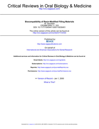
  • 9. Biomass production of L. acidophilus after incubation with Bis4GMA and UDMA for 40 h 120 100 0 ow 0 0 go - 40 20 I I 0 11c 00 A I -J r a I I Bis-GMA a N I_ L:'J*I UDMA Biomass production of L. aiophilus after incubation with EGDMA and TEGDMA for 40 h 140 (DPICL), and the contaminant triphenyl-stibane (TPSb) exhibited severe cytotoxic effects. Within the groups of (co)monomers and (co)initiators, high or moderate cytotoxic reactions were observed, whereas the evaluated decomposition/reaction products caused only moderate or slight effects (Table 1). The most important photo-initiator, camphoroquinone, which was found in significant amounts in aqueous extracts from resinbased materials, revealed moderate cytotoxic effects. This was confirmed by Atsumi et al. (1998) with permanent human submandibular-duct cells. Analysis of the data from various reports, taken together, indicates that, for most of the severely cytotoxic resin components, less toxic alternatives are available. In particular, the cytotoxicity of TEGDMA may be of great clinical interest. This relatively hydrophilic comonomer is released from many resin-based materials in considerable amounts (Spahl and Budzikiewicz, 1994; Geurtsen, 120 Blomass production of S. sobrinus after incubation with BIs.GMA and UDMA for 10 h in0 0 a 0 600 . v e -W B EGDMA TEGDMA 0 0 Figure 4a. Bis-GMA and UDMA significantly inhibited growth of L. 0 acidophilus (Hansel et al., 1998). Figure 4b. In contrast to base monomers, the relatively hydrophilic comonomers EGDMA and TEGDMA significantly promoted proliferation of L. acidophilus (Hansel et al., 1998). late (HEMA)I. These authors concluded that exposure time as well as interactions between and among various released substances significantly influence biocompatibility. The high cytotoxicity of the monomers Bis-GMA, UDMA, and TEGDMA was corroborated by other studies (Hanks et al., 1991; Lehmann et al., 1993; Yoshii, 1997). It has been reported that TEGDMA can induce lipid peroxidation of microsomes. Furthermore, this comonomer also exhibited surfactant-like potency which may cause detrimental solubilization of the lipid bilayer of cell membranes (Terakado et al., 1984; Fujisawa et al., 1988). Geurtsen et al. ( 1998a) investigated cytotoxic effects of 35 single monomers and additives of composite resins in permanent 3T3 cells and three primary human oral fibroblast cultures (Table 1). ED50 values varied significantly, from 0.0465 mM to > 5 mM. The tested inhibitor 2,6-di-t-butyl-4-methyl phenol (BHT), the photostabilizer 2-hydroxy-4-methoxy benzophenone (HMBP), the initiator diphenyliodonium chloride 340 A BIs4MA UDMA Biomass production of S. sobrinus after incubation with EGDMA and TEGDMA for 10 h 140 100..................i........., .. ........ I Il 0 ........ o0 .40 -! 0 B 011Id,IiI I Ii J." I I 20 EGDMA TEGDMA Figure 5a. In contrast to L. acidophilus, proliferation of S. sobrinus not affected by Bis-GMA (Hansel et al., 1998). was Figure 5b. EGDMA and TEGDMA also enhanced growth of S. sobrinus (Hansel et al., 1998). Crit Rev Oral Biol Med Crit Rev Oral Biot Med Downloaded from cro.sagepub.com at BIOLOGICAL LABS LIBRARY on December 27, 2013 For personal use only. No other uses without permission. fl(3)333 355 11(3):333-355 (2000)
  • 10. 1998; Spahi et al., 1998). Due to its high cytotoxic potency, this substance may significantly contribute to adverse local and systemic effects. The liberation of TEGDMA from resin restorations, therefore, should be minimized or prevented Furthermore, it must be emphasized that there is no cell line which is consistently more sensitive compared with others (Geurtsen et al., 1998a; MacDougall et al, 1998). Thus, each substance must be comprehensively screened for cytotoxicity by rneans of different permanent and primary cell cultures. The application of an Ii vitro pulp chamber device (dentin barrier assay) may help to simulate complex intra-oral interaction which can significantly modify cytotoxic effects in vivo (B) MICROBIAL EFFECTS There is currently a great controversy over whether pulpal irritation due to a resin restoration is mainly caused by bacteria proliferating between the cavity wall and the filling lBrannstrbm and Vojinovic, 1976; Qvist, 1993). Solid- as well as liquid-phase systems have been used to detect whether single-resin components can influence microbial growth (Updegraff et al., 1971; Hansel et cl., 1998). Proliferation of mutans streptoccoci (S. sobrinus) and lactobacilli (L. acidophilus) was either inhibited, promoted, or not influenced in a dose-dependent manner by the substance that was tested. Bis-GMA did not alter growth of S. sobrinus, whereas proliferation of L. acidophilus was significantly inhibited. UDMA reduced proliferation of both cariogenic pathogens. Generally, only very small amounts of these hydrophobic substances are released into an aqueous environment. Taken together, those large base monomers should not have microbial effects. The water-soluble comonomers EGDMA (ethylene glycol di-methacrylate) and TEGDMAA, however, promoted proliferation of S. sobrinus and L acidophilus (Figs. 4a, 4b, 5a 5b) iHansel et al., 1998). Analysis of these data clearly shows that the severely cytotoxic comonomers EGDMA and TEGDMA, which are present in considerable amounts in aqueous extracts of numerous resin-based materials, may significantly promote growth of cariogenic pathogens (Spahl et al 1998). Thus, cytotoxic properties and m icrobial growth promotion of these comon .mers may contribute to pulpal injury. (C) In ViVO STUDIES The LD,o concentrations (acute oral toxicity) of several substances have been evaluated in experimental animal studies The LD50 (per I kg body weight) was 950 mg for dibenzoyl-peroxide and 8400 mg for methyl-methacrylate (Deichmann. 941 1 Geurtsen, 1988). Due to the relatively low concentrations of components released from polymerized resin restorations in the oral cavity, acute oral (systemic) toxicity is not of great value in the assessment 11(3)):3 33-3 55 12000o) I 1 333 355 .OOOi of the biocompatibility of those materials. It has been reported that methyl methacrylate (MMA) may be teratogenic and can cause adverse cardiovascular effects in animals (Phillips et al., 1971, Singh et al., 1972; Karlsson et al., 1995). In addition, subcutaneous polymethyl-methacrlylate implants induced malignant tumors in rodents (Oppenheimer et al., 1955). Very few experiments have been performed to assess the acute local (pulp) toxicity of individual resin substances. It was found that the degree of pulpal alterations induced by those components correlates with cavity depth (Stanley, 1992). Two substances (dibenzoyl peroxide, 2-hydroxy-4-methoxy-benzophenone) caused pulpal inflammation in teeth of monkeys when applied to cavities without a protective lining (Stanley et a)., 1979)- (IV) Conventional and Polyacid-modified Composite Resins (A) COMPOSITION AND LEACHING OF COMPONENTS Conventional composite resins contain a polymerizable organic matrix, inorganic reinforcing fillers, and a silane coupling agent which bridges the organic and inorganic components (Ferracane, 1995). The organic matrix consists of several (co)monomers, e.g., Bis-GMA, UDMA, ethyleneglycol di-methacrylate compounds ( EGDMA, D/TEGDMA, etc.), and various additives ((co)initiators, stabilizer, inhibitorl (Table 1). In general, all organic ingredients of a composite resin are extractable by organic solvents, like methanol, after polymerization. A few components, however, are also leached into an aqueous medium. In particular, considerable amounts of TEGDMA may be released by polymerized composite resin into water. Bis-GMA, UDMA, EGDMA DEGDMA 1,6-hexanediol di-methacrylate, methyl methacrylate, camphorquinone, 4-N,N-dimethylamino-benzoic acid ethyl ester, and various other substances have been identified in minor concentrations in aqueous extracts (Geurtsen, 1998; Spahl et al., 1998). Several composite resins liberated formaldehyde into water over a long time period (11 5 days) This degradation product was especially noted in extracts from specimens with an unpolymerized oxygen-inhibited surface layer. The amount of extracted formaldehyde was partly in the order of magnitude which was found to cause local allergic reactions (Oysaed and Ruyter, 1988; Koch and Staehle, 1997). Inorganic substances (ions) like silicon, boron, sodium, and barium are also released from filler particles in trace amounts (0ysed et a)., 1988). 'Compomers' (polyacid-modified composite resins) have been developed to combine the fluoride release of glass-ionomer cements (GICs) with the mechanical prop- Crit Rev Oral Biol Med Crit Rev Oral Biol Med Downloaded from cro.sagepub.com at BIOLOGICAL LABS LIBRARY on December 27, 2013 For personal use only. No other uses without permission. 341 341
  • 11. erties of composite resins. Thus, these materials are composed of an ion-releasing glass, mainly calcium-aluminum-fluoro-silicate glass, and a polymerizable organic matrix. The fillers are partially silanized to couple the glass with the polymer network. In addition to conventional monomers (Bis-GMA, UDMA), the organic matrix of compomers contains bi-functional monomers with two carboxylic groups and two double-bond functions, e.g., TCB (tetracarboxylic butyl methacrylate), which is synthesized from butane-tetracarboxylic acid and 2hydroxyethyl methacrylate (HEMA) (Attin and Buchalla, 1998; Meyer et al., 1998). The bi-functional monomers can react with methacrylates by radical polymerization and by acid-base reaction to bring about release of ions from the glass in the presence of water. However, compomers do not contain water. Thus, no significant acid-base neutralization occurs during polymerization. Those materials, therefore, are much closer to composite resins than to glass-ionomer cements (Meyer et al., 1998). There are very few data on the release of substances from 'compomers'. The aqueous eluates of polyacidmodified composite resins were analyzed by gas chromatography/mass spectrometry (GC/MS) and high-performance liquid chromatography (HPLC). Ethylene glycol compounds (comonomers) and the hydrophilic monomer HEMA were the chief constituents identified in these aqueous extracts (Geurtsen et at., 1998b; Hamid et al., 1998). In addition, the following decomposition products of base monomers and several co-initiators were found: N-(2-cyanoethyl)-N-methylaniline (CEMA), 4N,N-dimethyl amino benzoic acid ethylester (DMABEE), and N,N-dimethyl amino ethyl methacrylate (DMAEMA) (Geurtsen et al., 1998b). Due to the ion-leaching glass fillers, compomers may also release fluoride, especially during the first few days after polymerization. But it must be emphasized that those materials leach out considerably smaller amounts of fluoride than do conventional and resin-modified glass-ionomer cements (Attin and Buchalla, 1998; Geurtsen et at., 1998c, 1999b). (B) CYTOTOXICITY Solid specimens and extracts of polymerized samples of composite resins have been tested in various cell cultures. The results of those studies have varied significantly, depending on the product tested (Lampert and Heidemann, 1980; Tronstadt and Wennberg, 1980). It was reported that polymerized specimens of one hybrid-type composite resin were moderately cytotoxic during a fouryear period in permanent and primary human oral cell cultures. Non-polymerized samples of this product, however, were severely cytotoxic and revealed genotoxic effects (Geurtsen, 1987a, 1988). Aqueous eluates of several hybrid-type composite resins induced only moderate cytotoxic reactions (Geurtsen, 1987b). 342 Nakamura et al. (1985) tested various consecutive extracts of light-curing products over six weeks in HeLa cells. The first eluates caused moderate to severe cellular alterations which were not observed with later extracts. In contrast, set material of light-cured and chemically cured composite resins revealed slight or no toxic effects when tested in an artificial pulp chamber made up of dentin slices placed between the materials and the cell cultures (Hanks et al., 1988). Polymerized composite resin separated from the extraction medium by etched dentin had toxic potency higher than that of specimens placed on unetched dentin (Hume, 1985). This observation indicates that the application of the 'total-etch technique' may be associated with a higher risk for pulp irritation due to composite resin restorations, even in combination with dentin bonding (Gerzina and Hume, 1996). Mohsen et al. (1998) investigated the influence of various parameters on the cytotoxic potential of several light-curable UDMA-based composite resins. Cytotoxicity decreased with increasing curing time and higher aging time. A reduction of cytotoxicity was also observed when polished samples without an oxygen-inhibited surface layer were tested. Furthermore, cell viability increased when specimens were extracted with water, ethanol, or other organic solvents prior to cell culture experiments (Mohsen et al., 1998; Rathbun et al., 1991). The cytotoxic potency of polyacid-modified composite resins varied considerably depending on the product tested (Pertot et al., 1997; Geurtsen et al., 1998b). Aqueous eluates of two polyacid-modified composite resins induced moderate injuries in permanent 3T3 fibroblast cultures, whereas one product (DyractTM Cem, De Trey Dentsply) caused severe cytotoxic effects. The severely toxic extract contained a very high concentration of TEGDMA, whereas the two moderately cytotoxic composite resins released only small amounts of comonomers, like HEMA, and various ethylene glycol compounds. In summary, then, cytotoxicity of composite resins varies depending on the product tested and especially upon the quantity of leachable components. An optimum polymerization, therefore, is of high importance for the cytocompatibility of those materials. Furthermore, extractable amounts of components should be reduced, e.g., by the use of less-water-soluble components. Severely cytotoxic substances should be replaced by less toxic alternatives, if available. (C) MICROBIAL EFFECTS Fresh specimens of several composite resins inhibited microbial growth (0rstavik and Hensten-Pettersen, 1978). But other authors found no significant influence on the proliferation of micro-organisms (Updegraff et al., 1971). Recently, it was reported that the water extract of a hybrid-type composite resin promoted the growth of Grit Rev Oral Biol Med Crit Rev Oral Biol Med Downloaded from cro.sagepub.com at BIOLOGICAL LABS LIBRARY on December 27, 2013 For personal use only. No other uses without permission. (2000) 11(3):333-355 (2000)
  • 12. treated dentin layers (Horsted ct al), 1986; Quist et al., 1989, Fujitani et at., 1992). When one considers the permeability of dentin, especially after acid pre-treatment, and the release of cytotoxic and hydrophilic the caries pathogen L. acidophilus. It was found that this eluate contained a high amount of TEGDMA. Aqueous extracts of another composite resin with a low concentration of TEGDMA, however, inhibited growth of this micro-organism. Thus, the release of high concentrations of TEGDMA may stimulate proliferation of cariogenic bacteria within marginal gaps which then may contribute to recurrent caries and pulpal irritation (Figs. 4a, 4b, 5a, 5b, 6a, 6b). Release of microbial growth-stimulating substances, therefore, should be minimized or prevented (Hansel et al., 1998). Alternatively, antibacterial monomers (like methacryloyloxydodecylpyridinium bromide, MDPB) or fillers, e.g., ApacideriM, based on apatite containing silver and zinc, have been added to composite resins. These experimental materials significantly inhibited proliferation of cariogenic bacteria (Syafiuddin et al., 1997. Imazato et a)., 1998c). (D) In ViVO STUDIES Local, non-specific toxicity of polymerized composite resins was determined after implantation into various tissues, e.g, muscle and bone. Generally, composite resins caused only slight to moderate tissue alterations which decreased over time (Chan et a)., 1972; Howden and Silver, 1980, Wennberg et al., 1983). Numerous pulp studies have been performed for the evaluation of local specific reactions. The results of these usage tests have yielded contradictory results. An absence of or only slight pulp irritations were reported by Retief et al. ( 1973) and Suzuki et al. ( 1995). Other investigators, however, observed moderate to severe reactions (Tronstadt and Spangberg, 1974; Valcke et al., 1980). It has been observed that several co-factors influence the pulp toxicity of composite resins. A protective cavity lining coupled with an increased thickness in remaining dentin reduced the risk of pulpal inflammation (Quist, 1975; Dalleske et al., 1978) On the other hand, previous damage to the pulp due to deep caries lesions and acid-etching of dentin on the cavity floor enhanced toxicity (Fiore-Donno and Baume, 1966; Stanley et a)., 1 975; Eriksen and Leidal, 1979). Several authors have noted a correlation between pulpal irritations of resin-restored teeth and bacteria (and their by-products), whereas material toxicity per se was of no significance (Brannstrom and Vojinovic, 1976; Torstenson et a/., 1982; Cox, 1992, Cox and Suzuki, 1994). It should be recognized, however, that comonomers released from a resin filling into marginal clefts may stimulate microbial growth and consequently may, at least indirectly, contribute to pulpal irritation caused by those bacteria (Hansel et a)., 1998). However, pulpal inflammation was also found in resin-restored teeth without bacterial microleakage, especially when low-viscous bonding agents had been applied to thin acid-pre111)31333 355 (2000) 100 33-355 120001 Cril Ree A B Figure 6a. This is a 20-year-old patient presenting with an upper right first molar with an occlusal amalgam restoration and a composite resin filling. The amalgam restoration reveals marginal gaps; the resin filing shows only a slight marginal discoloration. Figure 6b. The restorations were removed without excavation of carious dentin. The amalgam cavity is caries-free, whereas extensive recurrent caries is visible beneath the composite restoration. This caries recurrence may have been caused by the growth of cariogenic micro-organisms due to release of comonomers, e.g., EGDMA or TEGDMA (Hansel et a/., 1998). Oral Biol Med Crit Rcv Oral Biol Mcd Downloaded from cro.sagepub.com at BIOLOGICAL LABS LIBRARY on December 27, 2013 For personal use only. No other uses without permission. 343
  • 13. (co)monomers from polymerized composite resin, it is reasonable to suggest that those substances can contribute significantly to pulp injuries (Hume, 1985; Hamid et al., 1996; Eick et al., 1997; Marshall et al., 1997; Abou Hashieh et al., 1998; Gale and Darvell, 1999). On the other hand, it must be emphasized that the barrier function of dentin may protect the pulp from toxic influences. Thus, for example, diffusion of monomers decreases significantly with thicker dentin, and the penetration of bacterial by-products is also reduced by dentin (Pissiotis and Spangberg, 1992; Hamid et al., 1996; Hamid and Hume, 1997a). Several retrospective and prospective studies on the clinical long-term performance of composite resins have been reported (Mjbr et al., 1990; Wilder et al., 1991; Roberts et al., 1992). In general, evaluation of the fillings included determination of pulpal conditions. A four-year clinical study with 1209 Class I and Class 11 restorations revealed long-lasting post-operative pain or pulpitis as a cause for failure in only four cases. Glass-ionomer cement was applied as a base in all cavities. Additionally, areas with thin dentin were covered with a calcium hydroxide cement (Geurtsen and Schoeler, 1997). A meta-analysis of 16 long-term clinical studies of posterior composite resin fillings also indicated no increased risk of pulpal irritation (El-Mowafy et alc., 1994). In summary, there is sufficient evidence that various factors associated with composite resin restorations possess tissue-damaging potency. Furthermore, pulp inflammation/necrosis as a consequence of a composite resin filling is probably due to the interaction of several tissue-irritating parameters rather than to a single detrimental factor. Usage tests as well as long-term clinical studies indicate that composite resins do not pose a special risk for the dental pulp if they are properly applied. (V) Resin-based Pit and Fissure Sealants (A) COMPOSITION AND RELEASE OF SUBSTANCES Resinous pit and fissure sealants consist of an unfilled or filled organic matrix. Like composite resins, the matrix of dental sealants is composed of a mixture of various (co)monomers (mainly Bis-GMA, UDMA, and TEGDMA) and additives. Some sealants also contain fluoridereleasing components. Either clear, tinted, or opaque auto-polymerizing and visible-light-curable products are available (Waggoner and Siegal, 1996). Public concern about the safety of resin sealants was greatly increased by the report of Olea et al. (1996). Using the HPLC technique, these authors reported that the potentially estrogenic substance bisphenol-A was released from fissure sealants in vivo. It must be emphasized that this observation was mainly based on one 344 female patient who had received fissure sealants two years earlier. Bisphenol A, however, is rapidly released from polymerized sealants and is quickly excreted (Ashby, 1997). Thus, it is more than unlikely that dental sealants can serve as a long-term intra-oral source for the leaching of bisphenol A. Olea et al. (1996) also reported that resin oligomers may liberate bisphenol-A after storage in alkaline (pH 13) or acidic (pH 1) media at 100°C for 30 min. But these conditions do not exist in vivo. In contrast to the findings by Olea et al. (I1996), using HPLC or GC/MS methodology, a number of authors reported that the product investigated by Olea et al. (1996), and other sealants as well, did not release bisphenol-A into water or ethanol. TEGDMA, however, was released from several sealants (Hamid and Hume, 1997b; Nathanson et al., 1997; Geurtsen et al., 1999a). Interestingly, TEGDMA was not identified by Olea et al. in their analysis. This strongly suggests that Olea et al. (1996) may have misinterpreted TEGDMA peaks as indicative of bisphenol-A. Due to different mass spectra and retention times, bisphenol-A and TEGDMA can be easily distinguished from each other by GC/MS. Accordingly, no bisphenol-A was found with GC/MS (Geurtsen et al., 1999a). In addition to TEGDMA, monomers (like Bis-GMA, UDMA), comonomers (mainly ethylene glycol compounds), and additives [e.g., camphorquinone and DMABEE (4-N,N-Dimethylaminobenzoic acid ethyl ester)l leach out from sealants in minor quantities (Hamid and Hume, 1997b; Nathanson et al., 1997; Spahl et al., 1998; Geurtsen et al., 1999a). (B) BIOCOMPATIBILITY Few in vitro studies have been published about the cytotoxicity of resinous pit and fissure sealants. There are no in vivo data available except for one case report of an allergic reaction to the sealants (see below) (Hallstrbm, 1993). Water extracts of two sealants were investigated in human embryonic lung fibroblast cultures. It was observed that both materials were more cytotoxic than PMMA (polymethyl methacrylate) acrylic controls (Rawls et al., 1992). Aqueous eluates of four sealants caused moderate-to-severe inhibition of cell growth in 3T3 cultures. Considerable cytotoxic effects in permanent 3T3 fibroblasts were caused by one product which leached high amounts of TEGDMA into de-ionized water (Geurtsen et al., 1999a). In general, only sparse results on biocompatibility and release of substances from these materials are available. These limited data as well as the similarities in composition and degradation compounds indicate that leaching and biological behavior of resin-based pit and fissure sealants should be comparable with that of composite resins. Grit Rev Oral Biol Crit Rev Oral Biol Med Med Downloaded from cro.sagepub.com at BIOLOGICAL LABS LIBRARY on December 27, 2013 For personal use only. No other uses without permission. (2000) 11(3):333-355 (2000)
  • 14. (B) BIOCOMPATIBILITY OF DENTIN ADHESIVES (VI) Dentin Adhesives (A) COMPOSITION (1) In vitro studies AND RELEASE OF SUBSTANCES Rakich et al. (1998) investigated the effects of four components of dentin bonding agents on the mitochondrial activity (MTT assay) of macrophages which are important in wound healing and inflammatory reactions. HEMA, 4methacryloxyethyl-trimellitic anhydride 14-META), BisGMA, and UDMA were evaluated. The cytocompatibility varied significantly: HEMA (best compatibility) > 4META >>> Bis-GMA > UDMA. A similar ranking of toxicity was found in experiments with Balb/c 3T3 mouse fibroblasts (Ratanasathien et al., 1995). Aqueous extracts of five adhesives were tested in 3T3 fibroblast cultures. The cytotoxic potency of those products varied considerably. The most toxic material, which almost completely inhibited cell growth, released TEGDMA in high amounts. Two materials released high concentrations of HEMA and reduced cell proliferation considerably. Additionally, two products liberated small quantities of (co)monomers (EGDMA, TEGDMA, or HEMA) that affected cell growth only slightly (Geurtsen et l., 1999c). The influence of dentin on the cytotoxicity of four dentin adhesives was evaluated with L 929 fibroblasts. Each product was tested using dentin slices with high and low hydraulic conductance. Generally, all adhesives were significantly more cytotoxic when slices with high hydraulic conductance were applied (Abou-Hashieh et al., 1998). These observations confirm that the nature and quantity of released substances, as well as the barrier function of dentin, decisively influence the cytotoxic potential of dentin adhesives. Some products release high quantities of (co)monomers (TEGDMA, HEMA) which may cause pulpal alterations, especially when applied in areas with a thin dentin layer. Thus, only good cytocompatible dentin adhesives are recommended as a pulp protective cavity lining on thicker dentin layers. Members of the group of dentin adhesives differ considerably in their chemical composition, mode of action, and clinical handling. Dentin adhesives are mainly used to improve the bonding between dentin and composite resins. In addition, dentin bonding agents are applied as cavity liners and for treatment of hypersensitive teeth with exposed root surfaces (Cox and Suzuki, 1994; lain et al., 1997; Schuckar and Geurtsen, 1997) Depending on the product, dentin adhesives may consist of one to three components ('bottles'). Conditioners (etchants) containing acid (e.g., phosphoric acid, maleic acid) or EDTA are used for partial or total removal of the dentin smear layer and for the structural modification of the binding substrate (Eick et al., 1997). The dissolved smear layer and conditioner are removed by thorough rinsing with water. This results in the opening of most orifices of the dentinal tubules. Primers consist of a 'hydrophilic' monomer (e.g., HEMA) or a mixture of monomers dissolved in solvents like water, ethanol, or acetone. Primers are applied to expand or re-expand the conditioned demineralized collagen network and to increase the wettability of the hydrophilic substrate for the hydrophobic components of the bonding agent. Furthermore, some dentin primers (e.g., SyntaciM) contain glutaraldehyde for stabilization of demineralized collagen fibers. B3onding agents are mainly unfilled mixtures of various hydrophobic and/or hydrophilic co)monomers (e.g., Bis-GMA, UDMA, TEGDMA, HEMA) and additives. They are used to bind the conditioned and/or primed dentin surface with composite resin. Several moderr products combine conditioner and primer ('self-conditioning primers') or primer and bonding agent into one bottle. Very little is known about the release of components from dentin adhesives. Nor-polymerized specimens of various dentin bonding agents were extracted by acetone. Gas chromatography/mass spectrometry (GC/MS) analysis revealed that the eluates predominantly contained ethylene-glycol methacrylates, such as EGDMA and D/TEGDMA, as well as smaller amounts of H-EIMA and MMA (methyl methacrylate) (Kanerva et al., 1994a). Geurtsen et al. ( 1999c) analyzed aqueous extracts of five modern dentin adhesives by GC/MS The adhesives analyzed released several substances, predominantly EGDMA, TEGDMA, and HEMA, into de-ionized water. All materials released camphoroquinone Additionally, four adhesives also released other (co)initiators, e ., 4-N,N -dimethylaminobenzoic acid ethyl ester (DMABEE) and diphenyliodoniumchloride (DPICI). I1 1(31-333-355 i2OM l(3i 333 355 2000 (2) Microbial effects Several micro-organisms are associated with caries lesions, especially mutans streptococci (S. mutans S sobrinus) and lactobacilli (Emilson and Krasse, 1985). Bacteria and their by-products may also cause pulpal irritation. Therefore, dentin adhesives with antimicrobial properties would be of great clinical advantage for prophylaxis of recurrent caries as well as for prevention of pulpal alterations due to microbial effects. The experimental incorporation of the monomer, methacryloyloxy dodecylpyridinium bromide, into a selfetching primer significantly increased the antimicrobial potency, even after polymerization (Imazato et al., 1998a,b). A screening of seven adhesives revealed antimicrobial effects of conditioners/etchants (EDTA, phosphoric acid). Antibacterial properties were also observed with primers Med Biol Crit Rev Oral Biot Med Rev Oral Downloaded from cro.sagepub.com at BIOLOGICAL LABS LIBRARY on December 27, 2013 For personal use only. No other uses without permission. 345
  • 15. containing maleic acid or glutaraldehyde. The other primers and most bonding agents, however, did not show any antimicrobial property (Emilson and Bergenholtz, 1993). Similar observations with other self-conditioning primers have been reported (Imazato et al., 1998c). A polymerizing dentin adhesive containing glutaraldehyde (SyntacTM) had a strong antibacterial effect when tested on eight microbial cultures. But set and aged materials were not tested (Fraga et al., 1996). It can be concluded that most dentin adhesives exhibit antimicrobial effects only in the initial phase after application. Thus, generally speaking, no anti-cariogenic and pulp-protecting effect due to long-term antimicrobial potency of dentin adhesives can be expected. (3) In vivo studies Dentin bonding agents may irritate the dental pulp by three mechanisms: (I) by the conditioner/etchant, (11) direct cytotoxic effects due to released substances, and (111) by bacteria or microbial by-products. Additionally, it must be appreciated that dentin conditioning/etching ('total-etch technique') can significantly increase transport of released organic substances through dentin. Thinner dentin layers may enhance pulpward diffusion of monomers as well as the total leaching of substances from polymerized dentin adhesive (Hamid et al., 1996; Hamid and Hume, 1997a). Interestingly, it was observed that diffusion of HEMA and TEGDMA, in particular, through dentin is not prevented by positive hydrostatic pressure (Gerzina and Hume, 1995). The application of a HEMA-containing bonding resin in combination with a composite resin reduced TEGDMA diffusion through dentin only slightly in comparison with a composite resin that was used without an adhesive. But the dentin adhesive-composite resin combination resulted in additional pulpward diffusion of considerable quantities of released HEMA. Thus, dentin adhesives that leach high amounts of the cytotoxic (co)monomer may cause pulp alteration, especially when the 'total-etch technique' is used. No pulp-protective effects due to reduced diffusion of TEGDMA leached from composite resin can be expected. Several studies on the response of the pulp to dentin adhesives have been performed. The results of these studies are not easily comparable because of the differences in the composition of the adhesives and the techniques used. The application of a three-component product, according to the principles of the total-etch technique, did not cause significant pulpal irritation of monkey teeth over a 90-day period (Inokoshi et al., 1998). Similar observations were made by White et al. (1994) in adult rhesus monkeys. Evidence was presented that irreversible pulpal inflammation is not induced when dentin adhesives are applied in cavities with a thick dentin layer, particularly in caries-free dentin preparations extending just inside the dento-enamel junction (Goracci et al., 346 1995; Pameijer and Stanley, 1995; Gilpatrick et al., 1996). Several investigators have compared the reactions of exposed and non-exposed pulps after the application of dentin adhesives in combination with dentin acid-etching. No differences were found between exposed or nonexposed pulps and between the adhesives and the Ca(OH)2 group, which generally served as a control (Akimoto et al., 1998; Cox et al., 1998; Tarim et al., 1998). But detrimental effects of the total-etch technique with dentin adhesives in vital-pulp-capping of monkey teeth were reported by Pameijer and Stanley (1998). Interestingly, these authors also observed that micro-organisms were present in a large percentage of vital and non-vital teeth. Thus, bacteria had no significant influence on the reaction of the pulps after application of dentin adhesives. Additionally, it was found that pulps directly capped with dentin adhesive may develop a sub-acute foreign body response due to impacted resin particles. The authors concluded that the long-term effect of resin particles in pulpal tissue needs further clarification (Gwinnett and Tay, 1998). These contradictory results clearly show that there are insufficient data for a reasonable conclusion on the effects of dentin adhesives on the pulp. In particular, it must be noted that the abovementioned histological pulp studies covered a period of only about three months. Consequently, the long-term, in vivo, biological effects are not known. Although dentin adhesives may be of benefit to reduce marginal microleakage and to prevent pulpal hypersensitivity in cavities with a relatively thick dentin layer, dentin in deep cavities should be covered by a cavity liner for pulp protection. This is corroborated by in vitro observations indicating that HEMA and TEGDMA may cross dentin even under positive hydrostatic pressure. Exposed pulps should be directly capped with Ca(OH-)2 until there is enough scientific evidence in support of the long-term clinical efficiency and biocompatibility of various dentin adhesives. (VII) Resin-modified Glass-ionomer Cements (A) COMPOSITION AND LEACHING OF SUBSTANCES Resin-modified glass-ionomer cements (rmGIC) contain ion-releasing glass particles, water-soluble polyacrylic acids, light-curable monomers (e.g., HEMA), and additives. Various products are composed of photo-curable side-chains linked to water-soluble polymeric acids. In contrast to polyacid-modified composite resins (compomers), rmGICs contain water and thus exhibit an acidbase reaction with salt formation and a free-radical polymerization (Kakaboura et al., 1996; Nicholson, 1998). Due to their ease of application in comparison with conventional GICs, resin-modified systems are being increasingly used clinically, especially in pediatric dentistry Crit Rev Oral Biol Med Crit Rev Oral Biol Med Downloaded from cro.sagepub.com at BIOLOGICAL LABS LIBRARY on December 27, 2013 For personal use only. No other uses without permission. I1(3):333 355 11(3):333-355 (2000)
  • 16. (Vaikuntam, 1997). Several investigators evaluated the release of fluoride from rmGlCs )Forss, 1993; Ulukapi et al., 1996; Friedl et al., 1997; Geurtsen et al., 1998c). The release of fluoride varied significantly, depending on the product tested and the extraction medium. In general, less fluoride leached from rmGlCs than from conventional GICs (Geurtsen, 1998). Leaching of organic components from various rmGICs into distilled water was investigated by Hamid et al. 11998). HEMA was the only compound detected in the water extracts by HPLC. Aqueous eluates of two rmGlCs were analyzed with GC/MS (Geurtsen et al., 1998b). Both products released HEMA into de-ionized water. In addition, one material (Vitrebond'IMm) released the initiator, diphenyliodoniumchloride (DPICI), which was also present in one dentin adhesive (SyntaciM) (Geurtsen et al., 1999c). DPICI was identified by its GC-decomposition products chlorine-, iodine- and bromine benzene. It might be possible, however, that these highly toxic substances are generated during polymerization and are thereafter released into an aqueous environment. Our knowledge about the quality and amount of leachable components from rmGlCs is insufficient. Besides (co)monomers, (co)initiators, etc., reaction or degradation products, e.g., benzenes, are released which may then cause adverse local and/or systemic effects. These leachable reaction or degradation products, therefore, should be identified as soon as possible. (B) BIOCOMPATIBILITY OF RESIN-MODIFIED GICs The cytocompatibility of various conventional and resinmodified GICs was evaluated in cultures of primary human osteoblasts. All materials, except for one product, exhibited good cytocompatibility. Viable osteoblasts grew inito contact with the specimens. One resin-modified GIC (Vitremer"'Nl), however, was very cytotoxic. The authors concluded that these adverse reactions might be due to release of high amounts of HEMA (Oliva et a)., 1996). Comparable observations were reported by Consiglio et il, ('1998). Two rmGlCs (VitrebondiM and Vitremeri'( reduced protein synthesis of human gingival fibroblasts by 00°o, whereas the four tested conventional GlCs caused significantly less inhibition of protein synthesis. Severe cytotoxicity of Vitremert'N in perma nent 3T3 fibroblasts was also found by Kan et al. (1997). The other rmGIC (Fu'ji 11 LCiSII caused no or only slight cellulal alterations. Finally, the detrimental cellular effects of Vitrebond'%- and the mild cytotoxicity of Fuji 11 LC I' were corroborated in monolayers of 3T3 fibroblasts, primary human gingival fibroblasts, human peripheral lymphocytes, and in dentin barrier tests with fibroblasts (Leyhaulsen et al., 1998, Geurtsen et al., 1998c; Stea et a)., 1998, Schmalz tot a)., 1999) (Figs. Ia-c, 2a-c). It appears that the deleterious effects of Vitrebond'im may be partly due to the release of the highly cytotoxic initiator, diphenyliodoniumchloride, and/or its decomposition products (Ceurtsen et a)., 1998c). It is noteworthy that the abovementioned in vitro studies produced fairly similar results, even though different cell types and biological endpoints were used. Besides its cytotoxic effect, fresh Vitremerii' also revealed moderate to strong antibacterial effects in cultures of mutans streptoccoci and lactobacilli (Fraga et al., 1996, Fried) et al., 1997). Direct and indirect genotoxic reactions were caused by Vitrebond"'5' in procaryotic and eucaryotic in vitro assays, whereas other resin-modified and conventional GICs induced only questionable indirect genotoxicity or no such effect. These in vitro results were confirmed by in vivo assays (Heil et a), 1996; Stea etal., 1998). Taken together, there is strong evidence that some resin-modified GICs may be genotoxic and severely cytotoxic due to release of various substances, e.g., HEMA and DPICI. Consequently, rmClCs may cause adverse local and/or systemic effects. Thus, leaching of those components must be minimized or prevented. (VIII) Allergic Reactions Caused by Resin-modified Filling Materials The allergic or sensitizing potential of resin materials and components was evaluated in various in vivo studies, e.g., maximization tests with guinea pigs. A marked allergic potential was exhibited by impurities of the matrix monomer Bis-GMA (B)jrkner et al. 1984; B)brkner, 1984b). Aliphatic urethane (meth)acrylates, for dental application, were more potent sensitizers than aromatic urethane acrylates and (meth)acrylated aliphatic urethane (Bj6rkner, 1984a). HEMA also revealed a marked allergic effect. From 60 to 100% of tested guinea pigs reacted to high concentrations of this monomer in the maximization test (Clemmensen, 1985). In addition, mutual cross-sensitivity to methacrylates was found. Guinea pigs sensitized to various methacrylates also responded strongly to methacrylates which had not been used for sensitization (Chung and Giles, 1977). Similar observations have also been made in humans (Kanerva et al., 1992b; Richter and Geier, 1996). Skin reactions were provoked by some bonding systems in guinea pigs and Macaca mulatta monkeys (Altuna and Freeman, 1987; Katsuno et al., 1998). Patients, and especially dental personnel, are increasingly exhibiting local and systemic allergic reactions to components of resinous dental materials (Kanerva et al., 1994a,b; Richter and Geier, 1996; Tschernitschek et al., 1998). It must be emphasized, however, that the number of individuals allergic to resin34 11(3 3~ 35~2001 1 1(3)333-355 (2000) Cr Re Ora Bil Me Crit Rev Orail Biol Med Downloaded from cro.sagepub.com at BIOLOGICAL LABS LIBRARY on December 27, 2013 For personal use only. No other uses without permission. 347
  • 17. based dental materials is still low. In addition, etiologic factors other than dental materials may also induce intra-oral contact allergy, e.g., ingredients in food, beverages, and oral hygiene products (De Rossi and Greenberg, 1998). Kallus and Mjor (1991) investigated the incidence of adverse reactions to dental materials. Two verified longstanding effects to resin-based materials (dentures) were found in 13,325 patients. Only eight patients out of a total number of 31 1 persons who had been referred to a dental hospital for possible allergic reactions to dental materials suffered from a verified allergy to methacrylates (Tschernitschek et al., 1998). Adverse reactions to a Bis-GMA-based fissure sealant in a six-year-old girl have been described by Hallstrom (1993). One day after application of the material, the patient developed multiple allergic reactions: asthma, blisters on the gingiva adjacent to the resin sealants, swellings, and rashes on various areas of the body (arms, palms, legs, lips, ears). The symptoms completely disappeared within nine days after removal of the sealants. A verified extra-oral hypersensitivity to composite resin was observed in one patient (Nathanson and Lockhart, 1979). Other patients revealed an allergic contact dermatitis to composite resin. The reactions were caused by Bis-GMA and benzoyl peroxide (Carmichael et al., 1997; Kanerva et al., 1989). Seventeen oral lichenoid lesions on the mucosa in contact with composite resin restorations were described by Lind (1998). Total remission of the lichenoid reactions was observed after replacement of the resin fillings in four cases and partial remission in another five patients. The author suggested that contact of the oral mucosa with formaldehyde derived from the resin restoration may have been the main etiologic factor in these cases. There are several reports in the medical and dental literature regarding hypersensitivity or allergic reaction of patients to MMA (Kaaber, 1990; Richter and Geier, 1996; Hochman and Zalkind, 1997) (Table 1). Dental personnel often have manual contact with resinous materials. Many acrylates rapidly penetrate latex or vinyl gloves (Kanerva et al., 1993). Monomer vapors can be inhaled through face masks. Thus, devices like gloves and face masks provide only incomplete protection. Taken together, the incidence of allergy to resinbased materials in dental professionals (dentists, nurses, dental technicians) has increased. Lonnroth and Shahnavaz (1998b) found that 16-17% of the dental personnel in their study were hypersensitive to dental materials, in comparison with 0.5%-2% in the control group. Hand dermatitis and symptoms on the fingers were significantly more frequent among dental professionals than in the control group (Lonnroth and Shahnavaz, 1998a). There are some resin components which are especially important with respect to allergic reactions in 348 dental personnel. Dental nurses showed a high incidence of allergic reactions to glutaraldehyde and benzoyl peroxide, whereas dental technicians were frequently hypersensitive to HEMA, EGDMA, MMA, benzoyl peroxide, TEGDMA, and dihydroxy-ethyl-p-toluidine (Schnuch and Geier, 1994). Other investigators have reported that dental nurses and dentists developed allergic contact dermatitis to HEMA caused by handling of dentin adhesives (Kanerva et al., 1992a, 1994a). Six nurses and one dentist had positive patch tests to composite resins. Four of those individuals were hypersensitive to Bis-GMA, and three reacted to TEGDMA; two patients were also hypersensitive to MMA. All of these dental nurses had to stop the practice of their profession (Kanerva et al., 1989). It is very likely that the use of resin-based materials in dentistry will significantly increase in the future. Concomitantly, the incidence of allergies to dental resin components will probably also increase considerably, among patients and especially among dental personnel. The proper handling of those products, therefore, is of considerable importance. Specifically, contact with unpolymerized material must be avoided. In addition, adequate ventilation of rooms for processing of resinbased materials is recommended, to minimize the concentration of monomer vapors in the air. (IX) Genotoxicity of Resinbased Restorative Materials There is little information on the genotoxic effects of individual resin components. Hensten-Pettersen et al. (1978) screened single ingredients of composite resins (initiators, inhibitors, UV-absorbers) in the classic Ames test. None of the tested components revealed mutagenic activity. Miller et al. (1986), however, found weak mutagenic reactions due to one co-initiator. No mutagenicity was observed with Bis-GMA and UDMA in the Ames test (Geurtsen, 1988). Fourteen mono-substances were investigated by means of the umu test (procaryotic, in vitro), the DIT (DNA-synthesis inhibition test; eucaryotic, in vitro), and the AFE assay (in vivo) (Table 1) (Heil et al., 1996). Interestingly, almost all tested resin components were genotoxic in at least one test system. The effects ranged from 'borderline' to 'strong positive'. No positive results were observed with the monomers BEMA (benzyl methacrylate) and HEMA, whereas Bis-GMA (umu, AFE) and UDMA (AFE) were genotoxic in one or two assays. Recently, it has been reported that TEGDMA induced large DNA sequence deletions in the hprt gene of V79 cells. The authors concluded that the induction of such DNA sequence deletions might be common for acrylates and methacrylates (Schweikl and Schmalz, 1999). Mutagenic effects in the Salmonella typhimurium mutagenicity test system ('Ames test') were produced by glutaraldehyde as well as by dimethyl sulfoxide Crit Rev Oral Biol Med Crit Rev Oral Biol Med Downloaded from cro.sagepub.com at BIOLOGICAL LABS LIBRARY on December 27, 2013 For personal use only. No other uses without permission. 1 1(3):333-355 11(3):333-355 (2000)
  • 18. (DMSO) and physiologic saline eluates of a dentin adhesive. Two other products exhibited no mutagenicity. It was concluded that the mutagenic effects of one of those dentin adhesives (SyntacFmv) were induced by the genotoxic ingredient, glutaraldehyde (Schweikl et al., 1994). Dose-dependent mutagenicity was found in Ames tests with unpolymerized rnaterial of two dentin adhesives. Genotoxic effects were also caused by monomeric solutions used in orthodontics for cementation. However, no specific component was identified as the causative agent in those studies (Athas et al., 1979; Li et al., 1990). Scarce data are available about the genotoxicity of composite resins. DMSO and cell culture medium extracts of two hybrid-type composite resins were investigated in various assays. Genotoxic effects were produced by the DMSO eluates of one composite resin (SonoCem]N1) in the eucaryotic DIT (Heil et al., 1996) (Table 2). A resin-modified glass-ionomer cement (Vitrebond[M) exhibited strong genotoxic action. Extracts of this rmGlC induced sister chromatid exchange in human peripheral lymphocytes in vitro. Genotoxicity was verified by three different in vitro and in vivo assays: the procaryotic umu test, the eucaryotic DIT, and the in vivo NFE assay. Analysis of water extracts of this GIC indicates that leaching of the initiator, DPIGI, may contribute to this strong genotoxic action (Heil et al 1996; Stea et al., 1998) (Table 2). Taken together, it is clear that there are insufficient data documenting the genotoxicity of resin-based dental materials. In light of the evidence of the genotoxic potential of some of these materials, it is important that the research in this area be given sericus consideration. (X) Conclusions and Summary The biocompatibility of a resin-based dental restorative material is predominantly determined by the amount and nature of released organic substances. The cytotoxic potential of these components varies significantly. TABLE 2 Mutagenicity of Aqueous Eluates and DMSO Extracts from Two Composite Resins (cr) and Two Resin-modified Glass-ionomer Cements (GIC) Product Aqueous Extract DMSO Extract HerculiteTM (cr) No Sono-CeMTM (cr) lonosea ITM (GIC) VitrebondTM (GIC) No No Yes2 No Yes'-3 Yes' n.d.4 Results from umu test, IDIT, 3AFE assay IHeil et al., 1996); 4not determined. 000 3133355 (3):3 3 3- 3 5 5 (2000) Additionally, interactive effects of secreted organic and/or inorganic substances may occur, e.g., synergism, additive effects, and antagonism. Resin-based restorations release several (co)monomers (like ethylene glycol compounds, HEMA) and additives (mainly initiating substances) into the oral cavity which may cause adverse local and systemic effects in patients, e.g., lichenoid reactions of the oral mucosa or allergic reactions. There are sufficient data demonstrating that the liberated substances may diffuse pulpward through dentin in high quantities (TEGDMA HEMA), particularly through thin dentin layers or after acid-etching of dentin. It appears that high quantities of these components may result in pulpal inflammation. Although it is of considerable significance, it is not known if substances leached from resinous materials enter the blood stream, and whether they are transformed in the liver and/or accumulate in other tissues. Manual contact between organic components and unprotected skin during application of resin-based materials poses an increased risk for allergic reactions in dental personnel. There is strong evidence that some ingredients of resin-based materials (e.g., glutaraldehyde), as well as aqueous extracts of various products, are genotoxic in vitro and in vivo. Thus, a high priority should be given to clear identification of the genotoxic components in resin-based materials. Taken together, one can conclude that resin-based restorations may cause adverse local and systemic effects. Therefore, a careful handling of resin-based dental materials is critical in order to minimize adverse effects, e.g., optimum polymerization, cavity lining in areas with deep dentin, and no direct skin contact. The increasing usage of tooth-colored fillings in the posterior region as well as the development and clinical application of new adhesive techniques (ceramic inlays and crowns, etc.) indicate that the use of resinous materials will significantly increase in the future. This, in turn, will very likely increase the incidence of local and systemic side-effects. The following aspects, therefore, should be primary aims of future research *Development of in vitro biocompatibility tests which better simulate in vivo conditions *Determination of those chemical-biological interactions which are critical for the biocompatibility of resinous materials *Investigation of the systemic distribution, metabolism, and/or accumulation of leached components in the human body *Identification of severely cytotoxic and/or genotoxic compounds and replacement of these substances by better biocompatible components *Development of resinous materials with high monomer-polymer conversion and low amounts of leachable substances Cri Re Ora Bil Me Crit Rev Oral Biol Med Downloaded from cro.sagepub.com at BIOLOGICAL LABS LIBRARY on December 27, 2013 For personal use only. No other uses without permission. 34 349
  • 19. REFERENCES Abou Hashieh 1, Franquin IC, Cosset A, Dejou J, Camps I (1998). Relationship between dentine hydraulic conductance and the cytotoxicity of four dentine bonding resins in vitro. I Dent 26:473-477. Akimoto N, Momoi Y, Kohno A, Suzuki S, Otsuki M, Suzuki S, et al. (1998). Biocompatibility of Clearfil Liner Bond 2 and Clearfil AP-X system on nonexposed and exposed primate teeth. Quintessence Int 29:177-188. Altuna G, Freeman E (1987). The reaction of skin to primers used in the "single-step" bonding systems. Am I Orthod Dentofac Orthop 91:105-1 10. Ashby 1 (1997). Bisphenol-A dental sealants: the inappropriateness of continued reference to a single female patient. Env Health Persp 105:362. Athas WF, Gutzke GE, Kubinski ZO, Kubinski H (1979). In vitro studies on the carcinogenic potential of orthodontic bonding materials. Ecotox Environm 3:401-410. Atsumi T, Murata I, Kamiyanagi 1, Fujisawa S, Ueha T (1998). Cytotoxicity of photosensitizers camphorquinone and 9-fluorenone with visible light irradiation on a human submandibular-duct cell line in vitro. Arch Oral Biol 43:73-81. Attin T, Buchalla W (1998). Material-specific and clinical assessment of compomers. Dtsch Zahnirztl Z 53:766-774. Autian 1 (1970). The use of rabbit implants and tissue cell culture tests for the evaluation of dental material. Int Dent 1 20:481-490. Autian J (1974). General toxicity and screening tests for dental materials. Int Dent 1 24:235-250. Bjorkner B (1984a). Sensitizing potential of urethane (meth)acrylates in the guinea pig. Contact Dermatitis 11:115-119. Bjorkner B (1984b). The sensitizing capacity of multifunctional acrylates in the guinea pig. Contact Dermatitis 11:236-246. Bjorkner B, Niklasson B, Persson K (1984). The sensitizing potential of di-(meth) acrylates based on bisphenol A or epoxy resin in the guinea pig. Contact Dermatitis 10:236-304. Brannstrbm M, Vojinovic 0 (1976). Response of the dental pulp to invasion of bacteria around three filling materials. J Dent Child 43:83-89. Burpee VF, Hackenberg RW, Hillegass DV, Arconti RJ, Sharp WV (1978). Acid phosphatase activity as enzymatic assay of biomedical compatibility of polymers. I Biomed Mater Res 12:767-771. Carmichael Al, Gibson IJ, Walls AWG (1997). Allergic contact dermatitis to bisphenol-A-glycidyldimethacrylate (BIS-GMA) dental resin associated with sensitivity to epoxy resin. Br Dent 1 8:297-298. 350 Chan KC, Soni NM, Khowassah MAF (1972). Tissue reactions to two composite resins. 1 Prosthet Dent 27:176-180. Chung CW, Giles A (1977). Sensitization potentials of methyl, ethyl, and n-butyl methacrylates and mutual cross-sensitivity in guinea pigs. I Invest Derm 68:187-190. Clemmensen S (1985). Sensitizing potential of 2-hydroxyethylmethacrylate. Contact Dermatitis 12:203-208. Consiglio R, Rengo S, Liguoro D, Riccitiello F, Formisano S, Palumbo G, et al. (1998). Inhibition by glassionomer cements of protein synthesis by human gingival fibroblasts in continuous culture. Arch Oral Biol 43:65-71. Cox CF (1992). Effects of adhesive resins and various dental cements on the pulp. Oper Dent 5:165-176. Cox CF, Suzuki S (1994). Re-evaluating pulp protection: calcium hydroxide liners vs. cohesive hybridization. J Am Dent Assoc 125:823-831. Cox CF, Hafez AA, Akimoto N, Otsuki M, Suzuki S, Tarim B (1998). Biocompatibility of primer, adhesive and resin composite systems on non-exposed and exposed pulps of non-human primate teeth. Am I Dent I0:S55-S63. Dalleske RL, Stanley HR, Heyde lB (1978). Human pulp response to a new composite system. Vytol composite restorative and bonding agent. Oral Surg Oral Med Oral Pathol 46:418-426. De Rossi SS, Greenberg MS (1998). Intraoral contact allergy: a literature review and case reports. I Am Dent Assoc 129:1435-1441. Deichmann W (1941). Toxicity of methyl, ethyl and nbutyl methacrylate. I Indust Hyg Toxicol 23:343-351. Dejoux J, Remusat M, Franquin IC (1993). Biocompatibility testing of restorative materials influencing dentin and pulp. I Biomed Mater Res 27:877-884. Dunkin RT, Chambers DW (1983). Gingival response to class V composite resin restorations. I Am Dent Assoc 106:482-484. Eick JD, Gwinnett AJ, Pashley DH, Robinson SI (1997). Current concepts on adhesion to dentin. Crit Rev Oral Biol Med 8:306-335. El-Mowafy OM, Lewis DW, Benmergui C, Levinton C (1994). Meta-analysis on long-term clinical performance of posterior composite restorations. J Dent 22:33-43. Elbaum R, Remusat M, Brouillet IL (1992). Biocompatibility of an enamel-dentin adhesive. Quintessence Int 23:173-182. Emilson CG, Bergenholtz G (1993). Antibacterial activity of dentinal bonding agents. Quintessence Int 24:511-515. Emilson CG, Krasse B (1985). Support for and implications of the specific plaque hypothesis. Scand I Dent Res 93:96-104. Biol Med Crit Rev Oral Biol Med Downloaded from cro.sagepub.com at BIOLOGICAL LABS LIBRARY on December 27, 2013 For personal use only. No other uses without permission. 11(3) 333-355 (2000) 11(3):333-355 (2000)
  • 20. Eriksen HM, Leidal TI (1979). Monkey pulpal response to composite restorations in cavities treated with various cleansing agents. Scand I Dent Res 87:309-317. Ferracane IL (1994). Elution of leachable components from composites. J Oral Rehabil 21:441-452. Ferracane IL 11995). Current trends in dental composites. Crit Rev, Oral Biol Med 6.302-318. Fiore-Donno G, Baume LI (1966). Etude histologique de la reaction pulpaire a l'gard dlu nouveau materiaux d'obturation 3M Addent. Schweiz Mschr Zahnheilk 76 877-887. Forss H 11993). Release of fluoride and other elements from light-cured glass ionomers in neutral and acidic conditions, J Dent Res 72:1257-1262. Fraga RC, Siqueira IF, de Uzeda M (1996). In vitro evaluation of antibacterial effects of photo-cured glass ionomer liners and dentin bonding agents during setting. Prosthet Dent 76:483-486. Friedl KH, Schmalz G, Hiller KA, Shams M (1997). Resinmodified glass ionomer cements: fluoride release and influence on Streptococcus mutans growth. Eur J Oral Sci 105:81-85. Fujisawa SrImai Y, Kojima K, Masuhara E (1978). Studies on hemolytic activity of bisphenol A diglycidyl methacrylate (Bis-GMA). J Dent Res 57:98-102. Fujisawa S, Kadoma Y, Kadoma Y (1988). IH and '3C NMR studies of the interaction of eugenol, phenol, and triethyleneglycol dimethacrylate with phospholipid liposomes as a model system lor odontoblast membranes. Dent Res 67:1438- 1441. Fujitani M, Inokoshi S, Hosoda H (1992). Effect of acid etching on the dental pulp ir adhesive composite restorations. Int Dent J 42 3-11. Gale MS, Darvell BW (1999). Dentine permeability and tracer tests. Dent 27 1 -11. Gerzina TM, Hume WR (1995). Effect of hydrostatic pressure on the diffusion of monomers through dentin in 0itro. Dent Res 74 369-373. Gerzina TM, Hume WR (1996). Diffusion of monomers from bonding resin-resin composite combinations through dentine in vitro. J Dent 24:125-128. Geurtsen W 1987a) .Subcellular damage caused by the unfillecd component systems of a composite. Dtsch Zahntirztl Z 42 580-583. Geurtsen W (1987b). Studies on the cellular tolerance of posterior composites. Dtsch Zahndrztl Z 42:960-963. Geurtsen W 11988). Die zellulare Vertraglichkeit zahnarztlicher Komposite ICellular compatibility of dental composite resinsl. Munich, Germany Carl Hanser Publ. Comp., ISBN 3-446-15058-7. Geurtsen W (1998). Substances released from dental resin composites and glass ionomer cements. Eur J Oral Sci 106:687-695. Geurtsen V Iehausen G (1997). Biological aspects of 11131 333 35S I I( 3):333-355 20001 000) root canal filling materials-histocompatibility, cytoand mutagenicity. Clin Oral Invest 1:5-11. toxicity, Geurtsen W, Schoeler 1 (1997). A 4-year retrospective clinical study of class I and 11 composite fillings. Dent 25:229-232. Geurtsen W, Lehmann F, Spahl W, Leyhausen G (1998a). Cytotoxicity of 35 dental resin composite monomers/additives in permanent 3T3 and three human primary fibroblast cultures. J Biomed Mater Res 41:474-480. Geurtsen W, Spahl W, Leyhausen G (1998b). Residual monomer/additive release and variability cytotoxicom- pomers. I Dent Res 77:2012-2019. Geurtsen W, Bubeck P, Leyhausen G, Garcia-Godoy F (1998c). Effects of extraction media upon fluoride release from a resin-modified glass-ionomer cement. Clin Oral Invest 2:143-146. Geurtsen W, Spahl W, Leyhausen G (1999a). Variability of cytotoxicity and leaching of substances from four light-curing pit and fissure sealants. Biorned Mater Res 44:73-77. Geurtsen W, Leyhausen G, Garcia-Godoy F (I 1999b). Effect of storage media on the fluoride release and surface microstructure of four polyacid-modified composite resins ("compomers"). Dent Mater 415 196-201. Geurtsen W, Spahl W, Muller K, Leyhausen G (1999c). Variability of cytotoxicity and leaching of substances from five dentin adhesives. J Biomned Mater Res 48 772-777. Gilpatrick RO, Johnson W, Moore D, Turner 1 (1996). Pulpal response to dentin etched with 10% phosphoric acid. Am I Dent 9:125- 129. Gopferich A (1996). Mechanisms of polymer degradation and erosion. Biomaterials 17:103-114. Goracci G, Mori G, Bazzucchi M (1995). Marginal seal and biocompatibility of a fourth-generation bonding agent. Dent Mater 11:343-347. Gwinnett AJ, Tay FR (1998). Early and intermediate time response of the dental pulp to an acid etch technique in vivo. Am J Dent 10:S35-S44. Hallstrbm U (1993). Adverse reaction to a fissure sealant: report of case. Dent Child 60: 143-146. Hamid A, Hume R (1997a). The effect of dentin thickness on diffusion of resin monomers in vitro Oral Rehabil 24:20-25. Hamid A, Hume WR (1997b). A study of component release from resin pit and fissure sealants in vitro. Dent Mater 13:98-102. Hamid A, Sutton W, Hume WR (1996). Variation in phosphoric acid concentration and treatment time and HEMA diffusion through dentin. Am Dent 9:211-214. Hamid A, Okamoto A, lwaku M, Hume WR (1998). Component release from light-activated glass Crit Rev Oral Biol Med Crit Rev in city of light-curing glass-ionomer cements and Oral Biol Downloaded from cro.sagepub.com at BIOLOGICAL LABS LIBRARY on December 27, 2013 For personal use only. No other uses without permission. 3511 35
  • 21. ionomer and compomer cements. I Oral Rehabil 25:94-99. Hanks CT, Craig RG, Diehl ML, Pashley DH (1988). Cytotoxicity of dental composites and other materials in a new in vitro device. I Oral Pathol 17:396-403. Hanks CT, Strawn SE, Wataha IC, Craig RG (1991). Cytotoxic effects of resin components on cultured mammalian fibroblasts. J Dent Res 70:1450-1455. Hansel C, Leyhausen G, Mai UEH, Geurtsen W (1998). Effects of various resin composite (co)monomers and extracts on two caries-associated micro-organisms in vitro. I Dent Res 77:60-67. Henderson ID, Mullarky RH, Ryan DE (1987). Tissue biocompatibility of kevlar aramid fibers and polymethylmethacrylate composites in rabbits. J Biomed Mater Res 21:59-64. Heil J, Reifferscheid G, Waldmann P, Leyhausen G, Geurtsen W (1996). Genotoxicity of dental materials. Mutat Res 368:181-194. Hensten-Pettersen A, Jacobsen N, lansen 1 (1978). Mutagenic potential and influence on cell growth of single components of dental polymers (abstract). I Dent Res 57(Spec Iss):297. Hochman N, Zalkind M (1997). Hypersensitivity to methyl methacrylate: mode of treatment. J Prosthet Dent 77:93-96. Horsted P, Simonsen AM, Larsen MI (1986). Monkey pulp reactions to restorative materials. Scand I Dent Res 94:154-163. Howden GF, Silver IA (1980). The use of an improved rabbit ear chamber technique for the study of dental materials. Int Endodont J 13:3-16. Hume WR (1985). A new technique for screening chemical toxicity to the pulp from dental restorative materials and procedures. l Dent Res 64:1322-1325. Hume WR, Gerzina TM (1996). Bioavailability of components of resin-based materials which are applied to teeth. Crit Rev Oral Biol Med 7:172-179. Imazato S, Ehara A, Torii M, Ebisu S (1998a). Antibacterial activity of dentine primer containing MDPB after curing. J Dent 26:267-271. Imazato S, Imai T, Ebisu S (1998b). Antibacterial activity of proprietary self-etching primers. Am J Dent 11: 106-108. Imazato S, Imai T, Russell RRB, Torii M, Ebisu S (1998c). Antibacterial activity of cured dental resin incorporating the antibacterial monomer MDPB and an adhesion-promoting monomer. J Biomed Mater Res 39:511-515. Inokoshi S, Fujitani M, Otsuki M, Sonoda H, Kitasako Y, Shimada Y, et al. (1998). Monkey pulpal responses to conventional and adhesive luting cements. Oper Dent 23:21-29. lain P, Vargas MA, Denehy GE, Boyer DB (1997). Dentin desensitizing agents: SEM and x-ray microanalysis 352 assessment. Am I Dent 10:21-27. Jordan WP, Sherman WI, King SE (1979). Threshold responses in formaldehyde-sensitive subjects. J Am Acad Dermatol 1:44-48. Kaaber S (1990). Allergy to dental materials with special reference to the use of amalgam and polymethylmethacrylate. Int Dent 1 40:359-365. Kakaboura A, Eliades G, Palaghias G (1996). An F1TR study on the setting mechanism of resin-modified glass-ionomer restoratives. Dent Mater 12:173-178. Kallus T, Mjir I (1991). Incidence of adverse effects of dental materials. Scand I Dent Res 99:236-240. Kan KC, Messer LB, Messer HH (1997). Variability in cytotoxicity and fluoride release of resin-modified glassionomer cements. J Dent Res 76:1502-1507. Kanerva L, Estlander T, Jolanki R (1989). Allergic contact dermatitis from dental composite resins due to aromatic epoxy acrylates and aliphatic acrylates. Contact Dermatitis 20:201-211. Kanerva L, Estlander T, Jolanki R (1992a). Active sensitization caused by 2-hydroxyethyl methacrylate, 2-hydroxypropyl methacrylate, ethyleneglycol dimethacrylate and N,N-dimethylaminoethyl methacrylate. J Eur Acad Derm Venereol 1: 165-169. Kanerva L, Estlander T, Jolanki R (1992b). Double active sensitization caused by acrylics. Am I Contact Dermatitis 3:23-26. Kanerva L, lolanki R, Estlander T (1993). Dentist's occupational allergic contact dermatitis caused by coconut diethanolamide, N-ethyl-4-toluene sulfonamide and 4-tolyldiethanolamine. Acta Derm Venereol 73:126-129. Kanerva L, Henriks-Eckermann ML, Estlander T, Jolanki R, Tarvainen K (1994a). Occupational allergic contact dermatitis and composition of acrylates in dental bonding systems. I EurAcad Derm Venereol 3:157-168. Kanerva L, Estlander T, lolanki R (1994b). Occupational skin allergy in the dental profession. Dermatol Clinics 12:517-532. Kanerva L, Jolanki R, Leino T, Estlander T (1995). Occupational allergic contact dermatitis from 2- hydroxyethyl methacrylate and ethylene glycol dimethacrylate in a modified acrylic structural adhesive. Contact Dermatitis 33:84-89. Karlsson J, Wendling W, Chen D, Zelinsky J, Jeevanandam V, Hellman S, et al. (1995). Methylmethacrylate monomer produces direct relaxation of vascular smooth muscle in vitro. Am Anaesthesiol Scand 39:685-689. Katsuno K, Manabe A, Kurihara A, Itoh K, Hisamitsu H, Wakumoto S, et al. (I1998). The adverse effect of commercial dentine-bonding systems on the skin of guinea pigs. I Oral Rehabil 25:180-184. Koch Mi, Staehle Hl (1997). Formaldehyde release from dental materials. Dtsch Zahnarztl Z 5 2:778-782. Crit Rev Crit Rev Oral Biol Med Oral Biol Med Downloaded from cro.sagepub.com at BIOLOGICAL LABS LIBRARY on December 27, 2013 For personal use only. No other uses without permission. 1 l(3):333-355 1 1(3):333-355 (2000) (2000)
  • 22. Lampert F, Heidemann D 11980). Composite filling materials in cell cultures. Dtsch Zahnarztl Z 35:483-485. Lehmann F, Levhausen G, Spahl W, Geurtsen W (1993). Comparative cell culture studies of the cytotoxicity of composite resin components. Dtsch Zahndrztl Z 48:651 -653. Leyhausen G, Heil 1, Reifferscheid G, Geurtsen W (1995). The genotoxic potential of composite components. Dtsch Zahinrztl Z 50: 134-136. Leyhausen G, Abtahi M, Karbakhsch M, Sapotnick A, Geurtsen W (1998). Biocompatibility of various lightcuring and one conventional glass-ionomer cement. Bionmaterials 19:559-564. Li Y, Noblitt TW, Dunipace Al, Stookey GK (1990). Evaluation of mutagenicity af restorative dental materials using the Ames salmonella/microsome test. J Dent Res 69 1188-1192. Lind PO (1998). Oral lichenoid reactions related to composite restorations. Acta Odontol Scand 46:63-65. Lbnnroth EC Shahnavaz H (1 998a). Hand dermatitis and symptoms from the fingers among Swedish dental personnel. Swed Dent J 22:23-32. Lbnnroth EC, Shahnavaz H (1998b). Adverse health reactions in skin, eyes, and respiratory tract among dental personnel in Sweden. Swed Dent J 22:33-45. MacDougall M, Selden (K, Nydegger JR, Carnes DL 1998). Immortalized mouse odontoblast cell line MO6-G3 application for in vitro biocompatibility testing. Am Dent lO:SII -S16. Marshall GW Ir, Marshall SJ, Kinney JH, Balooch M (1997). The dentin substrate: structure and properties related to bonding J Dent 25:441 -458. McCluggage SG, Holmstedt OV, Malloy RB (1980). An in vivo model for evaluating the response of pulp to various biomaterials. J Bionied Mater Res 14:63 1-639. Meyer IM. Cattani-Lorente MA, Dupuis V (1998). Compomers between glass-ionomer cements and comrposites. Bliomaterials 19529- 539. Miller EGO Washington VH, Bowles WH, Zimmermann ER (1986). Mutagenic potential of some chemical components of dental materials. Dent Mater 2: 163-165. M'jr IA, Iokstadt A, Ovist V (1990). Longevity of posterior restorations. Int Dent J 40:11-17. Mohsen NM, Craig RG, Hanks CT 11998). Cytotoxicity of urethane dimethacrylate composites before and after aging and leaching. I Biomed Mater Res 39:252-260. Nakamura M. Imai K, Oshima H, Kudo T, Yoshioka S, Kawahara fH (1985). Biocompatibility test of lightcured composites in vitro. Dent Mater 1 4:231-237. Nathanson D. Lockhart P (1979). Delayed extraoral hypersensitivity to dental composite material. Oral Surc Oral Med Oral Pathol 47:329-333. Nathanson D, Lerpitayakun P, Lamkin MS, Edalatpour M, Chou t L 11997)1 In vitro elution of leachable components 1 1(3).-333-355 0000) from dental sealants. I Am Dent Assoc 128:151 7-1523. Nicholson JW (1998). Chemistry of glass-ionomer cements: a review. Biomaterials 19:484-494. Olea N, Pulpgar R, Perez P, Olea-Serrano F, Rivas A, Novillo-Fertrell A, et al. (1996). Estrogenicity of resinbased composites and sealants used in dentistry. Env Health Persp 104:298-305. Oliva A, Della Ragione F, Salerno A, Riccio V, Tartaro G, Cozzolino A, et al. (1996). Biocompatibility studies on glass ionomer cements by primary cultures of human osteoblasts. Biomaterials 17:1351- 1356. Oppenheimer BS, Oppenheimer ET, Danishefsky AP, Stout AP, Eirich FR (1955). Further studies of polymers as carcinogenic agents in animals. Cancer Res 15:333-340 Orstavik D, Hensten-Pettersen A (1978). Antibacterial activity of tooth-colored dental restorative materials. I Dent Res 57:171 174. 0ysaed H, Ruyter IE (1986). Water sorption and filler characteristics of composites for use in posterior teeth. I Dent Res 65:1315-1318. 0ysaed H, Ruyter IE, Kleven S (1988). Release of formaldehyde from dental composites. Dent Res 67:1289- 1294. Pameijer CH, Stanley HR (1995). Pulp reaction to a dentin bonding agent. Am I Dent 8 140-144. Pameijer CH, Stanley HR (1998). The disastrous effects of the "total etch" technique in vital pulp capping in primates. Am J Dent 11 :S45-S54. Pearson GJ, Longman CM (1989). Water sorption and solubility of resin-based materials following inadequate polymerization by a visible-light curing system. I Oral Rehabil 16:57-61. Pertot WJ, Stephan G, Tardieu C, Proust IP (1997). Comparison of the intraosseous biocompatibility of Dyract and Super EBA. J Endodont 23:315-319. Peumans M, Van Meerbeek B, Lambrechts P, Vanherle G, Quirynen M (1998). The influence of direct composite additions for the correction of tooth form and/or position on periodontal health. A retrospective study. Periodontol 69:422-427. Phillips H, Cole PV, Lettin AW (1971). Cardiovascular effects of implanted acrylic bone cement. Br Med J 3(772):460-461. Pissiotis E, Spangberg L (1992). Dentin as inhibitor of bacterial toxicity on pulpal cells in vitro. Endod 18:166- 171. Plant CG, Jones DW (1976). The damaging effects of restorative materials. Br Dent 1 140:406-412. Quist V (1975). Pulpal reactions in human teeth to tooth coloured filling materials. Scand J Dent Res 83:54-66. Ovist V (1993). Resin restorations leakage, bacteria, pulp Endodont Dent Traumatol 9 127- 152. Quist V, Stoltze K, Quist (1989). Human pulp reactions to resin restorations performed with different acid- Crit Rev Oral Biol Med Downloaded from cro.sagepub.com at BIOLOGICAL LABS LIBRARY on December 27, 2013 For personal use only. No other uses without permission. 353