4. Anatomy of the deep veins
Below the knee Above the knee
!Anterior tibial ! Popliteal Vein in popliteal
fossa
!Posterior tibial
! From confluence of 3 calf
!Peroneal veins
!Gastrocnemial ! To adductor canal
!Soleal
! (Superficial) Femoral
! Profunda Femoris joins
4cm below inguinal
!Variable ligament
!Paired ! Common Femoral
!Tricky! ! Long/ great saphenous
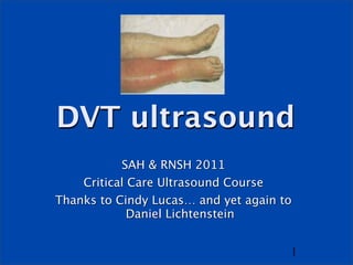
!Relevance of DVT?
5. Here’s the problem
It would be nice to scan But it’s hard!
the below knee veins ! Variable
!Incr sensitivity ! Paired
!Incr accuracy ! Tiny
! Tricky
!Variable ! And most of them don’t
ermbolize
!Paired
! (But some do…)
!Tricky!
!Relevance of DVT?
6. Previous top tip: just
look for above knee
Leave the calves to the
sonographers!
6
7. But Lichtenstein came up with a solution
! Except for the anterior tibials, the below
knee veins travel all in a line, a couple cm
below the interosseous membrane
! together with their arteries: 2 veins for
each artery = 6 vessels, all lined up
! We can see them from the front of the leg!
! Probe between the tibia & fibula
8. Now we have 2
options
1. Just above knee: leave the
calves to the sonographers!
2. Below knee (anterior
approach)
8
10. Probe & preset?
! Ideally linear probe / vein preset
! But curved probe / FAST preset works
too
! Don’t need Doppler
11. Compression US
! Probe in transverse position
! Just squash the vein!
! If it squashes easily & completely, there
is no DVT
! If it doesn’t, there’s a DVT
12.
13.
14. Normal veins
! Completely compressible
! Press hard enough to just indent the
artery
15. Features of DVT
! Gold standard sign: vein not completely
compressible
! You might see thrombus
! Vein might fail to augment on Doppler
26. Which sites can I
compress?
! Internal Jugular V
! Subclavian V
! IVC
! Saphenofemoral confluence (up fem)
! Lower (superf) femoral near adductor hiatus
! Long saphenous V
! Short saphenous V
! Popliteal vein & trifurcation
! Beloe knee veins
27. Which sites should I
compress?
! Up to you
! The more veins you scan, the more sensitive
you are… eg UL veins add 4% in PE
! The fewer you scan, the less irritating it is
! 3-point scan is reasonable
1. Upper femoral (confluence)
2. Lower femoral (near adductor hiatus)
3. Popliteal (irritating if supine) …or …below knee
(weird at first)
28. 1: Groin
! Probe in transverse position
! Start just below inguinal ligament
! ‘Mickey Mouse’ sign
! Femoral A
! Saphenofemoral confluence
! Then compress
36. 3: fem V just above knee
! Adductor hiatus
! Medial to the bone
! Hand behind, presses forward
37.
38.
39.
40.
41.
42.
43. 4: popliteal fossa
! Lie patient on side, or lift leg
! Popliteal vein
! Superficial to popliteal artery
! visualise bone beneath
! follow it to the trifurcation
44.
45.
46.
47.
48.
49. 5: below the knee
! Supine patient
! Probe transverse
! Between tibia & fibula
50.
51.
52.
53. Handy Hints as you go
down the leg
1. Decrease greyscale (dynamic range)
2. Decrease frequency
3. Increase depth as you go
4. Obese: change to curved probe
5. Sit with legs over bed / stand up
6. Valsalva (humming works)
7. Doppler …
54. Pitfalls
! Duplicate venous systems (duplex
popliteal up to 35%)
! Non occlusive thrombus
! LSV, SSV
! Ant tibial veins
! However … ‘90% = 100%’
55. One more time: Handy Hints
! You don’t need Doppler
! You don’t need linear probe
! But you won’t be 100%
! Below-knee isn’t that hard
! Sitting up / standing
! Valsalva (humming works)
56. DVT US: Summary
! Compression US
! Groin
! Just above knee
! Below knee